(Aug 2025)
Cancer Immunology,Immunotherapy : CII 74 9
GPX4 is a key ferroptosis regulator orchestrating T cells and CAR-T-cells sensitivity to ferroptosis
Induction of ferroptosis,an iron-dependent form of regulated cell death,holds promise as a strategy to overcome tumor resistance to conventional therapies and enhance immunotherapy responses. However,while the susceptibility of tumor cells to ferroptosis is extensively studied,limited data exists on the vulnerability of immune cells to disturbed iron balance and lipid peroxidation. Here,we found that T-cell stimulation rewires iron and redox homeostasis and by increasing levels of reactive oxygen species and labile iron promotes lipid peroxidation and T-cells’ ferroptosis. Upon stimulation,we detected changes in the balance of ferroptosis-suppressive proteins,including decrease of GPX4. Subsequently,we identified GPX4 as a master regulator orchestrating T/CAR-T-cells’ sensitivity to ferroptosis and observed that GPX4 inhibitors impair CAR-T cells’ antitumor functions. Our study demonstrated differential GPX4 expression and diverse susceptibility to ferroptosis between CD4⁺ and CD8⁺ T cells. Among analyzed subsets of naïve,central memory (CM),effector memory (EM),and terminally differentiated effector memory (TEMRA),CD8⁺ EM and CD8⁺ TEMRA cells exhibited the highest sensitivity to ferroptosis. We also showed that ferroptosis limited the anti-tumor efficacy of CAR-T cells,while ferroptosis inhibition improved their therapeutic effect,both in vitro and in vivo. Our findings are not only important to understand vulnerabilities of CAR-T cells but may also hold particular significance for their therapeutic development. In this context,future anticancer therapies should be carefully designed to selectively induce the ferroptosis of tumor cells without impeding cytotoxic cells’ antitumor efficacy. Additionally,we postulate that promoting less differentiated phenotype of CAR-T cells should be exploited therapeutically to create CAR-T products characterized by decreased sensitivity to ferroptosis within tumor microenvironment.Supplementary InformationThe online version contains supplementary material available at 10.1007/s00262-025-04133-w.
View Publication
产品类型:
产品号#:
17951
100-0695
17951RF
产品名:
EasySep™人T细胞分选试剂盒
EasySep™人T细胞分选试剂盒
RoboSep™ 人T细胞分选试剂盒
L. Yan et al. (nov 2022)
BMC emergency medicine 22 1 182
Role of CD8+ T cell exhaustion in the progression and prognosis of acute respiratory distress syndrome induced by sepsis: a prospective observational study.
BACKGROUND CD8+ T cells are important for protective immunity against intracellular pathogens. Excessive amounts of antigen and/or inflammatory signals often lead to the gradual deterioration of CD8+ T cell function,a state called exhaustion". However the association between CD8+ T cell exhaustion and acute respiratory distress syndrome (ARDS) has not been studied. This study was conducted to elucidate how CD8+ T cells and inhibitory receptors were related to the clinical prognosis of ARDS. METHODS A prospective observational study in an emergency department enrolled patients who were diagnosed with sepsis-associated ARDS according to the sepsis-3 criteria and Berlin definition. Peripheral blood samples were collected within 24??h post recruitment. CD8+ T cell count proliferation ratio cytokine secretion and the expression of coinhibitory receptors were assayed. RESULTS Sixty-two patients with ARDS met the inclusion criteria. CD8+ T cell counts and proliferation rates were dramatically decreased in non-surviving ARDS patients. Increasing programmed cell death 1 (PD-1) expression on the CD8+ T cell surface was seen in patients with worse organ function while an increasing level of T cell immunoglobulin mucin-3 (Tim-3) was associated with a longer duration of the shock. Kaplan-Meier analysis showed that low CD8+ T cell percentages and increased inhibitory molecule expression were significantly associated with a worse survival rate. CONCLUSIONS CD8+ T cells and coinhibitory receptors are promising independent prognostic markers of sepsis-induced ARDS and increased CD8+ T cell exhaustion is significantly correlated with poor prognosis."
View Publication
产品类型:
产品号#:
17953
17953RF
100-0710
产品名:
EasySep™人CD8+ T细胞分选试剂盒
RoboSep™ 人CD8+ T细胞分选试剂盒
EasySep™人CD8+ T细胞分选试剂盒
He X-S et al. (DEC 2004)
The Journal of clinical investigation 114 12 1812--9
T cell-dependent production of IFN-gamma by NK cells in response to influenza A virus.
The role of human NK cells in viral infections is poorly understood. We used a cytokine flow-cytometry assay to simultaneously investigate the IFN-gamma response of NK and T lymphocytes to influenza A virus (fluA). When PBMCs from fluA-immune adult donors were incubated with fluA,IFN-gamma was produced by both CD56(dim) and CD56(bright) subsets of NK cells,as well as by fluA-specific T cells. Purified NK cells did not produce IFN-gamma in response to fluA,while depletion of T lymphocytes reduced to background levels the fluA-induced IFN-gamma production by NK cells,which indicates that T cells are required for the IFN-gamma response of NK cells. The fluA-induced IFN-gamma production of NK cells was suppressed by anti-IL-2 Ab,while recombinant IL-2 replaced the helper function of T cells for IFN-gamma production by NK cells. This indicates that IL-2 produced by fluA-specific T cells is involved in the T cell-dependent IFN-gamma response of NK cells to fluA. Taken together,these results suggest that at an early stage of recurrent viral infection,NK-mediated innate immunity to the virus is enhanced by preexisting virus-specific T cells.
View Publication
产品类型:
产品号#:
15621
15661
15021
15061
15025
15065
产品名:
RosetteSep™人CD3去除抗体混合物
RosetteSep™人CD3去除抗体混合物
RosetteSep™人T细胞富集抗体混合物
RosetteSep™人T细胞富集抗体混合物
RosetteSep™人NK细胞富集抗体混合物
RosetteSep™人NK细胞富集抗体混合物
Sato K et al. (JAN 2006)
The Journal of experimental medicine 203 1 239--50
TRAIL-expressing T cells induce apoptosis of vascular smooth muscle cells in the atherosclerotic plaque.
Acute coronary syndromes (ACS) are precipitated by a rupture of the atherosclerotic plaque,often at the site of T cell and macrophage infiltration. Here,we show that plaque-infiltrating CD4 T cells effectively kill vascular smooth muscle cells (VSMC). VSMCs sensitive to T cell-mediated killing express the death receptor DR5 (TNF-related apoptosis-inducing ligand [TRAIL] receptor 2),and anti-TRAIL and anti-DR5 antibodies block T cell-mediated apoptosis. CD4 T cells that express TRAIL upon stimulation are expanded in patients with ACS and more effectively induce VSMC apoptosis. Adoptive transfer of plaque-derived CD4 T cells into immunodeficient mice that are engrafted with human atherosclerotic plaque results in apoptosis of VSMCs,which was prevented by coadministration of anti-TRAIL antibody. These data identify that the death pathway is triggered by TRAIL-producing CD4 T cells as a direct mechanism of VSMC apoptosis,a process which may lead to plaque destabilization.
View Publication
产品类型:
产品号#:
15022
15062
产品名:
RosetteSep™人CD4+ T细胞富集抗体混合物
RosetteSep™人CD4+ T细胞富集抗体混合物
Prodeus A et al. (SEP 2017)
JCI insight 2 18
VISTA.COMP - an engineered checkpoint receptor agonist that potently suppresses T cell-mediated immune responses.
V-domain immunoglobulin suppressor of T cell activation (VISTA) is a recently discovered immune checkpoint ligand that functions to suppress T cell activity. The therapeutic potential of activating this immune checkpoint pathway to reduce inflammatory responses remains untapped,largely due to the inability to derive agonists targeting its unknown receptor. A dimeric construct of the IgV domain of VISTA (VISTA-Fc) was shown to suppress the activation of T cells in vitro. However,this effect required its immobilization on a solid surface,suggesting that VISTA-Fc may display limited efficacy as a VISTA-receptor agonist in vivo. Herein,we have designed a stable pentameric VISTA construct (VISTA.COMP) by genetically fusing its IgV domain to the pentamerization domain from the cartilage oligomeric matrix protein (COMP). In contrast to VISTA-Fc,VISTA.COMP does not require immobilization to inhibit the proliferation of CD4+ T cells undergoing polyclonal activation. Furthermore,we show that VISTA.COMP,but not VISTA-Fc,functions as an immunosuppressive agonist in vivo capable of prolonging the survival of skin allografts in a mouse transplant model as well as rescuing mice from acute concanavalin-A-induced hepatitis. Collectively,we believe our data demonstrate that VISTA.COMP is a checkpoint receptor agonist and the first agent to our knowledge targeting the putative VISTA-receptor to suppress T cell-mediated immune responses.
View Publication
产品类型:
产品号#:
19852
19852RF
产品名:
EasySep™小鼠CD4+ T细胞分选试剂盒
RoboSep™ 小鼠CD4+ T细胞分选试剂盒
R. Montandon et al. (JUN 2013)
Proceedings of the National Academy of Sciences of the United States of America 110 24 E2199--208
Innate pro-B-cell progenitors protect against type 1 diabetes by regulating autoimmune effector T cells.
Diverse hematopoietic progenitors,including myeloid populations arising in inflammatory and tumoral conditions and multipotent cells,mobilized by hematopoietic growth factors or emerging during parasitic infections,display tolerogenic properties. Innate immune stimuli confer regulatory functions to various mature B-cell subsets but immature B-cell progenitors endowed with suppressive properties per se or after differentiating into more mature regulatory B cells remain to be characterized. Herein we provide evidence for innate pro-B cells (CpG-proBs) that emerged within the bone marrow both in vitro and in vivo upon Toll-like receptor-9 activation and whose adoptive transfer protected nonobese diabetic mice against type 1 diabetes (T1D). These cells responded to IFN-$\gamma$ released by activated effector T cells (Teffs),by up-regulating their Fas ligand (FasL) expression,which enabled them to kill Teffs through apoptosis. In turn,IFN-$\gamma$ derived from CpG-proBs enhanced IFN-$\gamma$ while dramatically reducing IL-21 production by Teffs. In keeping with the crucial pathogenic role played by IL-21 in T1D,adoptively transferred IFN-$\gamma$-deficient CpG-proBs did not prevent T1D development. Additionally,CpG-proBs matured in vivo into diverse pancreatic and splenic suppressive FasL(high) B-cell subsets. CpG-proBs may become instrumental in cell therapy of autoimmune diseases either on their own or as graft complement in autologous stem cell transplantation.
View Publication
产品类型:
产品号#:
产品名:
I. Koprivica et al. ( 2018)
Frontiers in immunology 9 3130
Ethyl Pyruvate Stimulates Regulatory T Cells and Ameliorates Type 1 Diabetes Development in Mice.
Type 1 diabetes (T1D) is an autoimmune disease in which a strong inflammatory response causes the death of insulin-producing pancreatic beta-cells,while inefficient regulatory mechanisms allow that response to become chronic. Ethyl pyruvate (EP),a stable pyruvate derivate and certified inhibitor of an alarmin-high mobility group box 1 (HMGB1),exerts anti-oxidant and anti-inflammatory properties in animal models of rheumatoid arthritis and encephalomyelitis. To test its therapeutic potential in T1D,EP was administered intraperitoneally to C57BL/6 mice with multiple low-dose streptozotocin (MLDS)-induced T1D. EP treatment decreased T1D incidence,reduced the infiltration of cells into the pancreatic islets and preserved beta-cell function. Apart from reducing HMGB1 expression,EP treatment successfully interfered with the inflammatory response within the local pancreatic lymph nodes and in the pancreas. Its effect was restricted to boosting the regulatory arm of the immune response through up-regulation of tolerogenic dendritic cells (CD11c+CD11b-CD103+) within the pancreatic infiltrates and through the enhancement of regulatory T cell (Treg) levels (CD4+CD25highFoxP3+). These EP-stimulated Treg displayed enhanced suppressive capacity reflected in increased levels of CTLA-4,secreted TGF-beta,and IL-10 and in the more efficient inhibition of effector T cell proliferation compared to Treg from diabetic animals. Higher levels of Treg were a result of increased differentiation and proliferation (Ki67+ cells),but also of the heightened potency for migration due to increased expression of adhesion molecules (CD11a and CD62L) and CXCR3 chemokine receptor. Treg isolated from EP-treated mice had the activated phenotype and T-bet expression more frequently,suggesting that they readily suppressed IFN-gamma-producing cells. The effect of EP on Treg was also reproduced in vitro. Overall,our results show that EP treatment reduced T1D incidence in C57BL/6 mice predominantly by enhancing Treg differentiation,proliferation,their suppressive capacity,and recruitment into the pancreas.
View Publication
产品类型:
产品号#:
17858
17858RF
15621
15661
100-0694
产品名:
EasySep™人CD14正选试剂盒II
RoboSep™ 人CD14正选试剂盒II
RosetteSep™人CD3去除抗体混合物
RosetteSep™人CD3去除抗体混合物
EasySep™人CD14正选试剂盒II
(Jan 2025)
PLOS Pathogens 21 1
Dynamics of tissue repair regulatory T cells and damage in acute Trypanosoma cruzi infection
Tissue-repair regulatory T cells (trTregs) comprise a specialized cell subset essential for tissue homeostasis and repair. While well-studied in sterile injury models,their role in infection-induced tissue damage and antimicrobial immunity is less understood. We investigated trTreg dynamics during acute Trypanosoma cruzi infection,marked by extensive tissue damage and strong CD8+ immunity. Unlike sterile injury models,trTregs significantly declined in secondary lymphoid organs and non-lymphoid target tissues during infection,correlating with systemic and local tissue damage,and downregulation of function-associated genes in skeletal muscle. This decline was linked to decreased systemic IL-33 levels,a key trTreg growth factor,and promoted by the Th1 cytokine IFN-γ. Early recombinant IL-33 treatment increased trTregs,type 2 innate lymphoid cells,and parasite-specific CD8+ cells at specific time points after infection,leading to reduced tissue damage,lower parasite burden,and improved disease outcome. Our findings not only provide novel insights into trTregs during infection but also highlight the potential of optimizing immune balance by modulating trTreg responses to promote tissue repair while maintaining effective pathogen control during infection-induced injury. Author summaryDuring Chagas’ disease,caused by the protozoan Trypanosoma cruzi,severe organ damage is generated by the interplay between the parasite and the immune response. In our investigation,we examined the role of tissue-repair regulatory T cells (trTregs) during the acute phase of T. cruzi infection in mice. Surprisingly,we observed a reduction in trTregs at the peak of tissue damage,contrary to their usual accumulation after injury in other contexts. This decline aligned with decreased levels of interleukin-33,a critical factor for trTreg survival,and was promoted by the effector cytokine IFN-γ. Administering interleukin-33 at early infection times not only boosted trTregs but also expanded other reparative and antiparasitic immune cells. Consequently,these treated mice exhibited reduced damage and lower parasite levels in tissues. Our findings provide new insights into how trTreg function during infection-related injury,paving the way for strategies that balance the immune response to support tissue repair without weakening the body’s ability to fight the infection. This approach could have broader implications for treating infectious diseases and conditions involving tissue damage.
View Publication
产品类型:
产品号#:
19852
19852RF
产品名:
EasySep™小鼠CD4+ T细胞分选试剂盒
RoboSep™ 小鼠CD4+ T细胞分选试剂盒
(Apr 2024)
Nature Communications 15
PD-L1- and IL-4-expressing basophils promote pathogenic accumulation of T follicular helper cells in lupus
Systemic lupus erythematosus (SLE) is an autoimmune disease characterized by anti-nuclear autoantibodies whose production is promoted by autoreactive T follicular helper (TFH) cells. During SLE pathogenesis,basophils accumulate in secondary lymphoid organs (SLO),amplify autoantibody production and disease progression through mechanisms that remain to be defined. Here,we provide evidence for a direct functional relationship between TFH cells and basophils during lupus pathogenesis,both in humans and mice. PD-L1 upregulation on basophils and IL-4 production are associated with TFH and TFH2 cell expansions and with disease activity. Pathogenic TFH cell accumulation,maintenance,and function in SLO were dependent on PD-L1 and IL-4 in basophils,which induced a transcriptional program allowing TFH2 cell differentiation and function. Our study establishes a direct mechanistic link between basophils and TFH cells in SLE that promotes autoantibody production and lupus nephritis. Basophils have been implicated in systemic lupus erythematosus (SLE),as evidenced by the fact that basophil-deficient mice do not develop the disease. Here,the authors demonstrate that PD-L1 and IL-4 expression in basophils promotes the pathogenic accumulation of follicular helper T cells in patients with SLE and murine models.
View Publication
产品类型:
产品号#:
19852
19844
19844RF
19852RF
产品名:
EasySep™小鼠CD4+ T细胞分选试剂盒
EasySep™小鼠Pan-B细胞分选试剂盒
RoboSep™ 小鼠Pan-B细胞分选试剂盒
RoboSep™ 小鼠CD4+ T细胞分选试剂盒
E. Vokali et al. (jan 2020)
Nature communications 11 1 538
Lymphatic endothelial cells prime na\ive CD8+ T cells into memory cells under steady-state conditions."
Lymphatic endothelial cells (LECs) chemoattract na{\{i}}ve T cells and promote their survival in the lymph nodes and can cross-present antigens to na{\"{i}}ve CD8+ T cells to drive their proliferation despite lacking key costimulatory molecules. However the functional consequence of LEC priming of CD8+ T cells is unknown. Here we show that while many proliferating LEC-educated T cells enter early apoptosis the remainders comprise a long-lived memory subset with transcriptional metabolic and phenotypic features of central memory and stem cell-like memory T cells. In vivo these memory cells preferentially home to lymph nodes and display rapid proliferation and effector differentiation following memory recall and can protect mice against a subsequent bacterial infection. These findings introduce a new immunomodulatory role for LECs in directly generating a memory-like subset of quiescent yet antigen-experienced CD8+ T cells that are long-lived and can rapidly differentiate into effector cells upon inflammatory antigenic challenge."""
View Publication
Lee JY et al. (DEC 2009)
Journal of leukocyte biology 86 6 1285--94
Dynamic alterations in chemokine gradients induce transendothelial shuttling of human T cells under physiologic shear conditions.
The active movement of cells from subendothelial compartments into the bloodstream (intravasation) has been recognized for several decades by histologic and physiologic studies,yet the molecular effectors of this process are relatively uncharacterized. For extravasation,studies based predominantly on static transwell assays support a general model,whereby transendothelial migration (TEM) occurs via chemoattraction toward increasing chemokine concentrations. However,this model of chemotaxis cannot readily reconcile how chemokines influence intravasation,as shear forces of blood flow would likely abrogate luminal chemokine gradient(s). Thus,to analyze how T cells integrate perivascular chemokine signals under physiologic flow,we developed a novel transwell-based flow chamber allowing for real-time modulation of chemokine levels above (luminal/apical compartment) and below (abluminal/subendothelial compartment) HUVEC monolayers. We routinely observed human T cell TEM across HUVEC monolayers with the combination of luminal CXCL12 and abluminal CCL5. With increasing concentrations of CXCL12 in the luminal compartment,transmigrated T cells did not undergo retrograde transendothelial migration (retro-TEM). However,when exposedto abluminal CXCL12,transmigrated T cells underwent striking retro-TEM and re-entered the flow stream [corrected]. This CXCL12 fugetactic (chemorepellant) effect was concentration-dependent,augmented by apical flow,blocked by antibodies to integrins,and reduced by AMD3100 in a dose-dependent manner. Moreover,CXCL12-induced retro-TEM was inhibited by PI3K antagonism and cAMP agonism. These findings broaden our understanding of chemokine biology and support a novel paradigm by which temporospatial modulations in subendothelial chemokine display drive cell migration from interstitial compartments into the bloodstream.
View Publication


 EasySep™小鼠TIL(CD45)正选试剂盒





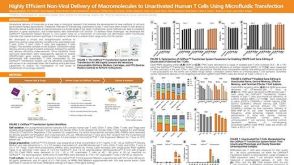 科学海报Highly Efficient Non-Viral Delivery of Macromolecules to Unactivated Human T Cells Using Microfluidic Transfection
 沪公网安备31010102008431号