S. Biswas et al. ( 2022)
Frontiers in immunology 13 875320
Pharmacological Inhibition of MALT1 Ameliorates Autoimmune Pathogenesis and Can Be Uncoupled From Effects on Regulatory T-Cells.
MALT1 forms part of a central signaling node downstream of immunoreceptor tyrosine-based activation motif (ITAM)-containing receptors,across a broad range of immune cell subsets,and regulates NF-$\kappa$B driven transcriptional responses via dual scaffolding-protease activity. Allosteric inhibition of MALT1 activity has demonstrated benefit in animal models of inflammation. However,development of MALT1 inhibitors to treat autoimmune and inflammatory diseases (A&ID) has been hindered by reports linking MALT1 inhibition and genetic loss-of-function to reductions in regulatory T-cell (Treg) numbers and development of auto-inflammatory syndromes. Using an allosteric MALT1 inhibitor,we investigated the consequence of pharmacological inhibition of MALT1 on proinflammatory cells compared to regulatory T-cells. Consistent with its known role in ITAM-driven responses,MALT1 inhibition suppressed proinflammatory cytokine production from activated human T-cells and monocyte-derived macrophages,and attenuated B-cell proliferation. Oral administration of a MALT1 inhibitor reduced disease severity and synovial cytokine production in a rat collagen-induced arthritis model. Interestingly,reduction in splenic Treg numbers was less pronounced in the context of inflammation compared with na{\{i}}ve animals. Additionally in the context of the disease model we observed an uncoupling of anti-inflammatory effects of MALT1 inhibition from Treg reduction with lower systemic concentrations of inhibitor needed to reduce disease severity compared to that required to reduce Treg numbers. MALT1 inhibition did not affect suppressive function of human Tregs in vitro. These data indicate that anti-inflammatory efficacy can be achieved with MALT1 inhibition without impacting the number or function of Tregs further supporting the potential of MALT1 inhibition in the treatment of autoimmune disease."
View Publication
产品类型:
产品号#:
100-0785
19654
19654RF
产品名:
ImmunoCult™ 人CD3/CD28/CD2 T细胞激活剂
EasySep™ Direct 人 PBMC 分选试剂盒
RoboSep™ Direct 人 PBMC 分选试剂盒
J. Wu et al. ( 2022)
Pathology oncology research : POR 28 1610555
Enhancing Natural Killer Cell-Mediated Cancer Immunotherapy by the Biological Macromolecule Nocardia rubra Cell-Wall Skeleton.
The biological macromolecule Nocardia rubra cell-wall skeleton (Nr-CWS) has well-established immune-stimulating and anti-tumor activities. However,the role of Nr-CWS on natural killer (NK) cells remains unclear. Here,we explore the function and related mechanisms of Nr-CWS on NK cells. Using a tumor-bearing model,we show that Nr-CWS has slightly effect on solid tumor. In addition,using a tumor metastasis model,we show that Nr-CWS suppresses the lung metastasis induced by B16F10 melanoma cells in mice,which indicates that Nr-CWS may up-regulate the function of NK cells. Further investigation demonstrated that Nr-CWS can increase the expression of TRAIL and FasL on spleen NK cells from Nr-CWS treated B16F10 tumor metastasis mice. The spleen index and serum levels of TNF-$\alpha$,IFN-$\gamma$,and IL-2 in B16F10 tumor metastasis mice treated with Nr-CWS were significantly increased. In vitro,the studies using purified or sorted NK cells revealed that Nr-CWS increases the expression of CD69,TRAIL,and FasL,decreases the expression of CD27,and enhances NK cell cytotoxicity. The intracellular expression of IFN-$\gamma$,TNF-$\alpha$,perforin (prf),granzyme-B (GrzB),and secreted TNF-$\alpha$,IFN-$\gamma$,IL-6 of the cultured NK cells were significantly increased after treatment with Nr-CWS. Overall,the findings indicate that Nr-CWS could suppress the lung metastasis induced by B16F10 melanoma cells,which may be exerted through its effect on NK cells by promoting NK cell terminal differentiation (CD27lowCD11bhigh),and up-regulating the production of cytokines and cytotoxic molecules.
View Publication
产品类型:
产品号#:
19855
19855RF
产品名:
EasySep™小鼠NK细胞分选试剂盒
RoboSep™ 小鼠NK细胞分选试剂盒
M. Soutto et al. ( 2019)
Nature communications 10 1 3039
Activation of STAT3 signaling is mediated by TFF1 silencing in gastric neoplasia.
TFF1,a secreted protein,plays an essential role in keeping the integrity of gastric mucosa and its barrier function. Loss of TFF1 expression in the TFF1-knockout (KO) mouse leads to a pro-inflammatory phenotype with a cascade of gastric lesions that include low-grade dysplasia,high-grade dysplasia,and adenocarcinomas. In this study,we demonstrate nuclear localization of p-STATY705,with significant overexpression of several STAT3 target genes in gastric glands from the TFF1-KO mice. We also show frequent loss of TFF1 with nuclear localization of STAT3 in human gastric cancers. The reconstitution of TFF1 protein in human gastric cancer cells and 3D gastric glands organoids from TFF1-KO mice abrogates IL6-induced nuclear p-STAT3Y705 expression. Reconstitution of TFF1 inhibits IL6-induced STAT3 transcription activity,suppressing expression of its target genes. TFF1 blocks IL6R$\alpha$-GP130 complex formation through interfering with binding of IL6 to its receptor IL6R$\alpha$. These findings demonstrate a functional role of TFF1 in suppressing gastric tumorigenesis by impeding the IL6-STAT3 pro-inflammatory signaling axis.
View Publication
Human dorsal forebrain organoids show differentiation-state-specific protein secretion
SummaryThe human brain microenvironment undergoes dynamic changes during development,which have been incompletely characterized in in vitro models including neural organoids. Here,we used liquid chromatography-mass spectrometry to investigate proteome and secretome changes in human dorsal forebrain organoids derived from three hiPSC lines at days 20,35,and 50 of differentiation. Proteome and immunohistochemical analysis revealed reduced proliferation and increased differentiation of progenitor cells gradually over time. In contrast,secretome analysis showed distinct characteristics at each timepoint — notably,at day 35,the numbers of cell adhesion molecules,synaptic proteins,and proteases were increased. Taken together,we present a resource describing the dynamic features of a neural organoid proteome and secretome across different genetic backgrounds. We describe the unique niche composition of neural organoids during the period of neurogenesis and suggest that synaptic proteins may play a role in guiding neurogenesis. Graphical abstract Highlights•Proteomic analysis of DFOs on three time points shows neural differentiation•Protein secretion increases during peak neurogenesis at D35 and D50•Cell adhesion molecules,synapse proteins,and metalloproteases are mainly secreted at D35•Extracellular matrix proteins are predominantly secreted at D50 Natural sciences; Biological sciences; Neuroscience; Tissue Engineering
View Publication
产品类型:
产品号#:
100-0276
100-1130
产品名:
mTeSR™ Plus
mTeSR™ Plus
(Feb 2024)
Communications Biology 7
Modular tissue-in-a-CUBE platform to model blood-brain barrier (BBB) and brain interaction
With the advent of increasingly sophisticated organoids,there is growing demand for technology to replicate the interactions between multiple tissues or organs. This is challenging to achieve,however,due to the varying culture conditions of the different cell types that make up each tissue. Current methods often require complicated microfluidic setups,but fragile tissue samples tend not to fare well with rough handling. Furthermore,the more complicated the human system to be replicated,the more difficult the model becomes to operate. Here,we present the development of a multi-tissue chip platform that takes advantage of the modularity and convenient handling ability of a CUBE device. We first developed a blood-brain barrier-in-a-CUBE by layering astrocytes,pericytes,and brain microvascular endothelial cells in the CUBE,and confirmed the expression and function of important tight junction and transporter proteins in the blood-brain barrier model. Then,we demonstrated the application of integrating Tissue-in-a-CUBE with a chip in simulating the in vitro testing of the permeability of a drug through the blood-brain barrier to the brain and its effect on treating the glioblastoma brain cancer model. We anticipate that this platform can be adapted for use with organoids to build complex human systems in vitro by the combination of multiple simple CUBE units. Development of platform to integrate multiple Tissue-in-a-CUBEs in a chip for tissue-tissue interaction,demonstrated by simulating the testing of the permeability and effect of a cancer drug in a BBB-Brain cancer model.
View Publication
产品类型:
产品号#:
100-0483
100-0484
100-0276
100-1130
产品名:
Hausser Scientificᵀᴹ 明线血球计数板
ReLeSR™
mTeSR™ Plus
mTeSR™ Plus
(Jan 2025)
Communications Biology 8
NLRP7 maintains the genomic stability during early human embryogenesis via mediating alternative splicing
Genomic instability is the main cause of abnormal embryo development and abortion. NLRP7 dysfunctions affect embryonic development and lead to Hydatidiform Moles,but the underlying mechanisms remain largely elusive. Here,we show that NLRP7 knockout affects the genetic stability,resulting in increased DNA damage in both human embryonic stem cells and blastoids,making embryonic cells in blastoids more susceptible to apoptosis. Mechanistically,NLRP7 can interact with factors related to alternative splicing and DNA damage response,including DDX39B,PRPF8,THRAP3 and PARP1. Moreover,NLRP7 dysfunction leads to abnormal alternative splicing of genes involved in homologous recombination in human embryonic stem cells,Such as Brca1 and Rad51. These results indicate that NLRP7-mediated Alternative splicing is potentially required for the maintenance of genome integrity during early human embryogenesis. Together,this study uncovers that NLRP7 plays an essential role in the maintenance of genetic stability during early human embryonic development by regulating alternative splicing of homologous recombination-related genes. NLRP7 plays an essential role in the maintenance of genetic stability during early human embryonic development by regulating alternative splicing of homologous recombination-related genes.
View Publication
产品类型:
产品号#:
34411
34415
34421
34425
34450
34460
85850
85857
产品名:
AggreWell™ 400 24孔板,1个
AggreWell™400 24孔板,5个
AggreWell™ 400 6孔板,1个
AggreWell™ 400 6孔板,5个
AggreWell™400 24孔板启动套装
AggreWell™ 400 6孔板启动套装
mTeSR™1
mTeSR™1
(May 2024)
Cell reports 43 6
Macrophages enhance contractile force in iPSC-derived human engineered cardiac tissue
SUMMARY Resident cardiac macrophages are critical mediators of cardiac function. Despite their known importance to cardiac electrophysiology and tissue maintenance,there are currently no stem-cell-derived models of human engineered cardiac tissues (hECTs) that include resident macrophages. In this study,we made an induced pluripotent stem cell (iPSC)-derived hECT model with a resident population of macrophages (iM0) to better recapitulate the native myocardium and characterized their impact on tissue function. Macrophage retention within the hECTs was confirmed via immunofluorescence after 28 days of cultivation. The inclusion of iM0s significantly impacted hECT function,increasing contractile force production. A potential mechanism underlying these changes was revealed by the interrogation of calcium signaling,which demonstrated the modulation of ?-adrenergic signaling in +iM0 hECTs. Collectively,these findings demonstrate that macrophages significantly enhance cardiac function in iPSC-derived hECT models,emphasizing the need to further explore their contributions not only in healthy hECT models but also in the contexts of disease and injury. In brief Lock and Graney et al. develop a human engineered cardiac tissue with an incorporated iPSC-derived macrophage population to better mimic the complex cell landscape of the native myocardium. Macrophage inclusion leads to increased contractile function of the tissue,which is attributed to macrophage stimulation of the cardiomyocyte ?-adrenergic signaling pathway. Graphical Abstract
View Publication
产品类型:
产品号#:
05310
100-0276
100-1130
产品名:
STEMdiff™ 造血试剂盒
mTeSR™ Plus
mTeSR™ Plus
(Jun 2025)
Cellular and Molecular Life Sciences: CMLS 82 1
The ADCY1-mediated cAMP signaling pathway mediates functional effects of montelukast treatment in brain organoids
Montelukast (MTK) is a drug widely used for treating allergic rhinitis and asthma. However,severe neuropsychiatric adverse events related to MTK have been reported,with limited understanding of the underlying mechanisms. Here we leveraged human forebrain organoids (hFOs) and showed that MTK exposure in hFOs downregulated the expression of genes associated with multiple neuronal functions and neuropsychiatric disorders. The following integrative analysis highlighted adenylate cyclase 1 (ADCY1),a main regulator of the cAMP signaling pathway,as a hub gene mediating the functional effects of MTK exposure. We also showed that MTK exposure resulted in a reduction of cAMP and neuroactivities,and caused neural maturation defects. These cellular phenotypes could be recapitulated by treating hFOs with ST034307,a selective ADCY1 inhibitor,or partially rescued by ADCY1 overexpression in hFOs. Together,this study underscored that MTK exposure caused neuropsychiatric effects through inhibiting the ADCY1-mediated cAMP signaling pathway.Supplementary InformationThe online version contains supplementary material available at 10.1007/s00018-025-05764-z.
View Publication
Laminin-associated integrins mediate Diffuse Intrinsic Pontine Glioma infiltration and therapy response within a neural assembloid model
Diffuse Intrinsic Pontine Glioma (DIPG) is a highly aggressive and fatal pediatric brain cancer. One pre-requisite for tumor cells to infiltrate is adhesion to extracellular matrix (ECM) components. However,it remains largely unknown which ECM proteins are critical in enabling DIPG adhesion and migration and which integrin receptors mediate these processes. Here,we identify laminin as a key ECM protein that supports robust DIPG cell adhesion and migration. To study DIPG infiltration,we developed a DIPG-neural assembloid model,which is composed of a DIPG spheroid fused to a human induced pluripotent stem cell-derived neural organoid. Using this assembloid model,we demonstrate that knockdown of laminin-associated integrins significantly impedes DIPG infiltration. Moreover,laminin-associated integrin knockdown improves DIPG response to radiation and HDAC inhibitor treatment within the DIPG-neural assembloids. These findings reveal the critical role of laminin-associated integrins in mediating DIPG progression and drug response. The results also provide evidence that disrupting integrin receptors may offer a novel therapeutic strategy to enhance DIPG treatment outcomes. Finally,these results establish DIPG-neural assembloid models as a powerful tool to study DIPG disease progression and enable drug discovery.Supplementary InformationThe online version contains supplementary material available at 10.1186/s40478-024-01765-4.
View Publication
产品类型:
产品号#:
34811
34815
34821
34825
34850
34860
100-0276
100-1130
产品名:
AggreWell™ 800 24孔板,1个
AggreWell™ 800 24孔板,5个
AggreWell™ 800 6孔板,1个
AggreWell™ 800 6孔板,5个
AggreWell™ 800 24孔板启动套装
AggreWell™ 800 6孔板启动套装
mTeSR™ Plus
mTeSR™ Plus
(Mar 2025)
Journal of Neuroinflammation 22
Bystander neuronal progenitors in forebrain organoids promote protective antiviral responses
Neurotropic viruses are the most common cause of infectious encephalitis and highly target neurons for infection. Our understanding of the intrinsic capacity of neuronal innate immune responses to mediate protective antiviral responses remains incomplete. Here,we evaluated the role of intercellular crosstalk in mediating intrinsic neuronal immunity and its contribution to limiting viral infection. We found that in the absence of viral antagonism,neurons transcriptionally induce robust interferon signaling and can effectively signal to uninfected bystander neurons. Yet,in two-dimensional cultures,this dynamic response did not restrict viral spread. Interestingly,this differed in the context of viral infection in three-dimensional forebrain organoids with complex neuronal subtypes and cellular organization,where we observed protective capacity. We showed antiviral crosstalk between infected neurons and bystander neural progenitors is mediated by type I interferon signaling. Using spatial transcriptomics,we then uncovered regions containing bystander neural progenitors that expressed distinct antiviral genes,revealing critical underpinnings of protective antiviral responses among neuronal subtypes. These findings underscore the importance of interneuronal communication in protective antiviral immunity in the brain and implicate key contributions to protective antiviral signaling.Supplementary InformationThe online version contains supplementary material available at 10.1186/s12974-025-03381-y.
View Publication


 EasySep™小鼠TIL(CD45)正选试剂盒






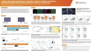 科学海报A Novel mRNA-LNP Platform to Rapidly Generate Functional Forebrain Neurons from hPSCs Using NGN2
 沪公网安备31010102008431号