M. Pardons et al. (nov 2019)
Cell reports 29 9 2783--2795.e5
Latency-Reversing Agents Induce Differential Responses in Distinct Memory CD4 T Cell Subsets in Individuals on Antiretroviral Therapy.
Latent proviruses persist in central (TCM),transitional (TTM),and effector (TEM) memory cells. We measured the levels of cellular factors involved in HIV gene expression in these subsets. The highest levels of acetylated H4,active nuclear factor $\kappa$B (NF-$\kappa$B),and active positive transcription elongation factor b (P-TEFb) were measured in TEM,TCM,and TTM cells,respectively. Vorinostat and romidepsin display opposite abilities to induce H4 acetylation across subsets. Protein kinase C (PKC) agonists are more efficient at inducing NF-$\kappa$B phosphorylation in TCM cells but more potent at activating PTEF-b in the TEM subset. We selected the most efficient latency-reversing agents (LRAs) and measured their ability to reverse latency in each subset. While ingenol alone has modest activities in the three subsets,its combination with a histone deacetylase inhibitor (HDACi) dramatically increases latency reversal in TCM cells. Altogether,these results indicate that cellular HIV reservoirs are differentially responsive to common LRAs and suggest that combination of compounds will be required to achieve latency reversal in all subsets.
View Publication
产品类型:
产品号#:
17955
17955RF
100-0960
产品名:
EasySep™人NK细胞分选试剂盒
RoboSep™ 人NK细胞分选试剂盒
EasySep™人NK细胞分离试剂盒
N. H. Overgaard et al. (JUN 2018)
Frontiers in immunology 9 1301
Genetically Induced Tumors in the Oncopig Model Invoke an Antitumor Immune Response Dominated by Cytotoxic CD8 T Cells and Differentiated T Cells Alongside a Regulatory Response Mediated by FOXP3+ T Cells and Immunoregulatory Molecules
In recent years,immunotherapy has shown considerable promise in the management of several malignancies. However,the majority of preclinical studies have been conducted in rodents,the results of which often translate poorly to patients given the substantial differences between murine and human immunology. As the porcine immune system is far more analogous to that of humans,pigs may serve as a supplementary preclinical model for future testing of such therapies. We have generated the genetically modified Oncopig with inducible tumor formation resulting from concomitant KRAS(G12D) and TP53(R167H) mutations under control of an adenoviral vector Cre-recombinase (AdCre). The objective of this study was to characterize the tumor microenvironment in this novel animal model with respect to T-cell responses in particular and to elucidate the potential use of Oncopigs for future preclinical testing of cancer immunotherapies. In this study,we observed pronounced intratumoral T-cell infiltration with a strong CD8$\beta$(+) predominance alongside a representation of highly differentiated $\gamma$$\delta$ T cells. The infiltrating CD8$\beta$(+) T cells displayed increased expression of the cytotoxic marker perforin when compared with the peripheral T-cell pool. Similarly,there was robust granzyme B staining localizing to the tumors; affirming the presence of cytotoxic immune cells within the tumor. In parallel with this antitumor immune response,the tumors displayed enrichment in FOXP3-expressing T cells and increased gene expression of indoleamine 2,3-dioxygenase 1 (IDO1),cytotoxic T-lymphocyte-associated protein 4 (CTLA4),and programmed death-ligand 1 (PDL1). Finally,we investigated the Oncopig immune system in mediating antitumor immunity. We observed pronounced killing of autologous tumor cells,which demonstrates the propensity of the Oncopig immune system to recognize and mount a cytotoxic response against tumor cells. Together,these findings suggest innate and adaptive recognition of the induced tumors with a concomitant in vivo suppression of T-cell effector functions. Combined,the data support that the Oncopig may serve as a valuable model for future preclinical testing of immunotherapies aimed at reactivating tumor-directed cytotoxicity in vivo.
View Publication
产品类型:
产品号#:
85415
85420
85450
85460
86415
86420
86450
86460
产品名:
SepMate™-15 (IVD)
SepMate™-15 (IVD)
SepMate™-50 (IVD)
SepMate™-50 (IVD)
SepMate™-15 (RUO)
SepMate™-15 (RUO)
SepMate™-50 (RUO)
SepMate™-50 (RUO)
Ortiz-Lazareno PC et al. ( 2008)
Immunology 124 4 534--541
MG132 proteasome inhibitor modulates proinflammatory cytokines production and expression of their receptors in U937 cells: involvement of nuclear factor-kappaB and activator protein-1.
In response to inflammatory stimuli,monocytes/macrophages secrete greater quantities of the proinflammatory cytokines tumour necrosis factor-alpha (TNF-alpha),interleukin-1beta (IL-1beta) and IL-6. The inflammatory process and the innate immune response are related to the activation of several transcription factors,such as nuclear factor kappaB (NF-kappaB) and activator protein 1 (AP-1). The proteasome is a multimeric protease complex,which plays a vital role in several cellular functions,including the regulation of transcription factors like NF-kappaB. In this study,we used the human monocyte cell line U937 stimulated with lipopolysaccharide (LPS) and phorbol 12-myristate 13-acetate (PMA) as a model to investigate the in vitro effects of MG132,a proteasome inhibitor,on the release of TNF-alpha,IL-1beta and IL-6 and on the expression of their membrane and soluble receptors TNF-R1,IL-1R1 and IL-6R. We also analysed the effects of MG132 on the activation of NF-kappaB and AP-1 and on the IkappaB molecule. MG132 significantly inhibited the secretion of those proinflammatory cytokines. MG132 increased the release of the soluble receptors TNF-R1 and IL-1R1 from U937 cells and decreased their cell-surface expression. MG132 also increased IL-6R cell-surface expression and decreased its release. Proteasome inhibition also led to an increase in LPS+PMA-induced AP-1 activation and the attenuation of LPS+PMA-induced IkappaB degradation,resulting in the abolition of NF-kappaB activation. Our experiments strongly suggest that the proteasome is an important factor in the regulation of proinflammatory cytokines and their receptors.
View Publication
产品类型:
产品号#:
73262
73264
产品名:
(S) -MG132
(S) -MG132
Benziane B et al. ( 2009)
American journal of physiology. Cell physiology 297 6 C1554--66
AMP-activated protein kinase activator A-769662 is an inhibitor of the Na(+)-K(+)-ATPase.
Muscle contraction and metabolic stress are potent activators of AMP-activated protein kinase (AMPK). AMPK restores energy balance by activating processes that produce energy while inhibiting those that consume energy. The role of AMPK in the regulation of active ion transport is unclear. Our aim was to determine the effect of the AMPK activator A-769662 on Na(+)-K(+)-ATPase function in skeletal muscle cells. Short-term incubation of differentiated rat L6 myotubes with 100 microM A-769662 increased AMPK and acetyl-CoA carboxylase (ACC) phosphorylation in parallel with decreased Na(+)-K(+)-ATPase alpha(1)-subunit abundance at the plasma membrane and ouabain-sensitive (86)Rb(+) uptake. Notably,the effect of A-769662 on Na(+)-K(+)-ATPase was similar in muscle cells that do not express AMPK alpha(1)- and alpha(2)-catalytic subunits. A-769662 directly inhibits the alpha(1)-isoform of the Na(+)-K(+)-ATPase,purified from rat and human kidney cells in vitro with IC(50) 57 microM and 220 microM,respectively. Inhibition of the Na(+)-K(+)-ATPase by 100 microM ouabain decreases sodium pump activity and cell surface abundance,similar to the effect of A-769662,without affecting AMPK and ACC phosphorylation. In conclusion,the AMPK activator A-769662 inhibits Na(+)-K(+)-ATPase activity and decreases the sodium pump cell surface abundance in L6 skeletal muscle cells. The effect of A-769662 on sodium pump is due to direct inhibition of the Na(+)-K(+)-ATPase activity,rather than AMPK activation. This AMPK-independent effect on Na(+)-K(+)-ATPase calls into question the use of A-769662 as a specific AMPK activator for metabolic studies.
View Publication
产品类型:
产品号#:
72922
72924
产品名:
A769662
A769662, 50 mg
Heinzel K et al. (JAN 2007)
Journal of immunology (Baltimore,Md. : 1950) 178 2 858--68
Bone marrow-derived hemopoietic precursors commit to the T cell lineage only after arrival in the thymic microenvironment.
T lymphocytes develop in the thymus from hemopoietic precursors that commit to the T cell lineage under the influence of Notch signals. In this study,we show by single cell analyses that the most immature hemopoietic precursors in the adult mouse thymus are uncommitted and specify to the T cell lineage only after their arrival in the thymus. These precursors express high levels of surface Notch receptors and rapidly lose B cell potential upon the provision of Notch signals. Using a novel culture system with complexed,soluble Notch ligands that allows the titration of T cell lineage commitment,we find that these precursors are highly sensitive to both Delta and Jagged ligands. In contrast,their phenotypical and functional counterparts in the bone marrow are resistant to Notch signals that efficiently induce T cell lineage commitment in thymic precursors. Mechanistically,this is not due to differences in receptor expression,because early T lineage precursors,bone marrow lineage marker-negative,Sca-1-positive,c-Kit-positive and common lymphoid progenitor cells,express comparable amounts of surface Notch receptors. Our data demonstrate that the sensitivity to Notch-mediated T lineage commitment is stage-dependent and argue against the bone marrow as the site of T cell lineage commitment.
View Publication
产品类型:
产品号#:
03231
产品名:
MethoCult™ M3231
Lehnertz B et al. (MAY 2010)
The Journal of experimental medicine 207 5 915--22
Activating and inhibitory functions for the histone lysine methyltransferase G9a in T helper cell differentiation and function.
Accumulating evidence suggests that the regulation of gene expression by histone lysine methylation is crucial for several biological processes. The histone lysine methyltransferase G9a is responsible for the majority of dimethylation of histone H3 at lysine 9 (H3K9me2) and is required for the efficient repression of developmentally regulated genes during embryonic stem cell differentiation. However,whether G9a plays a similar role in adult cells is still unclear. We identify a critical role for G9a in CD4(+) T helper (Th) cell differentiation and function. G9a-deficient Th cells are specifically impaired in their induction of Th2 lineage-specific cytokines IL-4,IL-5,and IL-13 and fail to protect against infection with the intestinal helminth Trichuris muris. Furthermore,G9a-deficient Th cells are characterised by the increased expression of IL-17A,which is associated with a loss of H3K9me2 at the Il17a locus. Collectively,our results establish unpredicted and complex roles for G9a in regulating gene expression during lineage commitment in adult CD4(+) T cells.
View Publication
产品类型:
产品号#:
21000
20119
20155
19752
19752RF
产品名:
RoboSep™- S
RoboSep™ 吸头组件抛光剂
RoboSep™分选管套装(9个塑料管)
J. Ingels et al. (Apr 2024)
Cell Reports Medicine 5 5
Neoantigen-targeted dendritic cell vaccination in lung cancer patients induces long-lived T cells exhibiting the full differentiation spectrum
Non-small cell lung cancer (NSCLC) is known for high relapse rates despite resection in early stages. Here,we present the results of a phase I clinical trial in which a dendritic cell (DC) vaccine targeting patient-individual neoantigens is evaluated in patients with resected NSCLC. Vaccine manufacturing is feasible in six of 10 enrolled patients. Toxicity is limited to grade 1–2 adverse events. Systemic T cell responses are observed in five out of six vaccinated patients,with T cell responses remaining detectable up to 19 months post vaccination. Single-cell analysis indicates that the responsive T cell population is polyclonal and exhibits the near-entire spectrum of T cell differentiation states,including a naive-like state,but excluding exhausted cell states. Three of six vaccinated patients experience disease recurrence during the follow-up period of 2 years. Collectively,these data support the feasibility,safety,and immunogenicity of this treatment in resected NSCLC.
View Publication
产品类型:
产品号#:
100-0785
10970
10990
产品名:
ImmunoCult™ 人CD3/CD28/CD2 T细胞激活剂
ImmunoCult™ 人CD3/CD28/CD2 T细胞激活剂
ImmunoCult™ 人CD3/CD28/CD2 T细胞激活剂
J. Robert et al. (May 2025)
Nature Communications 16
Expression of an interleukin-2 partial agonist enhances regulatory T cell persistence and efficacy in mouse autoimmune models
Regulatory T (Treg)-based cell therapy holds promise for autoimmune and inflammatory diseases,yet challenges remain regarding the functional stability and persistence of transferred Tregs. Here we engineer Tregs to express a partial agonist form of IL-2 (IL-2pa) to enhance persistence while avoiding toxicity from excessive signaling. Mouse Tregs expressing wild-type IL-2 (Tregs-IL2wt) have only a transient growth advantage,limited by toxicity from likely excessive signaling. By contrast,mouse Tregs-IL2pa exhibit sustained expansion,long-term survival in immunocompetent mice for over a year,and bystander expansion of endogenous Tregs. Tregs-IL2pa maintain a stable activated phenotype,Treg-specific demethylation,and a diverse TCR repertoire. In vivo,prophylactic transfer of Tregs-IL2pa ameliorates multi-organ autoimmunity in a Treg depletion-induced mouse autoimmune model. Lastly,compared with control Treg,human Tregs-IL2pa show enhanced survival in the IL-2-depleted environment of immune-deficient mice and improved control of xenogeneic graft-versus-host disease. Our results thus show that IL-2pa self-sufficiency enhances the stability,durability and efficacy of Treg therapies in preclinical settings. Subject terms: Cell delivery,Regulatory T cells,Autoimmune diseases,Interleukins
View Publication
产品类型:
产品号#:
100-0784
10971
10991
产品名:
ImmunoCult™ 人CD3/CD28 T细胞激活剂
ImmunoCult™ 人CD3/CD28 T细胞激活剂
ImmunoCult™ 人CD3/CD28 T细胞激活剂
A. Xu et al. (jan 2022)
Journal of immunology (Baltimore,Md. : 1950) 208 1 155--168
Prosurvival IL-7-Stimulated Weak Strength of mTORC1-S6K Controls T Cell Memory via Transcriptional FOXO1-TCF1-Id3 and Metabolic AMPK$\alpha$1-ULK1-ATG7 Pathways.
CD8+ memory T (TM) cells play a critical role in immune defense against infection. Two common $\gamma$-chain family cytokines,IL-2 and IL-7,although triggering the same mTORC1-S6K pathway,distinctly induce effector T (TE) cells and TM cells,respectively,but the underlying mechanism(s) remains elusive. In this study,we generated IL-7R-/and AMPK$\alpha$1-knockout (KO)/OTI mice. By using genetic and pharmaceutical tools,we demonstrate that IL-7 deficiency represses expression of FOXO1,TCF1,p-AMPK$\alpha$1 (T172),and p-ULK1 (S555) and abolishes T cell memory differentiation in IL-7R KO T cells after Listeria monocytogenesis rLmOVA infection. IL-2- and IL-7-stimulated strong and weak S6K (IL-2/S6Kstrong and IL-7/S6Kweak) signals control short-lived IL-7R-CD62L-KLRG1+ TE and long-term IL-7R+CD62L+KLRG1- TM cell formations,respectively. To assess underlying molecular pathway(s),we performed flow cytometry,Western blotting,confocal microscopy,and Seahorse assay analyses by using the IL-7/S6Kweak-stimulated TM (IL-7/TM) and the control IL-2/S6Kstrong-stimulated TE (IL-2/TE) cells. We determine that the IL-7/S6Kweak signal activates transcriptional FOXO1,TCF1,and Id3 and metabolic p-AMPK$\alpha$1,p-ULK1,and ATG7 molecules in IL-7/TM cells. IL-7/TM cells upregulate IL-7R and CD62L,promote mitochondria biogenesis and fatty acid oxidation metabolism,and show long-term cell survival and functional recall responses. Interestingly,AMPK$\alpha$1 deficiency abolishes the AMPK$\alpha$1 but maintains the FOXO1 pathway and induces a metabolic switch from fatty acid oxidation to glycolysis in AMPK$\alpha$1 KO IL-7/TM cells,leading to loss of cell survival and recall responses. Taken together,our data demonstrate that IL-7-stimulated weak strength of mTORC1-S6K signaling controls T cell memory via activation of transcriptional FOXO1-TCF1-Id3 and metabolic AMPK$\alpha$1-ULK1-ATG7 pathways. This (to our knowledge) novel finding provides a new mechanism for a distinct IL-2/IL-7 stimulation model in T cell memory and greatly impacts vaccine development.
View Publication
S. S. Leung et al. (sep 2022)
Diabetes 71 9 1994--2008
Soluble RAGE Prevents Type 1 Diabetes Expanding Functional Regulatory T Cells.
Type 1 diabetes is an autoimmune disease with no cure,where clinical translation of promising therapeutics has been hampered by the reproducibility crisis. Here,short-term administration of an antagonist to the receptor for advanced glycation end products (sRAGE) protected against murine diabetes at two independent research centers. Treatment with sRAGE increased regulatory T cells (Tregs) within the islets,pancreatic lymph nodes,and spleen,increasing islet insulin expression and function. Diabetes protection was abrogated by Treg depletion and shown to be dependent on antagonizing RAGE with use of knockout mice. Human Tregs treated with a RAGE ligand downregulated genes for suppression,migration,and Treg homeostasis (FOXP3,IL7R,TIGIT,JAK1,STAT3,STAT5b,CCR4). Loss of suppressive function was reversed by sRAGE,where Tregs increased proliferation and suppressed conventional T-cell division,confirming that sRAGE expands functional human Tregs. These results highlight sRAGE as an attractive treatment to prevent diabetes,showing efficacy and reproducibility at multiple research centers and in human T cells.
View Publication
产品类型:
产品号#:
17555
18000
17555RF
产品名:
EasySep™人初始CD4+ T细胞分选试剂盒II
EasySep™磁极
RoboSep™ 人初始CD4+ T细胞分选试剂盒II
E. Trolio et al. (Mar 2026)
STAR Protocols 7 2
Protocol for immunomagnetic enrichment of T cells from complex murine tissues
SummaryT cells are the central effectors and regulators of the adaptive immune response. This protocol provides a step-by-step approach for isolating and enriching total T cells by negative selection from complex murine tissues,including bone marrow (BM),liver,heart,and kidneys. We describe steps for tissue harvesting,preparation of single-cell suspensions,and immunomagnetic enrichment. We then outline procedures for flow cytometric assessment of cell purity and viability. This protocol enables efficient recovery of high-quality T cells for reliable downstream analyses. Graphical abstract Highlights•Isolation of leukocytes from murine BM,liver,heart and kidneys•Non-enzymatic dissociation of kidney and heart tissue•Protocol for immunomagnetic enrichment of T cells•Flow cytometry analysis of T cell purity and viability Publisher’s note: Undertaking any experimental protocol requires adherence to local institutional guidelines for laboratory safety and ethics. T cells are the central effectors and regulators of the adaptive immune response. This protocol provides a step-by-step approach for isolating and enriching total T cells by negative selection from complex murine tissues,including bone marrow (BM),liver,heart,and kidneys. We describe steps for tissue harvesting,preparation of single-cell suspensions,and immunomagnetic enrichment. We then outline procedures for flow cytometric assessment of cell purity and viability. This protocol enables efficient recovery of high-quality T cells for reliable downstream analyses.
View Publication


 EasySep™小鼠TIL(CD45)正选试剂盒





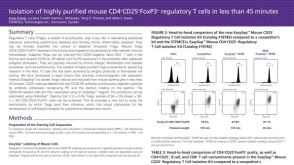 科学海报Isolation of Highly Purified Mouse CD4+CD25+Foxp3+ Regulatory T Cells in Less
 沪公网安备31010102008431号