Voo KS et al. (JUL 2014)
The Journal of Immunology 193 2 627--34
Targeting of TLRs inhibits CD4+ regulatory T cell function and activates lymphocytes in human peripheral blood mononuclear cells.
Accumulating evidence suggests elements within tumors induce exhaustion of effector T cells and infiltration of immunosuppressive regulatory T cells (Tregs),thus preventing the development of durable antitumor immunity. Therefore,the discovery of agents that simultaneously block Treg suppressive function and reinvigorate effector function of lymphocytes is key to the development of effective cancer immunotherapy. Previous studies have shown that TLR ligands (TLRLs) could modulate the function of these T cell targets; however,those studies relied on cell-free or accessory cell-based assay systems that do not accurately reflect in vivo responses. In contrast,we used a human PBMC-based proliferation assay system to simultaneously monitor the effect of TLRLs on T cells (CD4(+),CD8(+),Tregs),B cells,and NK cells,which gave different and even conflicting results. We found that the TLR7/8L:CL097 could simultaneously activate CD8(+) T cells,B cells,and NK cells plus block Treg suppression of T cells and B cells. The TLRLs TLR1/2L:Pam3CSK4,TLR5L:flagellin,TLR4L:LPS,and TLR8/7L:CL075 also blocked Treg suppression of CD4(+) or CD8(+) T cell proliferation,but not B cell proliferation. Besides CL097,TLR2L:PGN,CL075,and TLR9L:CpG-A,CpG-B,and CpG-C) were strong activators of NK cells. Importantly,we found that Pam3CSK4 could: 1) activate CD4(+) T cell proliferation,2) inhibit the expansion of IL-10(+) naturally occurring FOXP3(+) Tregs and induction of IL-10(+) CD4(+) Tregs (IL-10-producing type 1 Treg),and 3) block naturally occurring FOXP3(+) Tregs suppressive function. Our results suggest these agents could serve as adjuvants to enhance the efficacy of current immunotherapeutic strategies in cancer patients.
View Publication
产品类型:
产品号#:
19052
19052RF
19055
19055RF
产品名:
EasySep™人CD4+ T细胞富集试剂盒
RoboSep™ 人CD4+ T细胞富集试剂盒含滤芯吸头
EasySep™人NK细胞富集试剂盒
RoboSep™ 人NK细胞富集试剂盒含滤芯吸头
Garcia-Bates TM et al. (MAR 2016)
Journal of immunology (Baltimore,Md. : 1950) 196 6 2870--8
Enhanced Cytotoxic CD8 T Cell Priming Using Dendritic Cell-Expressing Human Papillomavirus-16 E6/E7-p16INK4 Fusion Protein with Sequenced Anti-Programmed Death-1.
The incidence of human papillomavirus (HPV)-related head and neck squamous cell carcinoma has increased in recent decades,though HPV prevention vaccines may reduce this rise in the future. HPV-related cancers express the viral oncoproteins E6 and E7. The latter inactivates the tumor suppressor protein retinoblastoma (Rb),which leads to the overexpression of p16(INK4) protein,providing unique Ags for therapeutic HPV-specific cancer vaccination. We developed potential adenoviral vaccines that express a fusion protein of HPV-16 E6 and E7 (Ad.E6E7) alone or fused with p16 (Ad.E6E7p16) and also encoding an anti-programmed death (PD)-1 Ab. Human monocyte-derived dendritic cells (DC) transduced with Ad.E6E7 or Ad.E6E7p16 with or without Ad.αPD1 were used to activate autologous CD8 CTL in vitro. CTL responses were tested against naturally HPV-infected head and neck squamous cell carcinoma cells using IFN-γ ELISPOT and [(51)Cr]release assay. Surprisingly,stimulation and antitumor activity of CTL were increased after incubation with Ad.E6E7p16-transduced DC (DC.E6E7p16) compared with Ad.E6E7 (DC.E6E7),a result that may be due to an effect of p16 on cyclin-dependent kinase 4 levels and IL-12 secretion by DC. Moreover,the beneficial effect was most prominent when anti-PD-1 was introduced during the second round of stimulation (after initial priming). These data suggest that careful sequencing of Ad.E6E7.p16 with Ad.αPD1 could improve antitumor immunity against HPV-related tumors and that p16 may enhance the immunogenicity of DC,through cyclin-dependent pathways,Th1 cytokine secretion,and by adding a nonviral Ag highly overexpressed in HPV-induced cancers.
View Publication
产品类型:
产品号#:
18058
18058RF
19158
19158RF
19053
19053RF
产品名:
EasySep™人CD8+ T细胞富集试剂盒
RoboSep™ 人CD8+ T细胞富集试剂盒含滤芯吸头
(Jun 2025)
Nature Communications 16 Suppl 2
Iron deficiency causes aspartate-sensitive dysfunction in CD8+ T cells
Iron is an irreplaceable co-factor for metabolism. Iron deficiency affects >1 billion people and decreased iron availability impairs immunity. Nevertheless,how iron deprivation impacts immune cell function remains poorly characterised. We interrogate how physiologically low iron availability affects CD8+ T cell metabolism and function,using multi-omic and metabolic labelling approaches. Iron limitation does not substantially alter initial post-activation increases in cell size and CD25 upregulation. However,low iron profoundly stalls proliferation (without influencing cell viability),alters histone methylation status,gene expression,and disrupts mitochondrial membrane potential. Glucose and glutamine metabolism in the TCA cycle is limited and partially reverses to a reductive trajectory. Previous studies identified mitochondria-derived aspartate as crucial for proliferation of transformed cells. Despite aberrant TCA cycling,aspartate is increased in stalled iron deficient CD8+ T cells but is not utilised for nucleotide synthesis,likely due to trapping within depolarised mitochondria. Exogenous aspartate markedly rescues expansion and some functions of severely iron-deficient CD8+ T cells. Overall,iron scarcity creates a mitochondrial-located metabolic bottleneck,which is bypassed by supplying inhibited biochemical processes with aspartate. These findings reveal molecular consequences of iron deficiency for CD8+ T cell function,providing mechanistic insight into the basis for immune impairment during iron deficiency. Iron has been shown to be necessary for the activation and differentiation of CD8+ T cells. Here the authors investigate changes in CD8+ T cell metabolism in iron limiting conditions and find that aspartate is increased yet downstream nucleotide synthesis is suppressed and addition of exogenous aspartate partially rescues T cell function.
View Publication
产品类型:
产品号#:
18000
18102
18103
20144
产品名:
EasySep™磁极
EasyPlate™ EasySep™磁极
EasyEights™EasySep™磁极
EasySep™缓冲液
W. Wang et al. (may 2019)
Nature 569 7755 270--274
CD8+ T cells regulate tumour ferroptosis during cancer immunotherapy.
Cancer immunotherapy restores or enhances the effector function of CD8+ T cells in the tumour microenvironment1,2. CD8+ T cells activated by cancer immunotherapy clear tumours mainly by inducing cell death through perforin-granzyme and Fas-Fas ligand pathways3,4. Ferroptosis is a form of cell death that differs from apoptosis and results from iron-dependent accumulation of lipid peroxide5,6. Although it has been investigated in vitro7,8,there is emerging evidence that ferroptosis might be implicated in a variety of pathological scenarios9,10. It is unclear whether,and how,ferroptosis is involved in T cell immunity and cancer immunotherapy. Here we show that immunotherapy-activated CD8+ T cells enhance ferroptosis-specific lipid peroxidation in tumour cells,and that increased ferroptosis contributes to the anti-tumour efficacy of immunotherapy. Mechanistically,interferon gamma (IFNgamma) released from CD8+ T cells downregulates the expression of SLC3A2 and SLC7A11,two subunits of the glutamate-cystine antiporter system xc-,impairs the uptake of cystine by tumour cells,and as a consequence,promotes tumour cell lipid peroxidation and ferroptosis. In mouse models,depletion of cystine or cysteine by cyst(e)inase (an engineered enzyme that degrades both cystine and cysteine) in combination with checkpoint blockade synergistically enhanced T cell-mediated anti-tumour immunity and induced ferroptosis in tumour cells. Expression of system xc- was negatively associated,in cancer patients,with CD8+ T cell signature,IFNgamma expression,and patient outcome. Analyses of human transcriptomes before and during nivolumab therapy revealed that clinical benefits correlate with reduced expression of SLC3A2 and increased IFNgamma and CD8. Thus,T cell-promoted tumour ferroptosis is an anti-tumour mechanism,and targeting this pathway in combination with checkpoint blockade is a potential therapeutic approach.
View Publication
产品类型:
产品号#:
17953
17953RF
19853
19853RF
100-0710
产品名:
EasySep™人CD8+ T细胞分选试剂盒
RoboSep™ 人CD8+ T细胞分选试剂盒
EasySep™小鼠CD8+ T细胞分选试剂盒
RoboSep™ 小鼠CD8+ T细胞分选试剂盒
EasySep™人CD8+ T细胞分选试剂盒
Krummey SM et al. (MAR 2016)
Journal of Immunology 196 6 2838--46
Low-Affinity Memory CD8+ T Cells Mediate Robust Heterologous Immunity.
Heterologous immunity is recognized as a significant barrier to transplant tolerance. Whereas it has been established that pathogen-elicited memory T cells can have high or low affinity for cross-reactive allogeneic peptide-MHC,the role of TCR affinity during heterologous immunity has not been explored. We established a model with which to investigate the impact of TCR-priming affinity on memory T cell populations following a graft rechallenge. In contrast to high-affinity priming,low-affinity priming elicited fully differentiated memory T cells with a CD45RB(hi) status. High CD45RB status enabled robust secondary responses in vivo,as demonstrated by faster graft rejection kinetics and greater proliferative responses. CD45RB blockade prolonged graft survival in low affinity-primed mice,but not in high affinity-primed mice. Mechanistically,low affinity-primed memory CD8(+) T cells produced more IL-2 and significantly upregulated IL-2Rα expression during rechallenge. We found that CD45RB(hi) status was also a stable marker of priming affinity within polyclonal CD8(+) T cell populations. Following high-affinity rechallenge,low affinity-primed CD45RB(hi) cells became CD45RB(lo),demonstrating that CD45RB status acts as an affinity-based differentiation switch on CD8(+) T cells. Thus,these data establish a novel mechanism by which CD45 isoforms tune low affinity-primed memory CD8(+) T cells to become potent secondary effectors following heterologous rechallenge. These findings have direct implications for allogeneic heterologous immunity by demonstrating that despite a lower precursor frequency,low-affinity priming is sufficient to generate memory cells that mediate potent secondary responses against a cross-reactive graft challenge.
View Publication
产品类型:
产品号#:
19853
19853RF
产品名:
EasySep™小鼠CD8+ T细胞分选试剂盒
RoboSep™ 小鼠CD8+ T细胞分选试剂盒
(Sep 2024)
Nature Communications 15
Local administration of regulatory T cells promotes tissue healing
Regulatory T cells (Tregs) are crucial immune cells for tissue repair and regeneration. However,their potential as a cell-based regenerative therapy is not yet fully understood. Here,we show that local delivery of exogenous Tregs into injured mouse bone,muscle,and skin greatly enhances tissue healing. Mechanistically,exogenous Tregs rapidly adopt an injury-specific phenotype in response to the damaged tissue microenvironment,upregulating genes involved in immunomodulation and tissue healing. We demonstrate that exogenous Tregs exert their regenerative effect by directly and indirectly modulating monocytes/macrophages (Mo/MΦ) in injured tissues,promoting their switch to an anti-inflammatory and pro-healing state via factors such as interleukin (IL)-10. Validating the key role of IL-10 in exogenous Treg-mediated repair and regeneration,the pro-healing capacity of these cells is lost when Il10 is knocked out. Additionally,exogenous Tregs reduce neutrophil and cytotoxic T cell accumulation and IFN-γ production in damaged tissues,further dampening the pro-inflammatory Mo/MΦ phenotype. Highlighting the potential of this approach,we demonstrate that allogeneic and human Tregs also promote tissue healing. Together,this study establishes exogenous Tregs as a possible universal cell-based therapy for regenerative medicine and provides key mechanistic insights that could be harnessed to develop immune cell-based therapies to enhance tissue healing. Regulatory T cells (Tregs) are known for suppressing inflammatory processes,but their full capacity for tissue regeneration is yet to be harnessed. Here,the authors demonstrate the efficiency of Tregs in facilitating tissue healing in mouse models of bone,muscle,and skin injury,with monocytes/macrophages and interleukin-10 playing a key mechanistic role in the process.
View Publication
Effector T Cells Abrogate Stroma-Mediated Chemoresistance in Ovarian Cancer.
Effector T cells and fibroblasts are major components in the tumor microenvironment. The means through which these cellular interactions affect chemoresistance is unclear. Here,we show that fibroblasts diminish nuclear accumulation of platinum in ovarian cancer cells,resulting in resistance to platinum-based chemotherapy. We demonstrate that glutathione and cysteine released by fibroblasts contribute to this resistance. CD8(+) T cells abolish the resistance by altering glutathione and cystine metabolism in fibroblasts. CD8(+) T-cell-derived interferon (IFN)γ controls fibroblast glutathione and cysteine through upregulation of gamma-glutamyltransferases and transcriptional repression of system xc(-) cystine and glutamate antiporter via the JAK/STAT1 pathway. The presence of stromal fibroblasts and CD8(+) T cells is negatively and positively associated with ovarian cancer patient survival,respectively. Thus,our work uncovers a mode of action for effector T cells: they abrogate stromal-mediated chemoresistance. Capitalizing upon the interplay between chemotherapy and immunotherapy holds high potential for cancer treatment.
View Publication
产品类型:
产品号#:
17953
17953RF
15022
15062
100-0710
产品名:
EasySep™人CD8+ T细胞分选试剂盒
RoboSep™ 人CD8+ T细胞分选试剂盒
RosetteSep™人CD4+ T细胞富集抗体混合物
RosetteSep™人CD4+ T细胞富集抗体混合物
EasySep™人CD8+ T细胞分选试剂盒
K. Yang et al. (dec 2018)
Scientific reports 8 1 17727
T cell-derived lymphotoxin limits Th1 response during HSV-1 infection.
Though lymphotoxin (LT) is highly expressed by type I helper T (Th1) cells,its contribution to CD4+ T cell differentiation during infections and diseases remains a mystery. In HSV-1 infection,we observed that LTbetaR signaling is required to limit the Th1 response. Using bone marrow chimeric mice,mixed-T-cell chimeric mice,and LTbetaR in vivo blockades,we unexpectedly observed that LT,especially T cell-derived LT,played an indispensable role in limiting the Th1 response. The LTbetaR-Ig blockade promoted the Th1 response by increasing infiltration of monocytes and monocyte-derived DCs and up-regulating IL-12 secretion in the lymphoid environment. Our findings identified a novel role for T cell-derived LT in manipulating Th1 differentiation.
View Publication
产品类型:
产品号#:
产品名:
(Apr 2025)
Communications Biology 8
Single-cell transcriptional responses of T cells during microsporidia infection
T cells have been reported to play critical roles in preventing of microsporidia dissemination. However,there roles and functions of each subset remain unclear. Here in the study,we performed a thorough analysis of murine splenic T-cell response analysis via single-cell RNA sequencing during microsporidia E. cuniculi infection. We demonstrated that Type I T helper (Th1) cells,T follicular helper (Tfh) cells,effector CD8 + T cells and proliferating CD8 + T cells were activated and expanded after infection. Activated Th1 cells and Tfh cells presented significantly upregulated gene expression of Ifng and Il21,respectively. A subcluster of Th1 cells with high Csf1 expression was detected after infection. Subsets of activated CD8 + T cells were markedly enriched with high expression of cytotoxic-function related genes such as Gzma and Gzmb,whereas some active CD8 T cells were enriched with proliferation-function related genes Mki67 and Stmn1. Other subsets of T cells including NK T cells,Myb+ T cells,γδ T cells and Cxcr6+ T cells,were also analyzed in this study yet no expansion was observed. In summary,our findings provide in-depth and comprehensive insights into T-cell responses during microsporidia infection,which will be valuable for further investigations. This study provides a comprehensive landscape of mouse T cells responses during microsporidia infection at a single-cell resolution reporting that Th1,Tfh,effector and proliferating CD8 + T cell subsets were activated and expanded upon infection.
View Publication
产品类型:
产品号#:
19851
19851RF
产品名:
EasySep™小鼠T细胞分选试剂盒
RoboSep™ 小鼠T细胞分选试剂盒
Benoist H et al. (JUL 2009)
Journal of leukocyte biology 86 1 103--14
Two structurally identical mannose-specific jacalin-related lectins display different effects on human T lymphocyte activation and cell death.
Plant lectins displaying similar single sugar-binding specificity and identical molecular structure might present various biological effects. To explore this possibility,the effects on human lymphocytes of two mannose-specific and structurally closely related lectins,Morniga M from Morus nigra and artocarpin from Artocarpus integrifolia were investigated. In silico analysis revealed that Morniga M presents a more largely open carbohydrate-binding cavity than artocarpin,probably allowing interactions with a broader spectrum of carbohydrate moieties. In vitro,Morniga M interacted strongly with the lymphocyte surface and was uptaken quickly by cells. Morniga M and artocarpin triggered the proliferation and activation of human T and NK lymphocytes. A minority of B lymphocytes was activated in artocarpin-treated culture,whereas Morniga M favored the emergence of CD4+ CD8+ T lymphocytes. Moreover,cell death occurred in activated PBMC,activated T lymphocytes,and Jurkat T leukemia cells incubated with Morniga M only. The biological effects of both lectins were dependent on carbohydrate recognition. The Morniga M-induced cell death resulted,at least in part,from caspase-dependent apoptosis and FADD-dependent receptor-mediated cell death. Finally,Morniga M,but not artocarpin,triggered AICD of T lymphocytes. In conclusion,both lectins trigger lymphocyte activation,but only Morniga M induces cell death. In spite of similar in vitro mannose-binding specificities and virtually identical structure,only Morniga M probably interacts with carbohydrate moieties bound to molecules able to induce cell death. The present data suggest that subtle alterations in N-glycans can distinguish activation and cell death molecules at the lymphocyte surface.
View Publication
产品类型:
产品号#:
15021
15061
产品名:
RosetteSep™人T细胞富集抗体混合物
RosetteSep™人T细胞富集抗体混合物
S. Roser-Page et al. (jul 2022)
JBMR plus 6 7 e10636
Cyclic Adenosine Monophosphate (cAMP)-Dependent Phosphodiesterase Inhibition Promotes Bone Anabolism Through CD8+ T Cell Wnt-10b Production in Mice.
Cyclic adenosine monophosphate (cAMP)-dependent phosphodiesterase (PDE) inhibitors such as pentoxifylline (PTX) suppress cAMP degradation and promote cAMP-dependent signal transduction. PDE inhibitors increase bone formation and bone mass in preclinical models and are used clinically to treat psoriatic arthritis by targeting inflammatory mediators including activated T cells. T cell activation requires two signals: antigen-dependent CD3-activation,which stimulates cAMP production; and CD28 co-stimulation,which downregulates cAMP-signaling,through PDE activation. PDE-inhibitors consequently suppress T cell activation by disrupting CD28 co-stimulation. Interestingly,we have reported that when CD8+ T cells are activated in the absence of CD28 co-stimulation,they secrete Wnt-10b,a bone anabolic Wnt ligand that promotes bone formation. In the present study,we investigated whether the bone anabolic activity of the PDE-inhibitor PTX,has an immunocentric basis,involving Wnt-10b production by CD8+ T cells. When wild-type (WT) mice were administered PTX,biochemical markers of both bone resorption and formation were significantly increased,with net bone gain in the axial skeleton,as quantified by micro-computed tomography ($\mu$CT). By contrast,PTX increased only bone resorption in T cell knockout (KO) mice,causing net bone loss. Reconstituting T cell-deficient mice with WT,but not Wnt-10b knockout (KO) CD8+ T cells,rescued bone formation and prevented bone loss. To study the role of cAMP signaling in Wnt-10b expression,reverse-transcription polymerase chain reaction (RT-PCR) and luciferase-reporter assays were performed using primary T cells. PDE inhibitors intensified Wnt-10b promoter activity and messenger RNA (mRNA) accumulation in CD3 and CD28 activated CD8+ T cells. In contrast,inhibiting the cAMP pathway mediators protein kinase A (PKA) and cAMP response element-binding protein (CREB),suppressed Wnt-10b expression by T cells activated in the absence of CD28 co-stimulation. In conclusion,the data demonstrate a key role for Wnt-10b production by CD8+ T cells in the bone anabolic response to PDE-inhibitors and reveal competing T cell-independent pro-resorptive properties of PTX,which dominate under T cell-deficient conditions. Selective targeting of CD8+ T cells by PDE inhibitors may be a beneficial approach for promoting bone regeneration in osteoporotic conditions. {\textcopyright} 2022 The Authors. JBMR Plus published by Wiley Periodicals LLC on behalf of American Society for Bone and Mineral Research.
View Publication


 EasySep™小鼠TIL(CD45)正选试剂盒





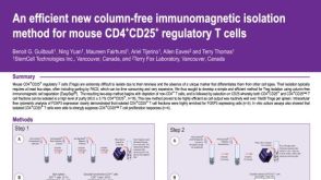 科学海报Immunomagnetic Isolation Method for Mouse CD4+CD25+ Regulatory T Cells
 沪公网安备31010102008431号