
 EasySep™小鼠TIL(CD45)正选试剂盒


搜索结果: 'EasySep'
-
-
 科学海报Column-Free Cell Enrichment of Human Th17 Cells from Peripheral Blood
产品类型:
Conference:
KEYSTONE 2012
产品号#:
产品名:
-
 科学海报Immunomagnetic Cell Isolation of Untouched Lymphocyte Populations Directly from Whole Blood
产品类型:
Conference:
AAI 2014
产品号#:
19655
19661
19662
19663
19664
19674
18000
19659
19655RF
89671
89671RF
89684
89684RF
19671
19671RF
19684
19684RF
产品名:
EasySep™ Direct人总淋巴细胞分选试剂盒
EasySep™ Direct人T细胞分选试剂盒
EasySep™ Direct人CD4+ T细胞分选试剂盒
EasySep™ Direct人CD8+ T细胞分选试剂盒
EasySep™ Direct人B-CLL细胞分选试剂盒
EasySep™ Direct人B细胞分选试剂盒
EasySep™磁极
EasySep™ Direct人Pan-粒细胞分选试剂盒
RoboSep™ Direct人总淋巴细胞分选试剂盒
EasySep™ Direct HLA交叉配型T细胞分选试剂盒
RoboSep™ Direct HLA交叉配型T细胞分选试剂盒
EasySep™ Direct HLA交叉配型B细胞分选试剂盒
RoboSep™ Direct HLA交叉配型B细胞分选试剂盒
-
 科学海报Isolation of Plasmacytoid Dendritic Cells from Human Peripheral Blood
产品类型:
Conference:
AAI 2009
产品号#:
19062
19062RF
21000
20155
20119
产品名:
EasySep™人浆细胞样DC富集试剂盒
RoboSep™ 人浆细胞样DC富集试剂盒含滤芯吸头
RoboSep™- S
RoboSep™分选管套装(9个塑料管)
RoboSep™ 吸头组件抛光剂
-
 科学海报Isolation of CD4+CD25+ from Whole Blood
产品类型:
Conference:
AAI 2003
产品号#:
15862
15862RF
18231
产品名:
-
 科学海报Pre-Enrichment of Dendritic Cells from Human Peripheral Blood Samples
产品类型:
Conference:
ISCT 2008
产品号#:
19251
19251RF
21000
20155
20119
产品名:
EasySep™人Pan-DC预富集试剂盒
RoboSep™ 人Pan-DC预富集试剂盒含滤芯吸头
RoboSep™- S
RoboSep™分选管套装(9个塑料管)
RoboSep™ 吸头组件抛光剂
-
 科学海报Fully Automated Magnetic Labeling and Separation of Hematopoietic Cells from Multiple Samples
产品类型:
Conference:
BSI 2005
产品号#:
18000
产品名:
EasySep™磁极
-
 科学海报Procedure for Negative Enrichment of Monocytes from Mouse Blood and Bone Marrow
产品类型:
Conference:
BSI 2008
产品号#:
21000
20155
20119
19761
19761RF
产品名:
RoboSep™- S
RoboSep™分选管套装(9个塑料管)
RoboSep™ 吸头组件抛光剂
-
 科学海报Column-Free Isolation of Highly Purified and Functional Human Regulatory T Cells
产品类型:
Conference:
ASI 2010
产品号#:
19232
19232RF
产品名:
EasySep™人CD4+ CD127low CD49d-调节性T细胞富集试剂盒
RoboSep™ CD4+ CD127low CD49d-调节性T细胞富集试剂盒
-
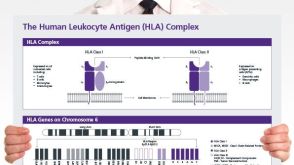 挂图The Human Leukocyte Antigen (HLA) Complex Provides an overview of the human leukocyte antigen (HLA) complex and nomenclature of HLA alleles
 -
 技术窍门组织解离方法指南
 -

产品类型:
产品号#:
18056
18056RF
产品名:

 沪公网安备31010102008431号