Soluble CD14 promotes Th17 expansion and differentiation through gamma-aminobutyric acid and expands infidel innate lymphoid cells
Interleukin-17 (IL-17) plays a central role in the pathogenesis of various autoimmune diseases. Soluble CD14 (sCD14),a marker of innate immune activation,is elevated in several inflammatory conditions. However,its influence on IL-17 production and the differentiation of Th17 cells remains poorly understood. We found that sCD14 enhances Th17-associated cytokine production and up-regulates critical transcription factors such as STAT3 and RORC. Notably,sCD14's effect on Th17 polarization was mediated indirectly through autologous sCD14-treated peripheral blood mononuclear cell (PBMC) supernatant (sCD14-PBMC-Sup). Additionally,we identified a distinct cytokine profile enriched for pro-inflammatory cytokines and chemokines in sCD14-treated T cells,further reinforcing the Th17-promoting role of sCD14. Interestingly,gamma-aminobutyric acid (GABA),a metabolite elevated in sCD14-treated monocytes,was identified as a potential contributor to Th17 polarization. GABA supplementation in T-cell cultures enhanced IL-17A secretion,indicating its role as a signaling molecule in T-cell differentiation. Our findings also revealed the expansion of innate lymphoid cell (ILC)2/3-like cells in T-cell cultures exposed to sCD14-PBMC-Sup and GABA,highlighting the potential role of monocytes in Th17-mediated immunity. Furthermore,while sCD14 promoted Th17 polarization,it simultaneously impaired T-cell activation and proliferation,suggesting an immunosuppressive effect mediated by soluble factors released from monocytes. These results underscore the dual role of sCD14 in modulating T-cell responses,promoting Th17 differentiation while suppressing T-cell effector functions. This study identifies a previously unrecognized role for sCD14 in promoting Th17 induction,highlighting its contribution to immune regulation and its potential as a therapeutic target in Th17-driven autoimmune conditions.
View Publication
产品类型:
产品号#:
19059
19059RF
产品名:
EasySep™人单核细胞富集试剂盒
RoboSep™ 人单核细胞富集试剂盒含滤芯吸头
M. Huyghe et al. (Oct 2024)
Frontiers in Immunology 15 3
Comparative analysis of iPSC-derived NK cells from two differentiation strategies reveals distinct signatures and cytotoxic activities
The ability to generate natural killer (NK) cells from induced pluripotent stem cells (iPSCs) has given rise to new possibilities for the large-scale production of homogeneous immunotherapeutic cellular products and opened new avenues towards the creation of “off-the-shelf” cancer immunotherapies. However,the differentiation of NK cells from iPSCs remains poorly understood,particularly regarding the ontogenic landscape of iPSC-derived NK (iNK) cells produced in vitro and the influence that the differentiation strategy employed may have on the iNK profile. To investigate this question,we conducted a comparative analysis of two sets of iNK cells generated from the same iPSC line using two different protocols: (i) a short-term,clinically compatible feeder-free protocol corresponding to primitive hematopoiesis,and (ii) a lymphoid-based protocol representing the definitive hematopoietic step. Our work demonstrated that both protocols are capable of producing functional iNK cells. However,the two sets of resulting iNKs exhibited distinct phenotypes and transcriptomic profiles. The lymphoid-based differentiation approach generated iNKs with a more mature and activated profile,which demonstrated higher cytotoxicity against cancer cell lines compared to iNK cells produced under short-term feeder-free conditions suggesting that the differentiation strategy must be considered when designing iNK cell–based adoptive immunotherapies.
View Publication
产品类型:
产品号#:
15025
15065
产品名:
RosetteSep™人NK细胞富集抗体混合物
RosetteSep™人NK细胞富集抗体混合物
(Jun 2024)
iScience 27 7
Atypical KCNQ1/Kv7 channel function in a neonatal diabetes patient: Hypersecretion preceded the failure of pancreatic ?-cells
SummaryKCNQ1/Kv7,a low-voltage-gated K+ channel,regulates cardiac rhythm and glucose homeostasis. While KCNQ1 mutations are associated with long-QT syndrome and type2 diabetes,its function in human pancreatic cells remains controversial. We identified a homozygous KCNQ1 mutation (R397W) in an individual with permanent neonatal diabetes melitus (PNDM) without cardiovascular symptoms. To decipher the potential mechanism(s),we introduced the mutation into human embryonic stem cells and generated islet-like organoids (SC-islets) using CRISPR-mediated homology-repair. The mutation did not affect pancreatic differentiation,but affected channel function by increasing spike frequency and Ca2+ flux,leading to insulin hypersecretion. With prolonged culturing,the mutant islets decreased their secretion and gradually deteriorated,modeling a diabetic state,which accelerated by high glucose levels. The molecular basis was the downregulated expression of voltage-activated Ca2+ channels and oxidative phosphorylation. Our study provides a better understanding of the role of KCNQ1 in regulating insulin secretion and ?-cell survival in hereditary diabetes pathology. Graphical abstract Highlights•A permanent neonatal diabetes melitus patient carries a homozygous KCNQ1 mutation•KCNQ1R397W is loss of function and shows atypical electrophysiology in hESC-islets•Under high glucose,elevated Ca2+ flux leads to insulin hypersecretion•Mutant cells gradually switch phenotype,deteriorate,accelerated by high glucose Biological sciences; Endocrinology; Endocrinology; Health sciences; Internal medicine; Medical specialty; Medicine; Natural sciences; Physiology
View Publication
产品类型:
产品号#:
85850
85857
产品名:
mTeSR™1
mTeSR™1
(Mar 2025)
Nature Communications 16
An obesogenic FTO allele causes accelerated development, growth and insulin resistance in human skeletal muscle cells
Human GWAS have shown that obesogenic FTO polymorphisms correlate with lean mass,but the mechanisms have remained unclear. It is counterintuitive because lean mass is inversely correlated with obesity and metabolic diseases. Here,we use CRISPR to knock-in FTOrs9939609-A into hESC-derived tissue models,to elucidate potentially hidden roles of FTO during development. We find that among human tissues,FTOrs9939609-A most robustly affect human muscle progenitors’ proliferation,differentiation,senescence,thereby accelerating muscle developmental and metabolic aging. An edited FTOrs9939609-A allele over-stimulates insulin/IGF signaling via increased muscle-specific enhancer H3K27ac,FTO expression and m6A demethylation of H19 lncRNA and IGF2 mRNA,with excessive insulin/IGF signaling leading to insulin resistance upon replicative aging or exposure to high fat diet. This FTO-m6A-H19/IGF2 circuit may explain paradoxical GWAS findings linking FTOrs9939609-A to both leanness and obesity. Our results provide a proof-of-principle that CRISPR-hESC-tissue platforms can be harnessed to resolve puzzles in human metabolism. Human GWAS paradoxically linked FTO SNPs to both lean mass and sarcopenia/obesity. Here,Guang et al used CRISPR-edited stem cells to reveal that an obesogenic FTO SNP accelerates both muscle development and aging,by increasing RNA m6A demethylation.
View Publication
产品类型:
产品号#:
85850
85857
产品名:
mTeSR™1
mTeSR™1
(Sep 2024)
Frontiers in Immunology 15
SARS-CoV-2 spike protein induces the cytokine release syndrome by stimulating T cells to produce more IL-2
IntroductionCytokine release syndrome (CRS) is one of the leading causes of mortality in patients with COVID-19 caused by the SARS-CoV-2 coronavirus. However,the mechanism of CRS induced by SARS-CoV-2 is vague.MethodsUsing spike protein combined with IL-2,IFN-γ,and TNF-α to stimulate human peripheral blood mononuclear cells (PBMCs) to secrete CRS-related cytokines,the content of cytokines in the supernatant was detected,and the effects of NK,T,and monocytes were analyzed.ResultsThis study shows that dendritic cells loaded with spike protein of SARS-CoV-2 stimulate T cells to release much more interleukin-2 (IL-2,) which subsequently cooperates with spike protein to facilitate PBMCs to release IL-1β,IL-6,and IL-8. These effects are achieved via IL-2 stimulation of NK cells to release tumor necrosis factor-α (TNF-α) and interferon-γ (IFN-γ),as well as T cells to release IFN-γ Mechanistically,IFN-γ and TNF-α enhance the transcription of CD40,and the interaction of CD40 and its ligand stabilizes the membrane expression of toll-like receptor 4 (TLR4) that serves as a receptor of spike protein on the surface of monocytes. As a result,there is a constant interaction between spike protein and TLR4,leading to continuous activation of nuclear factor-κ-gene binding (NF-κB). Furthermore,TNF-α also activates NF-κB signaling in monocytes,which further cooperates with IFN-γ and spike protein to modulate NF-κB–dependent transcription of CRS-related inflammatory cytokines.DiscussionTargeting TNF-α/IFN-γ in combination with TLR4 may represent a promising therapeutic approach for alleviating CRS in individuals with COVID-19.
View Publication
产品类型:
产品号#:
19359
17951
100-0695
17951RF
100-0697
19359RF
产品名:
EasySep™人单核细胞分选试剂盒
EasySep™人T细胞分选试剂盒
EasySep™人T细胞分选试剂盒
RoboSep™ 人T细胞分选试剂盒
EasySep™人单核细胞分选试剂盒
RoboSep™ 人单核细胞分选试剂盒
D. Masyithah Darlan et al. (aug 2020)
Medicinski glasnik : official publication of the Medical Association of Zenica-Doboj Canton,Bosnia and Herzegovina 17 2 408--413
In vitro regulation of IL-6 and TGF-\ss by mesenchymal stem cells in systemic lupus erythematosus patients.
Aim To analyse the ability of mesenchymal stem cells (MSCs) to regulate interleukin 6 (IL-6) and transforming growth factor (TGF-$\beta$) expression in vitro under co-culture conditions in human systemic lupus erythematosus (SLE). Method This study used a post-test group design that used peripheral blood mononuclear cells (PBMCs) from SLE patients at Kariadi Hospital,Semarang,Indonesia,and MSCs from a human umbilical cord. The cells were divided into two groups. The control group of PBMCs was treated with a standard medium,and the treatment group was co-cultured with the MSCs at a 1:40 ratio. Following 24 h incubation,the levels of IL-6 and TGF-$\beta$ released in the culture medium were measured using a specific ELISA assay. Results This study showed a significant decrease in IL-6 level (p{\textless}0.05) and a significant increase in TGF-$\beta$ level (p{\textless}0.001) following 24 h of co-culture incubation of human SLE PBMCs cells and MSCs. Conclusion The PBMCs-to-MSCs ratio of 1:40 can regulate the IL-6 and TGF-$\beta$ levels in human SLE PBMCs.
View Publication
产品类型:
产品号#:
05465
产品名:
MesenCult™ 成骨细胞分化试剂盒 (人)
Z. Wang et al. (Apr 2023)
Redox biology 60 102618
FUT2-dependent fucosylation of HYOU1 protects intestinal stem cells against inflammatory injury by regulating unfolded protein response.
The intestinal epithelial repair after injury is coordinated by intestinal stem cells (ISCs). Fucosylation catalyzed by fucosyltransferase 2 (FUT2) of the intestinal epithelium is beneficial to mucosal healing but poorly defined is the influence on ISCs. The dextran sulfate sodium (DSS) and lipopolysaccharide (LPS) model were used to assess the role of FUT2 on ISCs after injury. The apoptosis,function,and stemness of ISCs were analyzed using intestinal organoids from WT and Fut2?ISC (ISC-specific Fut2 knockout) mice incubated with LPS and fucose. N-glycoproteomics,UEA-1 chromatography,and site-directed mutagenesis were monitored to dissect the regulatory mechanism,identify the target fucosylated protein and the corresponding modification site. Fucose could alleviate intestinal epithelial damage via upregulating FUT2 and ?-1,2-fucosylation of ISCs. Oxidative stress,mitochondrial dysfunction,and cell apoptosis were impeded by fucose. Meanwhile,fucose sustained the growth and proliferation capacity of intestinal organoids treated with LPS. Contrarily,FUT2 depletion in ISCs aggravated the epithelial damage and disrupted the growth and proliferation capacity of ISCs via escalating LPS-induced endoplasmic reticulum (ER) stress and initiating the IRE1/TRAF2/ASK1/JNK branch of unfolded protein response (UPR). Fucosylation of the chaperone protein HYOU1 at the N-glycosylation site of asparagine (Asn) 862 mediated by FUT2 was identified to facilitate ISCs survival and self-renewal,and improve ISCs resistance to ER stress and inflammatory injury. Our study highlights a fucosylation-dependent protective mechanism of ISCs against inflammation,which may provide a fascinating strategy for treating intestinal injury disorders.
View Publication
Dorsomorphin, a selective small molecule inhibitor of BMP signaling, promotes cardiomyogenesis in embryonic stem cells.
BACKGROUND Pluripotent embryonic stem (ES) cells,which have the capacity to give rise to all tissue types in the body,show great promise as a versatile source of cells for regenerative therapy. However,the basic mechanisms of lineage specification of pluripotent stem cells are largely unknown,and generating sufficient quantities of desired cell types remains a formidable challenge. Small molecules,particularly those that modulate key developmental pathways like the bone morphogenetic protein (BMP) signaling cascade,hold promise as tools to study in vitro lineage specification and to direct differentiation of stem cells toward particular cell types. METHODOLOGY/ PRINCIPAL FINDINGS We describe the use of dorsomorphin,a selective small molecule inhibitor of BMP signaling,to induce myocardial differentiation in mouse ES cells. Cardiac induction is very robust,increasing the yield of spontaneously beating cardiomyocytes by at least 20 fold. Dorsomorphin,unlike the endogenous BMP antagonist Noggin,robustly induces cardiomyogenesis when treatment is limited to the initial 24-hours of ES cell differentiation. Quantitative-PCR analyses of differentiating ES cells indicate that pharmacological inhibition of BMP signaling during the early critical stage promotes the development of the cardiomyocyte lineage,but reduces the differentiation of endothelial,smooth muscle,and hematopoietic cells. CONCLUSIONS/ SIGNIFICANCE Administration of a selective small molecule BMP inhibitor during the initial stages of ES cell differentiation substantially promotes the differentiation of primitive pluripotent cells toward the cardiomyocytic lineage,apparently at the expense of other mesodermal lineages. Small molecule modulators of developmental pathways like dorsomorphin could become versatile pharmacological tools for stem cell research and regenerative medicine.
View Publication
产品类型:
产品号#:
72102
100-0246
产品名:
Dorsomorphin
白消安(Busulfan)
Goessling W et al. (MAR 2009)
Cell 136 6 1136--47
Genetic interaction of PGE2 and Wnt signaling regulates developmental specification of stem cells and regeneration.
Interactions between developmental signaling pathways govern the formation and function of stem cells. Prostaglandin (PG) E2 regulates vertebrate hematopoietic stem cells (HSC). Similarly,the Wnt signaling pathway controls HSC self-renewal and bone marrow repopulation. Here,we show that wnt reporter activity in zebrafish HSCs is responsive to PGE2 modulation,demonstrating a direct interaction in vivo. Inhibition of PGE2 synthesis blocked wnt-induced alterations in HSC formation. PGE2 modified the wnt signaling cascade at the level of beta-catenin degradation through cAMP/PKA-mediated stabilizing phosphorylation events. The PGE2/Wnt interaction regulated murine stem and progenitor populations in vitro in hematopoietic ES cell assays and in vivo following transplantation. The relationship between PGE2 and Wnt was also conserved during regeneration of other organ systems. Our work provides in vivo evidence that Wnt activation in stem cells requires PGE2,and suggests the PGE2/Wnt interaction is a master regulator of vertebrate regeneration and recovery.
View Publication
产品类型:
产品号#:
72372
产品名:
16,16-二甲基前列腺素E2
Chen Y et al. (MAY 2009)
Journal of immunology (Baltimore,Md. : 1950) 182 10 6031--43
IgM antibodies to apoptosis-associated determinants recruit C1q and enhance dendritic cell phagocytosis of apoptotic cells.
Natural Abs,which arise without known immune exposure,have been described that specifically recognize cells dying from apoptosis,but their role in innate immunity remains poorly understood. Herein,we show that the immune response to neoantigenic determinants on apoptotic thymocytes is dominated by Abs to oxidation-associated Ags,phosphorylcholine (PC),a head group that becomes exposed during programmed cell death,and malondialdehyde (MDA),a reactive aldehyde degradation product of polyunsaturated lipids produced following exposure to reactive oxidation species. While natural Abs to apoptotic cells in naive adult mice were dominated by PC and MDA specificities,the amounts of these Abs were substantially boosted by treatment of mice with apoptotic cells. Moreover,the relative amounts of PC and MDA Abs was affected by V(H) gene inheritance. Ab interactions with apoptotic cells also mediated the recruitment of C1q,which enhanced apoptotic cell phagocytosis by immature dendritic cells. Significantly,IgM Abs to both PC and MDA were primary factors in determining the efficiency of serum-dependent apoptotic cell phagocytosis. Hence,we demonstrate a mechanism by which certain natural Abs that recognize neoantigens on apoptotic cells,in naive mice and those induced by immune exposure to apoptotic cells,can enhance the functional capabilities of immature dendritic cells for phagocytic engulfment of apoptotic cells.
View Publication


 EasySep™小鼠TIL(CD45)正选试剂盒





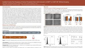 科学海报Scalable Enzyme-Free Passaging of Human Pluripotent Stem Cells Cultured in mTeSR™1 or TeSR™-E8™ Without Scraping
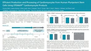 科学海报Efficient Production and Processing of Cardiomyocytes from Human Pluripotent Stem Cells Using STEMdiff™ Cardiomyocyte Products
 沪公网安备31010102008431号