
 EasySep™小鼠TIL(CD45)正选试剂盒


搜索结果: 'immunocult'
-
 冻存的SF培养的人外周血来源的M1巨噬细胞 冻存的人原代细胞
 -
 人外周血来源M0型巨噬细胞,无血清培养,冻存型 冻存的人原代细胞
 -
 冻存的SF培养的人外周血来源的M2a巨噬细胞 冻存的人原代细胞
 -
 冻存的ACF培养的人外周血来源的未成熟树突状细胞 冻存的人原代细胞
 -

-
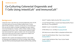 技术公告Co-Culturing Colorectal Organoids and T Cells using IntestiCult™ and ImmunoCult™
产品类型:
细胞类型:
T细胞,肠道细胞
产品号#:
产品名:
发布日期: 04/01/2024 -
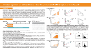 科学海报Activation, Expansion, and Culture of Human T Cells Using ImmunoCult™ cGMP-Compliant Ancillary Reagents
产品类型:
Conference:
ISCT 2024
产品号#:
产品名:
-
 实验方案How to Measure NK Cell-Mediated Antibody-Dependent Cellular Cytotoxicity Using Flow Cytometry-Based Assays
产品类型:
研究方向:
免疫学,细胞治疗开发
产品号#:
产品名:
-

产品类型:
产品号#:
07801
07811
07851
07861
10971
10991
15021
15061
18060
18061
产品名:
Lymphoprep™
Lymphoprep™
ImmunoCult™ 人CD3/CD28 T细胞激活剂
ImmunoCult™ 人CD3/CD28 T细胞激活剂
RosetteSep™人T细胞富集抗体混合物
RosetteSep™人T细胞富集抗体混合物
Lymphoprep™
Lymphoprep™
-
产品类型:
产品号#:
10981
15023
17898
85450
15063
17898RF
85460
产品名:
ImmunoCult™ XF 人T细胞扩增培养基,500 mL
RosetteSep™人CD8+ T细胞富集抗体混合物
EasySep™人CD45去除试剂盒II
SepMate™-50 (IVD)
RosetteSep™人CD8+ T细胞富集抗体混合物
RoboSep™ 人CD45去除试剂盒II
SepMate™-50 (IVD)

 沪公网安备31010102008431号