mTOR inhibition impacts the flagellin-augmented inflammatory and antimicrobial response of human airway epithelial cells to Pseudomonas aeruginosa
The airway epithelium provides a first line of defense against pathogens by release of antimicrobial factors and neutrophil-attracting chemokines. Pseudomonas (P.) aeruginosa,a Gram-negative bacterium that expresses flagellin as an important virulence factor,is a common cause of injurious airway inflammation. The aim of our study was to determine the contribution of flagellin to the inflammatory,antimicrobial,and metabolic responses of the airway epithelium to P. aeruginosa . Furthermore,as we previously showed that targeting mTOR limited the glycolytic and inflammatory response induced by flagellin,we assessed the effect of rapamycin on human bronchial epithelial (HBE) cells stimulated with flagellated and non-flagellated P. aeruginosa. Primary pseudostratified HBE cells,cultured on an air-liquid-interface,were treated on the basolateral side with medium,vehicle or rapamycin,exposed on the apical side with flagellated or flagellin-deficient P. aeruginosa,and analyzed for their inflammatory,antimicrobial,and glycolytic responses. Flagellin augmented the P. aeruginosa -induced expression of antimicrobial factors and secretion of chemokines by HBE cells but did not further increase the glycolytic response. Treatment of HBE cells with rapamycin inhibited mTOR activation in general and flagellin-augmented mTOR activation in particular,but did not affect the glycolytic response. Rapamycin,however,diminished the flagellin-augmented inflammatory and antimicrobial response induced by Pseudomonas . These results demonstrate that flagellin is a significant factor that augments the inflammatory and antimicrobial response of human airway epithelial cells upon exposure to P. aeruginosa and suggest that mTOR inhibition by rapamycin in the airway epithelium diminishes these exaggerated responses.
View Publication
产品类型:
产品号#:
05001
05021
05022
产品名:
PneumaCult™-ALI 培养基
PneumaCult™-ALI 培养基含12 mm Transwell®插件
PneumaCult™-ALI 培养基含6.5 mm Transwell®插件
(Apr 2024)
International Journal of Stem Cells 17 2
Energy Metabolism in Human Pluripotent Stem and Differentiated Cells Compared Using a Seahorse XF96 Extracellular Flux Analyzer
Evaluating cell metabolism is crucial during pluripotent stem cell (PSC) differentiation and somatic cell reprogramming as it affects cell fate. As cultured stem cells are heterogeneous,a comparative analysis of relative metabolism using existing metabolic analysis methods is difficult,resulting in inaccuracies. In this study,we measured human PSC basal metabolic levels using a Seahorse analyzer. We used fibroblasts,human induced PSCs,and human embryonic stem cells to monitor changes in basal metabolic levels according to cell number and determine the number of cells suitable for analysis. We evaluated normalization methods using glucose and selected the most suitable for the metabolic analysis of heterogeneous PSCs during the reprogramming stage. The response of fibroblasts to glucose increased with starvation time,with oxygen consumption rate and extracellular acidification rate responding most effectively to glucose 4 hours after starvation and declining after 5 hours of starvation. Fibroblasts and PSCs achieved appropriate responses to glucose without damaging their metabolism 2?4 and 2?3 hours after starvation,respectively. We developed a novel method for comparing basal metabolic rates of fibroblasts and PSCs,focusing on quantitative analysis of glycolysis and oxidative phosphorylation using glucose without enzyme inhibitors. This protocol enables efficient comparison of energy metabolism among cell types,including undifferentiated PSCs,differentiated cells,and cells undergoing cellular reprogramming,and addresses critical issues,such as differences in basal metabolic levels and sensitivity to normalization,providing valuable insights into cellular energetics.
View Publication
产品类型:
产品号#:
05990
产品名:
用于hESC/hiPSC维持培养的TeSR™-E8™
(May 2024)
STAR Protocols 5 2
Generation and enrichment of cerebellar GABAergic interneurons from human induced pluripotent stem cells and intracellular calcium measurements
SummaryGABAergic interneurons are inhibitory neurons of the CNS,playing a fundamental role in neural circuitry and activity. Here,we provide a robust protocol for the successful enrichment of human cerebellar GABAergic interneurons from human induced pluripotent stem cells (iPSCs) and measuring intracellular calcium transients. We describe in detail steps for culturing iPSCs; generating embryoid bodies; and differentiating and enriching for cerebellar GABAergic neurons (cGNs),with precise steps for their molecular characterization. We then detail the procedure for adeno-associated virus-mediated transduction of cGNs with genetically encoded calcium indicators,followed by intracellular calcium imaging and analyses.For complete details on the use and execution of this protocol,please refer to Pilotto et al.1 Graphical abstract Highlights•Steps described for generating GABAergic neurons from human iPSCs•Instructions for the enrichment of cerebellar GABAergic interneurons (cGNs)•Guide to calcium imaging of cGNs using genetically encoded calcium indicators Publisher’s note: Undertaking any experimental protocol requires adherence to local institutional guidelines for laboratory safety and ethics. GABAergic interneurons are inhibitory neurons of the CNS,playing a fundamental role in neural circuitry and activity. Here,we provide a robust protocol for the successful enrichment of human-cerebellar GABAergic interneurons from human induced pluripotent stem cells (iPSCs) and measuring intracellular calcium transients. We describe in detail steps for culturing iPSCs,and generating embryoid bodies,differentiating and enriching for cerebellar GABAergic neurons (cGNs),with precise steps for their molecular characterization. We then detail the procedure for adeno-associated virus-mediated transduction of cGNs with genetically encoded calcium indicators,followed by intracellular calcium imaging and analyses.
View Publication
产品类型:
产品号#:
85850
85857
产品名:
mTeSR™1
mTeSR™1
J. Yao et al. ( 2020)
Stem cells international 2020 6489396
Human Supernumerary Teeth-Derived Apical Papillary Stem Cells Possess Preferable Characteristics and Efficacy on Hepatic Fibrosis in Mice.
Dental tissue has been acknowledged as an advantaged source for high-quality dental pulp stem cell (DPSC) preparation. However,despite the accomplishment of the separation of DPSCs from permanent teeth and supernumerary teeth,the deficiency of rigorous and systematic clarification on the signatures and efficacy will hinder their prospects in regenerative medicine. In this study,we primitively isolated permanent teeth-derived DPSCs and supernumerary teeth-derived apical papillary stem cells (SCAP-Ss) with parental consent. Immunophenotype of DPSCs and SCAP-Ss was determined by a flow cytometry assay,and the cell viability was verified by multidimensional detections including cell proliferation,cell cycle,apoptosis,and senescence. The migration and clonogenic capacity were examined by a wound healing test and crystal violet staining,respectively. The multilineage differentiation potential was quantitated by utilizing Oil Red O staining and Alizarin Red staining,together with real-time PCR analysis. The efficacy on a mouse hepatic fibrosis model was evaluated by using histologic sections and liver function tests. Herein,we showed that SCAP-Ss exhibited comparable immunophenotype and adipogenic differentiation capacity as DPSCs. However,different from DPSCs,SCAP-Ss exhibited superiority in cell viability and osteogenic differentiation. Simultaneously,injection of DPSCs and SCAP-Ss significantly reduced inflammatory infiltration,enhanced liver-associated gene expression,and finally relieved symptoms of hepatic fibrosis. In conclusion,SCAP-Ss possess preferable characteristics and efficacy on hepatic fibrosis in mice. Our findings suggest that SCAP-Ss are an easily accessible postnatal stem cell source with multifaceted characteristics for regenerative medicine.
View Publication
Daga A et al. (MAY 2000)
Experimental hematology 28 5 569--74
The retroviral transduction of HOXC4 into human CD34(+) cells induces an in vitro expansion of clonogenic and early progenitors.
OBJECTIVE: +HOX genes are expressed in the hematopoietic system and increasing data point to their involvement in the control of proliferation and/or differentiation. Genes belonging to the C cluster are preferentially expressed in developing and differentiated lymphoid lineages. However,recent studies demonstrated,by RT-PCR,that the HOXC4 gene is also actively transcribed in the most undifferentiated hematopoietic cells (CD34(+)38(low)) and in more mature myeloid and erythroid progenitors. We evaluated the expression of HOXC4 protein on human CD34(+) cells and the in vitro effect of its overexpression on proliferation and differentiation. MATERIALS AND METHODS: We assessed the expression of HOXC4 on human CD34(+) cells using a polyclonal antibody raised against the C-terminal portion of the protein expressed using the baculovirus system. Overexpression of HOXC4 in human CD34(+) cells was obtained by retroviral gene transfer; its effect on clonogenic (CFU-GM,BFU-E,and CFU-GEMM) and early progenitors (LTC-IC) was evaluated. RESULTS: The HOXC4 protein is indeed expressed in human CD34(+) cells,and its overexpression in human CD34(+) cells increases the proliferation potential of clonogenic and early progenitors. CFU-GM showed a median threefold expansion (range: 1.1-19.4; p textless 0.002) compared with control transduced with the vector alone. The increment of BFU-E was higher (median ninefold,range 2.5-35; p textless 0. 0009) and erythroid colonies presented a larger size with normal morphology. An even more marked effect was observed on LTC-IC (median 13,onefold; range 4.1-102.1,p textless 0.0001). CONCLUSION: We demonstrate that HOXC4 is expressed in CD34(+) cells and that its overexpression induces an in vitro expansion of committed as well as very early hematopoietic progenitors. The most striking effect was obtained on LTC-IC with an expansion of 13.1-fold. The enforced expression of HOXC4 induced a significant increase (p textless 0.009) in the number of erythroid colonies compared with CFU-GM,although without perturbing,at least in vitro,the maturation program of the cells. On the other hand,the effect of the gene overexpression did not induce any skewing in the colony types derived from the myeloid lineage.
View Publication
产品类型:
产品号#:
04536
产品名:
MethoCult™ SF H4536
Wood ER et al. ( 2004)
Cancer research 64 18 6652--6659
A unique structure for epidermal growth factor receptor bound to GW572016 (Lapatinib): relationships among protein conformation, inhibitor off-rate, and receptor activity in tumor cells.
GW572016 (Lapatinib) is a tyrosine kinase inhibitor in clinical development for cancer that is a potent dual inhibitor of epidermal growth factor receptor (EGFR,ErbB-1) and ErbB-2. We determined the crystal structure of EGFR bound to GW572016. The compound is bound to an inactive-like conformation of EGFR that is very different from the active-like structure bound by the selective EGFR inhibitor OSI-774 (Tarceva) described previously. Surprisingly,we found that GW572016 has a very slow off-rate from the purified intracellular domains of EGFR and ErbB-2 compared with OSI-774 and another EGFR selective inhibitor,ZD-1839 (Iressa). Treatment of tumor cells with these inhibitors results in down-regulation of receptor tyrosine phosphorylation. We evaluated the duration of the drug effect after washing away free compound and found that the rate of recovery of receptor phosphorylation in the tumor cells reflected the inhibitor off-rate from the purified intracellular domain. The slow off-rate of GW572016 correlates with a prolonged down-regulation of receptor tyrosine phosphorylation in tumor cells. The differences in the off-rates of these drugs and the ability of GW572016 to inhibit ErbB-2 can be explained by the enzyme-inhibitor structures.
View Publication
Lambert AA et al. (AUG 2008)
Blood 112 4 1299--307
The C-type lectin surface receptor DCIR acts as a new attachment factor for HIV-1 in dendritic cells and contributes to trans- and cis-infection pathways.
The dynamic interplay between dendritic cells (DCs) and human immunodeficiency virus type-1 (HIV-1) is thought to result in viral dissemination and evasion of antiviral immunity. Although initial observations suggested that the C-type lectin receptor (CLR) DC-SIGN was responsible for the trans-infection function of the virus,subsequent studies demonstrated that trans-infection of CD4(+) T cells with HIV-1 can also occur through DC-SIGN-independent mechanisms. We demonstrate that a cell surface molecule designated DCIR (for DC immunoreceptor),a member of a recently described family of DC-expressing CLRs,can participate in the capture of HIV-1 and promote infection in trans and in cis of autologous CD4(+) T cells from human immature monocyte-derived DCs. The contribution of DCIR to these processes was revealed using DCIR-specific siRNAs and a polyclonal antibody specific for the carbohydrate recognition domain of DCIR. Data from transfection experiments indicated that DCIR acts as a ligand for HIV-1 and is involved in events leading to productive virus infection. Finally,we show that the neck domain of DCIR is important for the DCIR-mediated effect on virus binding and infection. These results point to a possible role for DCIR in HIV-1 pathogenesis by supporting the productive infection of DCs and promoting virus propagation.
View Publication
产品类型:
产品号#:
18058
18058RF
19052
19052RF
产品名:
EasySep™人CD4+ T细胞富集试剂盒
RoboSep™ 人CD4+ T细胞富集试剂盒含滤芯吸头
(Apr 2025)
Cells 14 8
LFA-1/ICAM-1 Interactions Between CD8+ and CD4+ T Cells Promote CD4+ Th1-Dominant Differentiation and CD8+ T Cell Cytotoxicity for Strong Antitumor Immunity After Cryo-Thermal Therapy
CD4+ T cells have been well-regarded as “helper” cells in activating the cytotoxicity of CD8+ T cells for effective tumor eradication,while few studies have focused on whether CD8+ T cells regulate CD4+ T cells. Our previous studies provided evidence for an interaction between CD4+ and CD8+ T cells after cryo-thermal therapy,but the mechanism remains unclear,especially pertaining to how CD8+ T cells promote the Th1 differentiation of CD4+ T cells. This study revealed that activated CD4+ and CD8+ T cells are critical for CTT-induced antitumor immunity,and the interaction between activated T cells is enhanced. The reciprocal regulation of activated CD8+ and CD4+ T cells was through LFA-1/ICAM-1 interactions,in which CD8+ T cells facilitate Notch1-dependent CD4+ Th1-dominant differentiation and promote IL-2 secretion of CD4+ T cells. Meanwhile,IL-2 derived from CD4+ T cells enhances the cytotoxicity of CD8+ T cells and establishes a positive feedback loop via increasing the expression of LFA-1 and ICAM-1 on T cells. Clinical analyses further validated that LFA-1/ICAM interactions between CD4+ and CD8+ T cells are correlated with clinical outcomes. Our study extends the functions of the LFA-1/ICAM-1 adhesion pathway,indicating its novel role in the interaction of CD4+ and CD8+ T cells.
View Publication
产品类型:
产品号#:
18953
18952
18952RF
18953RF
产品名:
EasySep™小鼠CD8a正选试剂盒II
EasySep™小鼠CD4正选试剂盒II
RoboSep™ 小鼠CD4正选试剂盒II
RoboSep™ 小鼠CD8a正选试剂盒II
R. J. Napier et al. ( 2020)
Nature communications 11 1 5406
T cell-intrinsic role for Nod2 in protection against Th17-mediated uveitis.
Mutations in nucleotide-binding oligomerization domain-containing protein 2 (NOD2) cause Blau syndrome,an inflammatory disorder characterized by uveitis. The antimicrobial functions of Nod2 are well-established,yet the cellular mechanisms by which dysregulated Nod2 causes uveitis remain unknown. Here,we report a non-conventional,T cell-intrinsic function for Nod2 in suppression of Th17 immunity and experimental uveitis. Reconstitution of lymphopenic hosts with Nod2-/- CD4+ T cells or retina-specific autoreactive CD4+ T cells lacking Nod2 reveals a T cell-autonomous,Rip2-independent mechanism for Nod2 in uveitis. In naive animals,Nod2 operates downstream of TCR ligation to suppress activation of memory CD4+ T cells that associate with an autoreactive-like profile involving IL-17 and Ccr7. Interestingly,CD4+ T cells from two Blau syndrome patients show elevated IL-17 and increased CCR7. Our data define Nod2 as a T cell-intrinsic rheostat of Th17 immunity,and open new avenues for T cell-based therapies for Nod2-associated disorders such as Blau syndrome.
View Publication
产品类型:
产品号#:
18952
19765
19767
19852
18952RF
19765RF
19767RF
19852RF
产品名:
EasySep™小鼠CD4正选试剂盒II
EasySep™小鼠Naïve CD4+ T细胞分选试剂盒
EasySep™小鼠记忆CD4+ T细胞分选试剂盒
EasySep™小鼠CD4+ T细胞分选试剂盒
RoboSep™ 小鼠CD4正选试剂盒II
RoboSep™ 小鼠Naïve CD4+ T细胞分选试剂盒
RoboSep™ 小鼠记忆CD4+ T细胞分选试剂盒
RoboSep™ 小鼠CD4+ T细胞分选试剂盒
Kimura T et al. (JUN 2004)
Blood 103 12 4478--86
The sphingosine 1-phosphate receptor agonist FTY720 supports CXCR4-dependent migration and bone marrow homing of human CD34+ progenitor cells.
The novel immunosuppressant FTY720 activates sphingosine 1-phosphate receptors (S1PRs) that affect responsiveness of lymphocytes to chemokines such as stromal cell-derived factor 1 (SDF-1),resulting in increased lymphocyte homing to secondary lymphoid organs. Since SDF-1 and its receptor CXCR4 are also involved in bone marrow (BM) homing of hematopoietic stem and progenitor cells (HPCs),we analyzed expression of S1PRs and the influence of FTY720 on SDF-1/CXCR4-mediated effects in human HPCs. By reverse transcriptase-polymerase chain reaction (RT-PCR),S1PRs were expressed in mobilized CD34+ HPCs,particularly in primitive CD34+/CD38- cells. Incubation of HPCs with FTY720 resulted in prolonged SDF-1-induced calcium mobilization and actin polymerization,and substantially increased SDF-1-dependent in vitro transendothelial migration,without affecting VLA-4,VLA-5,and CXCR4 expression. In nonobese diabetic-severe combined immunodeficient (NOD/SCID) mice,the number of CD34+/CD38- cells that homed to the BM after 18 hours was significantly raised by pretreatment of animals and cells with FTY720,tending to result in improved engraftment. In addition,in vitro growth of HPCs (week-5 cobblestone area-forming cells [CAFCs]) was 2.4-fold increased. We conclude that activation of S1PRs by FTY720 increases CXCR4 function in HPCs both in vitro and in vivo,supporting homing and proliferation of HPCs. In the hematopoietic microenvironment,S1PRs are involved in migration and maintenance of HPCs by modulating the effects of SDF-1.
View Publication

 EasySep™小鼠TIL(CD45)正选试剂盒


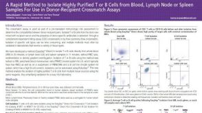 科学海报A Rapid Method to Isolate Highly Purified T or B Cells from Blood, Lymph Node or Spleen Samples For Use in Donor-Recipient Crossmatch Assays
 沪公网安备31010102008431号