Birbrair A et al. (JAN 2013)
Experimental cell research 319 1 45--63
Skeletal muscle neural progenitor cells exhibit properties of NG2-glia.
Reversing brain degeneration and trauma lesions will depend on cell therapy. Our previous work identified neural precursor cells derived from the skeletal muscle of Nestin-GFP transgenic mice,but their identity,origin,and potential survival in the brain are only vaguely understood. In this work,we show that Nestin-GFP+ progenitor cells share morphological and molecular markers with NG2-glia,including NG2,PDGFRα,O4,NGF receptor (p75),glutamate receptor-1(AMPA),and A2B5 expression. Although these cells exhibit NG2,they do not express other pericyte markers,such as α-SMA or connexin-43,and do not differentiate into the muscle lineage. Patch-clamp studies displayed outward potassium currents,probably carried through Kir6.1 channels. Given their potential therapeutic application,we compared their abundance in tissues and concluded that skeletal muscle is the richest source of predifferentiated neural precursor cells. We found that these cells migrate toward the neurogenic subventricular zone displaying their typical morphology and nestin-GFP expression two weeks after brain injection. For translational purposes,we sought to identify these neural progenitor cells in wild-type species by developing a DsRed expression vector under Nestin-Intron II control. This approach revealed them in nonhuman primates and aging rodents throughout the lifespan.
View Publication
产品类型:
产品号#:
05700
05701
05702
05703
05704
05715
产品名:
NeuroCult™ 基础培养基(小鼠和大鼠)
NeuroCult™ 扩增添加物(小鼠和大鼠)
NeuroCult™扩增试剂盒(小鼠和大鼠)
NeuroCult™ 分化添加物(小鼠和大鼠)
NeuroCult™ 分化试剂盒(小鼠和大鼠)
NeuroCult™成年中枢神经系统(CNS)组织酶解试剂盒(小鼠和大鼠)
Santos T et al. (DEC 2012)
ACS nano 6 12 10463--74
Polymeric nanoparticles to control the differentiation of neural stem cells in the subventricular zone of the brain.
Herein,we report the use of retinoic acid-loaded polymeric nanoparticles as a potent tool to induce the neuronal differentiation of subventricular zone neural stem cells. The intracellular delivery of retinoic acid by the nanoparticles activated nuclear retinoic acid receptors,decreased stemness,and increased proneurogenic gene expression. Importantly,this work reports for the first time a nanoparticle formulation able to modulate in vivo the subventricular zone neurogenic niche. The work further compares the dynamics of initial stages of differentiation between SVZ cells treated with retinoic acid-loaded polymeric nanoparticles and solubilized retinoic acid. The nanoparticle formulation developed here may ultimately offer new perspectives to treat neurodegenerative diseases.
View Publication
产品类型:
产品号#:
05707
产品名:
NeuroCult™化学解离试剂盒(小鼠)
Evans MJ et al. (JAN 2013)
Journal of Nuclear Medicine 54 1 90--95
Imaging Tumor Burden in the Brain with 89Zr-Transferrin
UNLABELLED A noninvasive technology that indiscriminately detects tumor tissue in the brain could substantially enhance the management of primary or metastatic brain tumors. Although the documented molecular heterogeneity of diseases that initiate or eventually deposit in the brain may preclude identifying a single smoking-gun molecular biomarker,many classes of brain tumors are generally avid for transferrin. Therefore,we reasoned that applying a radiolabeled derivative of transferrin ((89)Zr-labeled transferrin) may be an effective strategy to more thoroughly identify tumor tissue in the brain,regardless of the tumor's genetic background. METHODS Transferrin was radiolabeled with (89)Zr,and its properties with respect to human models of glioblastoma multiforme were studied in vivo. RESULTS In this report,we show proof of concept that (89)Zr-labeled transferrin ((89)Zr-transferrin) localizes to genetically diverse models of glioblastoma multiforme in vivo. Moreover,we demonstrate that (89)Zr-transferrin can detect an orthotopic lesion with exceptional contrast. Finally,the tumor-to-brain contrast conferred by (89)Zr-transferrin vastly exceeded that observed with (18)F-FDG,currently the most widely used radiotracer to assess tumor burden in the brain. CONCLUSION The results from this study suggest that (89)Zr-transferrin could be a broadly applicable tool for identifying and monitoring tumors in the brain,with realistic potential for near-term clinical translation.
View Publication
产品类型:
产品号#:
05750
05751
产品名:
NeuroCult™ NS-A 基础培养基(人)
NeuroCult™ NS-A 扩增试剂盒(人)
Ehnman M et al. (APR 2013)
Cancer Research 73 7 2139--2149
Distinct Effects of Ligand-Induced PDGFR and PDGFR Signaling in the Human Rhabdomyosarcoma Tumor Cell and Stroma Cell Compartments
Platelet-derived growth factor receptors (PDGFR) α and β have been suggested as potential targets for treatment of rhabdomyosarcoma,the most common soft tissue sarcoma in children. This study identifies biologic activities linked to PDGF signaling in rhabdomyosarcoma models and human sample collections. Analysis of gene expression profiles of 101 primary human rhabdomyosarcomas revealed elevated PDGF-C and -D expression in all subtypes,with PDGF-D as the solely overexpressed PDGFRβ ligand. By immunohistochemistry,PDGF-CC,PDGF-DD,and PDGFRα were found in tumor cells,whereas PDGFRβ was primarily detected in vascular stroma. These results are concordant with the biologic processes and pathways identified by data mining. While PDGF-CC/PDGFRα signaling associated with genes involved in the reactivation of developmental programs,PDGF-DD/PDGFRβ signaling related to wound healing and leukocyte differentiation. Clinicopathologic correlations further identified associations between PDGFRβ in vascular stroma and the alveolar subtype and with presence of metastases. Functional validation of our findings was carried out in molecularly distinct model systems,where therapeutic targeting reduced tumor burden in a PDGFR-dependent manner with effects on cell proliferation,vessel density,and macrophage infiltration. The PDGFR-selective inhibitor CP-673,451 regulated cell proliferation through mechanisms involving reduced phosphorylation of GSK-3α and GSK-3β. Additional tissue culture studies showed a PDGFR-dependent regulation of rhabdosphere formation/cancer cell stemness,differentiation,senescence,and apoptosis. In summary,the study shows a clinically relevant distinction in PDGF signaling in human rhabdomyosarcoma and also suggests continued exploration of the influence of stromal PDGFRs on sarcoma progression.
View Publication
产品类型:
产品号#:
05750
05751
产品名:
NeuroCult™ NS-A 基础培养基(人)
NeuroCult™ NS-A 扩增试剂盒(人)
Snuderl M et al. (FEB 2013)
Cell 152 5 1065--76
Targeting placental growth factor/neuropilin 1 pathway inhibits growth and spread of medulloblastoma.
Medulloblastoma is the most common pediatric malignant brain tumor. Although current therapies improve survival,these regimens are highly toxic and are associated with significant morbidity. Here,we report that placental growth factor (PlGF) is expressed in the majority of medulloblastomas,independent of their subtype. Moreover,high expression of PlGF receptor neuropilin 1 (Nrp1) correlates with poor overall survival in patients. We demonstrate that PlGF and Nrp1 are required for the growth and spread of medulloblastoma: PlGF/Nrp1 blockade results in direct antitumor effects in vivo,resulting in medulloblastoma regression,decreased metastasis,and increased mouse survival. We reveal that PlGF is produced in the cerebellar stroma via tumor-derived Sonic hedgehog (Shh) and show that PlGF acts through Nrp1-and not vascular endothelial growth factor receptor 1-to promote tumor cell survival. This critical tumor-stroma interaction-mediated by Shh,PlGF,and Nrp1 across medulloblastoma subtypes-supports the development of therapies targeting PlGF/Nrp1 pathway.
View Publication
产品类型:
产品号#:
05700
05701
05702
产品名:
NeuroCult™ 基础培养基(小鼠和大鼠)
NeuroCult™ 扩增添加物(小鼠和大鼠)
NeuroCult™扩增试剂盒(小鼠和大鼠)
Xu G et al. (MAY 2013)
Neuroscience 238 195--208
Functional analysis of platelet-derived growth factor receptor-β in neural stem/progenitor cells
Activation of neural stem/progenitor cells (NSPCs) is a potential therapeutic strategy of neurological disorders. In this study,NSPCs of subventricular zone were isolated and cultured from platelet-derived growth factor-β-receptor-knockout (PDGFR-β(-/-)) mice of postnatal day 1 (P1) and P28,and the roles of PDGFR-β were examined in these cells. In PDGFR-β-preserving control NSPCs,stem cell activities,such as numbers and diameters of secondary neurospheres,cell proliferation and survival rates,were significantly higher in P1 NSPCs than those in P28 NSPCs. In PDGFR-β(-/-) NSPCs,most of these parameters were decreased as compared with age-matched controls. Among them,the decrease of secondary neurosphere formation was most striking in P1 and P28 PDGFR-β(-/-) NSPCs and in P28 control NSPCs as compared with P1 control NSPCs. PCR-array and following quantitative real-time PCR (qRT-PCR) analyses demonstrated that expressions of fibroblast growth factor-2 (FGF2) and exons IV-IX of brain-derived neurotrophic factor (BDNF) were decreased,and noggin was increased in P1 PDGFR-β(-/-) as compared with P1 controls. Addition of BDNF rescued the number and diameter of secondary neurospheres in P1 PDGFR-β(-/-) NSPCs to similar levels as controls. The expressions of PDGFs and PDGFRs in control NSPCs were increased along with the differentiation-induction,where phosphorylated PDGFR-β was co-localized with neuronal and astrocyte differentiation markers. In controls,the neuronal differentiation was decreased,and the glial differentiation was increased from P1 to P28 NSPCs. Compared with P1 controls,neuronal differentiation was reduced in P1 PDGFR-β(-/-) NSPCs,whereas glial differentiation was comparable between the two genotypes. These results suggest that PDGFR-β signaling is important for the self-renewal and multipotency of NSPCs,particularly in neonatal NSPCs. BDNF,FGF2,and noggin may be involved in the effects of PDGFR-β signaling in these cells. Accordingly,the activation of PDGFR-β in NSPCs may be a novel therapeutic strategy of neurological diseases.
View Publication
产品类型:
产品号#:
05700
05701
05702
产品名:
NeuroCult™ 基础培养基(小鼠和大鼠)
NeuroCult™ 扩增添加物(小鼠和大鼠)
NeuroCult™扩增试剂盒(小鼠和大鼠)
Y. Kim et al. (May 2020)
FASEB Journal 34 6965-6983
Microtubule-associated protein 2 mediates induction of long-term potentiation in hippocampal neurons
Microtubule-associated protein (MAP) 2 has been perceived as a static cytoskeletal protein enriched in neuronal dendritic shafts. Emerging evidence indicates dynamic functions for various MAPs in activity-dependent synaptic plasticity. However,it is unclear how MAP2 is associated with synaptic plasticity mechanisms. Here,we demonstrate that specific silencing of high-molecular-weight MAP2 in vivo abolished induction of long-term potentiation (LTP) in the Schaffer collateral pathway of CA1 pyramidal neurons and in vitro blocked LTP-induced surface delivery of AMPA receptors and spine enlargement. In mature hippocampal neurons,we observed rapid translocation of a subpopulation of MAP2,present in dendritic shafts,to spines following LTP stimulation. Time-lapse confocal imaging showed that spine translocation of MAP2 was coupled with LTP-induced spine enlargement. Consistently,immunogold electron microscopy revealed that LTP stimulation of the Schaffer collateral pathway promoted MAP2 labeling in spine heads of CA1 neurons. This translocation depended on NMDA receptor activation and Ras-MAPK signaling. Furthermore,LTP stimulation led to an increase in surface-expressed AMPA receptors specifically in the neurons with MAP2 spine translocation. Altogether,this study indicates a novel role for MAP2 in LTP mechanisms and suggests that MAP2 participates in activity-dependent synaptic plasticity in mature hippocampal networks.
View Publication
产品类型:
产品号#:
05711
100-1281
产品名:
NeuroCult™ SM1 神经添加物
NeuroCult™ SM1 神经添加物
Q. Li et al. ( 2018)
Scientific reports 8 1 3531
Scalable Culturing of Primary Human Glioblastoma Tumor-Initiating Cells with a Cell-Friendly Culture System.
Glioblastoma is the most aggressive and deadly brain cancer. There is growing interest to develop drugs that specifically target to glioblastoma tumor-initiating cells (TICs). However,the cost-effective production of large numbers of high quality glioblastoma TICs for drug discovery with current cell culturing technologies remains very challenging. Here,we report a new method that cultures glioblastoma TICs in microscale alginate hydrogel tubes (or AlgTubes). The AlgTubes allowed long-term culturing ({\~{}}50 days,10 passages) of glioblastoma TICs with high growth rate ({\~{}}700-fold expansion/14 days),high cell viability and high volumetric yield ({\~{}}3.0 × 10⁸ cells/mL) without losing the stem cell properties,all offered large advancements over current culturing methods. This method can be applied for the scalable production of glioblastoma TICs at affordable cost for drug discovery.
View Publication

 EasySep™小鼠TIL(CD45)正选试剂盒


 实验方案Enzymatic Dissociation of Adult Mouse or Rat CNS Tissue into Single-Cell Suspension
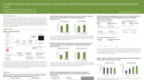 科学海报Complete Serum-Free Culture Kit and Protocols for Culturing High Yields of Functional Mature Neurons from Primary Embryonic Mouse CNS Tissues

 沪公网安备31010102008431号