Atkinson RL et al. ( 2013)
Breast cancer research : BCR 15 5 R77
Cancer stem cell markers are enriched in normal tissue adjacent to triple negative breast cancer and inversely correlated with DNA repair deficiency.
INTRODUCTION We hypothesized that cells present in normal tissue that bear cancer stem cell markers may represent a cancer cell of origin or a microenvironment primed for tumor development,and that their presence may correlate with the clinically defined subtypes of breast cancer that show increased tumorigenicity and stem cell features. methods: Normal tissues sampled at least 5 cm from primary tumors (normal adjacent tissue) were obtained from 61 chemotherapy-naive patients with breast cancer treated with mastectomy. Samples were stained simultaneously with immunofluorescence for CD44/CD49f/CD133/2 stem cell markers. We assessed the association between CD44+CD49f+CD133/2+ staining in normal adjacent tissue and breast cancer receptor subtype (defined by the expression of the estrogen (ER),progesterone (PR),or human epidermal growth factor-2 (Her2) receptors). We also examined the correlation between CD44+CD49f+CD133/2+ immunofluorescence and each of two previously published gene signatures,one derived from stem-cell enriched tissue and one from BRCA mutated tissue expected to have defective DNA repair. RESULTS Patients with triple negative breast cancer (ER–/PR–/HER2–) expressed CD44+CD49f+CD133/2+ in 9 of 9 normal adjacent tissue samples compared with 7 of 52 ER+ and/or Her2+ tumors (P textless 0.001). Further,expression of CD44+CD49f+CD133/2+ by normal adjacent tissue correlated positively with a stem cell-derived tumorigenic signature (P textless0.001) and inversely with a defective DNA-repair signature (P textless0.001). CONCLUSION Normal cells bearing cancer stem cell markers are associated with the triple negative receptor subtype of breast cancer. This study suggests stem cell staining and gene expression signatures from normal breast tissues represent novel tissue-based risk biomarkers for triple negative breast cancer. Validation of these results in additional studies of normal tissue from cancer-free women could lay the foundation for future targeted triple negative breast cancer prevention strategies.
View Publication
产品类型:
产品号#:
01700
01705
01702
产品名:
ALDEFLUOR™ 试剂盒
ALDEFLUOR™ DEAB试剂, 1.5 mM, 1 mL
ALDEFLUOR™检测缓冲液
Varga E et al. (OCT 2016)
Stem cell research 17 3 482--484
Generation of Mucopolysaccharidosis type II (MPS II) human induced pluripotent stem cell (iPSC) line from a 1-year-old male with pathogenic IDS mutation.
Peripheral blood was collected from a 1-year-old male patient with an X-linked recessive mutation of Iduronate 2-sulfatase (IDS) gene (NM000202.7(IDS):c.85CtextgreaterT) causing MPS II (OMIM 309900). Peripheral blood mononuclear cells (PBMCs) were reprogrammed by lentiviral delivery of a self-silencing hOKSM polycistronic vector. The pluripotency of the iPSC line was confirmed by the expression of pluripotency-associated markers and in vitro spontaneous differentiation towards the 3 germ layers. The iPSC line showed normal karyotype. The cell line offers a good platform to study MPS II pathophysiology,for drug testing,early biomarker discovery and gene therapy studies.
View Publication
产品类型:
产品号#:
05850
05857
05870
05875
85850
85857
85870
85875
产品名:
mTeSR™1
mTeSR™1
Ma D et al. (JAN 2017)
Stem cell research 18 51--53
Development of a human induced pluripotent stem cell (iPSC) line from a Parkinson's disease patient carrying the N551K variant in LRRK2 gene.
Peripheral blood mononuclear cells (PBMCs) were collected from a clinically diagnosed 64-year old male Parkinson's disease (PD) patient with N551K variant in the LRRK2 gene. The PMBCs were reprogrammed with the human OSKM transcription factors using the Sendai-virus reprogramming system. The transgene-free iPSC showed pluripotency confirmed by immunofluorescent staining for pluripotency markers and differentiated into the 3 germ layers in vivo. The iPSC line also showed normal karyotype. This cellular model can complement in vivo PD models for pathophysiological studies and drug screening.
View Publication
产品类型:
产品号#:
05850
05857
05870
05875
85850
85857
85870
85875
产品名:
mTeSR™1
mTeSR™1
Neben S et al. (MAR 1993)
Experimental hematology 21 3 438--43
Quantitation of murine hematopoietic stem cells in vitro by limiting dilution analysis of cobblestone area formation on a clonal stromal cell line.
Murine hematopoietic stem cells with varying proliferative capacity can be assayed by limiting dilution analysis of cobblestone area" (CA) formation on stromal layers in microlong-term bone marrow cultures. Cobblestone area forming cell (CAFC) frequency determined at early time points (day 7) correlates with mature stem cells measured as day 8 CFU-S�
View Publication
Della Chiesa M et al. (FEB 2010)
International immunology 22 2 91--100
GPR56 as a novel marker identifying the CD56dull CD16+ NK cell subset both in blood stream and in inflamed peripheral tissues.
To define novel human NK cell markers,we generated two mAbs specific for G-protein-coupled receptor 56 (GPR56),a surface glycoprotein that appears to be involved in cell-to-cell and cell-to-matrix interactions. GPR56 has been described in selected normal tissues,and in certain tumors,while,as yet,its expression on leukocytes is unknown. In this study,we show that anti-GPR56 mAbs,among leukocytes,prevalently recognize NK cells. In particular,these mAbs brightly stain CD56(dull) CD16(+) NK cells while react poorly with CD56(bright) CD16(+/-) NK cells. Consistently,we found that GPR56 was expressed on NK cells populating inflamed peripheral tissues while it was absent in lymph node-derived NK cells. We also show that activating stimuli,such as cytokines or exposure to monocyte-derived dendritic cell,down-regulate NK cell expression of GPR56 both at the protein and at the transcriptional level. Interestingly,IL-18,known to induce de novo expression of CCR7 on CD56(dull) CD16(+) NK cells,displayed the highest capability of modulating GPR56. Thus,together with the identification of GPR56 as a novel marker capable of discriminating different NK cells subsets,our data suggest that GPR56 may take part to the mechanisms regulating NK cell migration through the blood stream,peripheral tissues and lymph nodes.
View Publication
产品类型:
产品号#:
15025
15065
产品名:
RosetteSep™人NK细胞富集抗体混合物
RosetteSep™人NK细胞富集抗体混合物
Sikandar SS et al. (FEB 2010)
Cancer research 70 4 1469--78
NOTCH signaling is required for formation and self-renewal of tumor-initiating cells and for repression of secretory cell differentiation in colon cancer.
NOTCH signaling is critical for specifying the intestinal epithelial cell lineage and for initiating colorectal adenomas and colorectal cancers (CRC). Based on evidence that NOTCH is important for the maintenance and self-renewal of cancer-initiating cells in other malignancies,we studied the role of NOTCH signaling in colon cancer-initiating cells (CCIC). Tumors formed by CCICs maintain many properties of the primary CRCs from which they were derived,such as glandular organization,cell polarity,gap junctions,and expression of characteristic CRC molecular markers. Furthermore,CCICs have the property of self-renewal. In this study,we show that NOTCH signaling is 10- to 30-fold higher in CCIC compared with widely used colon cancer cell lines. Using small-molecule inhibition and short hairpin RNA knockdown,we show that NOTCH prevents CCIC apoptosis through repression of cell cycle kinase inhibitor p27 and transcription factor ATOH1. NOTCH is also critical to intrinsic maintenance of CCIC self-renewal and the repression of secretory cell lineage differentiation genes such as MUC2. Our findings describe a novel human cell system to study NOTCH signaling in CRC tumor initiation and suggest that inhibition of NOTCH signaling may improve CRC chemoprevention and chemotherapy.
View Publication
产品类型:
产品号#:
01700
01705
01702
产品名:
ALDEFLUOR™ 试剂盒
ALDEFLUOR™ DEAB试剂, 1.5 mM, 1 mL
ALDEFLUOR™检测缓冲液
Law JH et al. (JAN 2010)
PloS one 5 9
Molecular decoy to the Y-box binding protein-1 suppresses the growth of breast and prostate cancer cells whilst sparing normal cell viability.
The Y-box binding protein-1 (YB-1) is an oncogenic transcription/translation factor that is activated by phosphorylation at S102 whereby it induces the expression of growth promoting genes such as EGFR and HER-2. We recently illustrated by an in vitro kinase assay that a novel peptide to YB-1 was highly phosphorylated by the serine/threonine p90 S6 kinases RSK-1 and RSK-2,and to a lesser degree PKCα and AKT. Herein,we sought to develop this decoy cell permeable peptide (CPP) as a cancer therapeutic. This 9-mer was designed as an interference peptide that would prevent endogenous YB-1(S102) phosphorylation based on molecular docking. In cancer cells,the CPP blocked P-YB-1(S102) and down-regulated both HER-2 and EGFR transcript level and protein expression. Further,the CPP prevented YB-1 from binding to the EGFR promoter in a gel shift assay. Notably,the growth of breast (SUM149,MDA-MB-453,AU565) and prostate (PC3,LNCap) cancer cells was inhibited by ∼90% with the CPP. Further,treatment with this peptide enhanced sensitivity and overcame resistance to trastuzumab in cells expressing amplified HER-2. By contrast,the CPP had no inhibitory effect on the growth of normal immortalized breast epithelial (184htert) cells,primary breast epithelial cells,nor did it inhibit differentiation of hematopoietic progenitors. These data collectively suggest that the CPP is a novel approach to suppressing the growth of cancer cells while sparing normal cells and thereby establishes a proof-of-concept that blocking YB-1 activation is a new course of cancer therapeutics.
View Publication
产品类型:
产品号#:
05601
18056
18056RF
04435
04445
产品名:
EpiCult™-B 人培养基
MethoCult™ H4435 Enriched
MethoCult™ H4435 Enriched
Pineda JR et al. (APR 2013)
EMBO Molecular Medicine 5 4 548--562
Vascular-derived TGF-β increases in the stem cell niche and perturbs neurogenesis during aging and following irradiation in the adult mouse brain
Neurogenesis decreases during aging and following cranial radiotherapy,causing a progressive cognitive decline that is currently untreatable. However,functional neural stem cells remained present in the subventricular zone of high dose-irradiated and aged mouse brains. We therefore investigated whether alterations in the neurogenic niches are perhaps responsible for the neurogenesis decline. This hypothesis was supported by the absence of proliferation of neural stem cells that were engrafted into the vascular niches of irradiated host brains. Moreover,we observed a marked increase in TGF-β1 production by endothelial cells in the stem cell niche in both middle-aged and irradiated mice. In co-cultures,irradiated brain endothelial cells induced the apoptosis of neural stem/progenitor cells via TGF-β/Smad3 signalling. Strikingly,the blockade of TGF-β signalling in vivo using a neutralizing antibody or the selective inhibitor SB-505124 significantly improved neurogenesis in aged and irradiated mice,prevented apoptosis and increased the proliferation of neural stem/progenitor cells. These findings suggest that anti-TGF-β-based therapy may be used for future interventions to prevent neurogenic collapse following radiotherapy or during aging.
View Publication
Bö et al. (DEC 2005)
Journal of Immunological Methods 307 1-2 13--23
Establishment of a strategy for the rapid generation of a monoclonal antibody against the human protein SNEV (hNMP200) by flow-cytometric cell sorting
The screening for antigen-specific hybridoma cells with adequate production rates is still a time-,labour- and money-consuming procedure. A reduction in cell culture testing by specifically selecting those fused cells that produce antibody could therefore make hybridoma technology more attractive,even for small research groups or for newly discovered proteins at an early stage of research. Additional problems,such as the requirement to produce sufficient amounts of the unknown protein at a purity that allows specific immunisation of mice and testing of the resulting hybridoma clones,also need to be overcome. Here we present a new strategy to isolate rapidly and efficiently monoclonal antibodies against new proteins,for which only sequence information at the DNA level is known. The strategy consists of fusion of the protein to a hexa-His-tag to allow easy purification,production in yeast and insect cells to reduce background immunisation with host cell proteins and the selection of IgG-producing hybridoma cells by flow-cytometric cell sorting using the affinity matrix secretion assay technique. ?? 2005 Elsevier B.V. All rights reserved.
View Publication


 EasySep™小鼠TIL(CD45)正选试剂盒





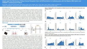 科学海报Simple Label-free Monitoring of Robust Human Pluripotent Stem Cell Differentiation to Cardiomyocytes with the Maestro MEA System and STEMdiff™ Cardiomyocyte Products
 小鼠IL-5 ELISA试剂盒 用于小鼠白细胞介素5的检测和测定
 重组人 IL-12 白介素12
 沪公网安备31010102008431号