Jain AK et al. (JAN 2012)
PLoS Biology 10 2 e1001268
P53 regulates cell cycle and micrornas to promote differentiation of human embryonic stem cells
Multiple studies show that tumor suppressor p53 is a barrier to dedifferentiation; whether this is strictly due to repression of proliferation remains a subject of debate. Here,we show that p53 plays an active role in promoting differentiation of human embryonic stem cells (hESCs) and opposing self-renewal by regulation of specific target genes and microRNAs. In contrast to mouse embryonic stem cells,p53 in hESCs is maintained at low levels in the nucleus,albeit in a deacetylated,inactive state. In response to retinoic acid,CBP/p300 acetylates p53 at lysine 373,which leads to dissociation from E3-ubiquitin ligases HDM2 and TRIM24. Stabilized p53 binds CDKN1A to establish a G(1) phase of cell cycle without activation of cell death pathways. In parallel,p53 activates expression of miR-34a and miR-145,which in turn repress stem cell factors OCT4,KLF4,LIN28A,and SOX2 and prevent backsliding to pluripotency. Induction of p53 levels is a key step: RNA-interference-mediated knockdown of p53 delays differentiation,whereas depletion of negative regulators of p53 or ectopic expression of p53 yields spontaneous differentiation of hESCs,independently of retinoic acid. Ectopic expression of p53R175H,a mutated form of p53 that does not bind DNA or regulate transcription,failed to induce differentiation. These studies underscore the importance of a p53-regulated network in determining the human stem cell state.
View Publication
产品类型:
产品号#:
05850
05857
05870
05875
07923
85850
85857
85870
85875
产品名:
Dispase (1 U/mL)
mTeSR™1
mTeSR™1
Naujok O and Lenzen S (SEP 2012)
Stem Cell Reviews and Reports 8 3 779--791
A critical re-evaluation of CD24-positivity of human embryonic stem cells differentiated into pancreatic progenitors.
Differentiation of embryonic stem cells (ESCs) into insulin-producing cells for cell replacement therapy of diabetes mellitus comprises the stepwise recapitulation of in vivo developmental stages of pancreatic organogenesis in an in vitro differentiation protocol. The chemical compounds IDE-1 and (-)-indolactam-V can be used to direct mouse and human ESCs through these stages to form definitive endoderm via an intermediate mesendodermal stage and finally into pancreatic endoderm. Cells of the pancreatic endoderm express the PDX1 transcription factor and contribute to all pancreatic cell types upon further in vitro or in vivo differentiation. Even though this differentiation approach is highly effective and reproducible,it generates heterogeneous populations containing PDX1-expressing pancreatic progenitors amongst other cell types. Thus,a technique to separate PDX1-expressing cells from this mixture is very desirable. Recently it has been reported that PDX1-positive pancreatic progenitors,derived from human embryonic stem cells,express the surface marker CD24. Therefore were subjected mouse and human ESCs to a small molecule differentiation approach and the expression of the surface marker CD24 was monitored in undifferentiated cells,cells committed to the definitive endoderm and cells reminiscent of the pancreatic endoderm. We observed that both mouse and human ESCs expressed CD24 in the pluripotent state,during the whole process of endoderm formation and upon further differentiation towards pancreatic endoderm. Thus CD24 is not a suitable cell surface marker for identification of PDX1-positive progenitor cells.
View Publication
产品类型:
产品号#:
05850
05857
05870
05875
07923
85850
85857
85870
85875
产品名:
Dispase (1 U/mL)
mTeSR™1
mTeSR™1
Miyazaki T et al. ( 2012)
Nature communications 3 1236
Laminin E8 fragments support efficient adhesion and expansion of dissociated human pluripotent stem cells.
Human embryonic stem cells (hESCs) and induced pluripotent stem cells (hiPSCs) have the potential to provide an infinite source of tissues for regenerative medicine. Although defined xeno-free media have been developed,culture conditions for reliable propagation of hESCs still require considerable improvement. Here we show that recombinant E8 fragments of laminin isoforms (LM-E8s),which are the minimum fragments conferring integrin-binding activity,promote greater adhesion of hESCs and hiPSCs than do Matrigel and intact laminin isoforms. Furthermore,LM-E8s sustain long-term self-renewal of hESCs and hiPSCs in defined xeno-free media with dissociated cell passaging. We successfully maintained three hESC and two hiPSC lines on LM-E8s in three defined media for 10 passages. hESCs maintained high level expression of pluripotency markers,had a normal karyotype after 30 passages and could differentiate into all three germ layers. This culture system allows robust proliferation of hESCs and hiPSCs for therapeutic applications.
View Publication
产品类型:
产品号#:
05850
05857
05870
05875
85850
85857
85870
85875
产品名:
mTeSR™1
mTeSR™1
Talavera-Adame D et al. (NOV 2016)
Diabetologia 59 11 2378--2386
Effective endothelial cell and human pluripotent stem cell interactions generate functional insulin-producing beta cells.
AIMS/HYPOTHESIS Endothelial cells (ECs) play an essential role in pancreatic organogenesis. We hypothesise that effective in vitro interactions between human microvascular endothelial cells (HMECs) and human pluripotent stem cells (hPSCs) results in the generation of functional pancreatic beta cells. METHODS Embryoid bodies (EBs) derived from hPSCs were cultured alone (controls) or with ECs in collagen gels. Subsequently,cells were analysed for pancreatic beta cell markers,and then isolated and expanded. Insulin secretion in response to glucose was evaluated in vitro by static and dynamic (perifusion) assays,and in vivo by EB transplantation into immunodeficient mice. RESULTS Co-cultured EBs had a higher expression of mature beta cells markers and enhanced insulin secretion in vitro,compared with controls. In mice,transplanted EBs had higher levels of human C-peptide secretion with a significant reduction in hyperglycaemia after the selective destruction of native pancreatic beta cells. In addition,there was significant in vitro upregulation of bone morphogenetic proteins 2 and 4 (BMP-2,4) in co-cultured cells,compared with controls. CONCLUSIONS/INTERPRETATION ECs provide essential signalling in vitro,such as activation of the BMP pathway,for derivation of functional insulin-producing beta cells from hPSCs.
View Publication
产品类型:
产品号#:
05850
05857
05870
05875
85850
85857
85870
85875
产品名:
mTeSR™1
mTeSR™1
Wang W et al. (MAR 2017)
Stem cells and development 26 6 394--404
Development of Islet Organoids from H9 Human Embryonic Stem Cells in Biomimetic 3D Scaffolds.
Success in the differentiating human embryonic stem cells (hESCs) into insulin-secreting β cells raises new hopes for diabetes treatment. In this work,we demonstrated the feasibility of developing islet organoids from hESCs within biomimetic 3D scaffolds. We showed that such a 3D microenvironment is critical to the generation of pancreatic endoderm and endocrine from hESCs. The organoids formed consisted of pancreatic α,β,δ,and pancreatic polypeptide (PP) cells. A high-level co-expression of PDX1,NKX6.1,and NGN3 in these cells suggests the characteristics of pancreatic β cells. More importantly,most insulin-secreting cells generated did not express glucagon,somatostatin,or PP. The expression of mature β cell marker genes such as Pdx1,Ngn3,Insulin,MafA,and Glut2 was detected in these 3D-induced cell clusters. A high-level expression of C-peptide confirmed the de novo endogenous insulin production in these 3D induced cells. Insulin-secretory granules,an indication of β cell maturity,were detected in these cells as well. Glucose challenging experiments suggested that these cells are sensitive to glucose levels due to their elevated maturity. Exposing the cells to a high concentration of glucose induced a sharp increase in insulin secretion.
View Publication
S. Schmidt et al. (nov 2019)
Pathogens (Basel,Switzerland) 8 4
Distinct Effects of Immunosuppressive Drugs on the Anti-Aspergillus Activity of Human Natural Killer Cells.
As the prognosis of invasive aspergillosis remains unacceptably poor in patients undergoing hematopoietic stem cell transplantation (HSCT),there is a growing interest in the adoptive transfer of antifungal effector cells,such as Natural Killer (NK) cells. Because immunosuppressive agents are required in most HSCT recipients,knowledge of the impact of these compounds on the antifungal activity of NK cells is a prerequisite for clinical trials. We,therefore,assessed the effect of methylprednisolone (mPRED),cyclosporin A (CsA) and mycophenolic acid (MPA) at different concentrations on proliferation,apoptosis/necrosis,and the direct and indirect anti-Aspergillus activity of human NK cells. Methylprednisolone decreased proliferation and increased apoptosis of NK cells in a significant manner. After seven days,a reduction of viable NK cells was seen for all three immunosuppressants,which was significant for MPA only. Cyclosporin A significantly inhibited the direct hyphal damage by NK cells in a dose-dependent manner. None of the immunosuppressive compounds had a major impact on the measured levels of interferon-$\gamma$,granulocyte-macrophage colony-stimulating factor and RANTES (regulated on activation,normal T cell expressed and secreted; CCL5). Our data demonstrate that commonly used immunosuppressive compounds have distinct effects on proliferation,viability and antifungal activity of human NK cells,which should be considered in designing studies on the use of NK cells for adoptive antifungal immunotherapy.
View Publication
产品类型:
产品号#:
19055
19055RF
19059
19059RF
产品名:
EasySep™人NK细胞富集试剂盒
RoboSep™ 人NK细胞富集试剂盒含滤芯吸头
EasySep™人单核细胞富集试剂盒
RoboSep™ 人单核细胞富集试剂盒含滤芯吸头
J.-H. Kim et al. (mar 2020)
International journal of molecular sciences 21 6
Thymosin $\beta$4-Enhancing Therapeutic Efficacy of Human Adipose-Derived Stem Cells in Mouse Ischemic Hindlimb Model.
Thymosin $\beta$4 (T$\beta$4) is a G-actin sequestering protein that contributes to diverse cellular activities,such as migration and angiogenesis. In this study,the beneficial effects of combined cell therapy with T$\beta$4 and human adipose-derived stem cells (hASCs) in a mouse ischemic hindlimb model were investigated. We observed that exogenous treatment with T$\beta$4 enhanced endogenous TMSB4X mRNA expression and promoted morphological changes (increased cell length) in hASCs. Interestingly,T$\beta$4 induced the active state of hASCs by up-regulating intracellular signaling pathways including the PI3K/AKT/mTOR and MAPK/ERK pathways. Treatment with T$\beta$4 significantly increased cell migration and sprouting from microbeads. Moreover,additional treatment with T$\beta$4 promoted the endothelial differentiation potential of hASCs by up-regulating various angiogenic genes. To evaluate the in vivo effects of the T$\beta$4-hASCs combination on vessel recruitment,dorsal window chambers were transplanted,and the co-treated mice were found to have a significantly increased number of microvessel branches. Transplantation of hASCs in combination with T$\beta$4 was found to improve blood flow and attenuate limb or foot loss post-ischemia compared to transplantation with hASCs alone. Taken together,the therapeutic application of hASCs combined with T$\beta$4 could be effective in enhancing endothelial differentiation and vascularization for treating hindlimb ischemia.
View Publication
产品类型:
产品号#:
05411
产品名:
MesenCult™ 增殖试剂盒(人)
J. Han et al. (jan 2009)
Cytotechnology 59 1 45--53
Anti-proliferative and apoptotic effects of oleuropein and hydroxytyrosol on human breast cancer MCF-7 cells.
Olive oil intake has been shown to induce significant levels of apoptosis in various cancer cells. These anti-cancer properties are thought to be mediated by phenolic compounds present in olive. These beneficial health effects of olive have been attributed,at least in part,to the presence of oleuropein and hydroxytyrosol. In this study,oleuropein and hydroxytyrosol,major phenolic compound of olive oil,was studied for its effects on growth in MCF-7 human breast cancer cells using assays for proliferation (MTT assay),cell viability (Guava ViaCount assay),cell apoptosis,cellcycle (flow cytometry). Oleuropein or hydroxytyrosol decreased cell viability,inhibited cell proliferation,and induced cell apoptosis in MCF-7 cells. Result of MTT assay showed that 200 mug/mL of oleuropein or 50 mug/mL of hydroxytyrosol remarkably reduced cell viability of MCF-7 cells. Oleuropein or hydroxytyrosol decrease of the number of MCF-7 cells by inhibiting the rate of cell proliferation and inducing cell apoptosis. Also hydroxytyrosol and oleuropein exhibited statistically significant block of G(1) to S phase transition manifested by the increase of cell number in G(0)/G(1) phase.
View Publication
Taubert I et al. (APR 2011)
Cytotherapy 13 4 459--66
Characterization of hematopoietic stem cell subsets from patients with multiple myeloma after mobilization with plerixafor.
BACKGROUND AIMS: Previous studies have demonstrated that the combination of granulocyte-colony-stimulating factor (G-CSF) + plerixafor is more efficient in mobilizing CD34(+) hematopoietic stem cells (HSC) into the peripheral blood than G-CSF alone. In this study we analyzed the impact of adding plerixafor to G-CSF upon the mobilization of different HSC subsets. METHODS: We characterized the immunophenotype of HSC subsets isolated from the peripheral blood of eight patients with multiple myeloma (MM) before and after treatment with plerixafor. All patients were supposed to collect stem cells prior to high-dose chemotherapy and consecutive autologous stem cell transplantation,and therefore received front-line mobilization with 4 days of G-CSF followed by a single dose of plerixafor. Samples of peripheral blood were analyzed comparatively by flow cytometry directly before and 12 h after administration of plerixafor. RESULTS: The number of aldehyde dehydrogenase (ALDH)(bright) and CD34(+) cells was significantly higher after plerixafor treatment (1.2-5.0 and 1.5-6.0 times; both P textless 0.01) and an enrichment of the very primitive CD34(+) CD38(-) and ALDH(bright) CD34(+) CD38(-) HSC subsets was detectable. Additionally,two distinct ALDH(+) subsets could be clearly distinguished. The small ALDH(high) subset showed a higher number of CD34(+) CD38(-) cells in contrast to the total ALDH(bright) subpopulation and probably represented a very primitive subpopulation of HSC. CONCLUSIONS: A combined staining of ALDH,CD34 and CD38 might represent a powerful tool for the identification of a very rare and primitive hematopoietic stem cell subset. The addition of plerixafor mobilized not only more CD34(+) cells but was also able to increase the proportion of more primitive stem cell subsets.
View Publication


 EasySep™小鼠TIL(CD45)正选试剂盒





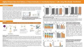 科学海报Highly Efficient Non-Viral Delivery of Macromolecules to Unactivated Human T Cells Using Microfluidic Transfection

 沪公网安备31010102008431号