X. Liu et al. ( 2017)
Nature Protocols 12 2 439--451
Conditional reprogramming and long-term expansion of normal and tumor cells from human biospecimens
Historically,it has been difficult to propagate cells in vitro that are derived directly from human tumors or healthy tissue. However,in vitro preclinical models are essential tools for both the study of basic cancer biology and the promotion of translational research,including drug discovery and drug target identification. This protocol describes conditional reprogramming (CR),which involves coculture of irradiated mouse fibroblast feeder cells with normal and tumor human epithelial cells in the presence of a Rho kinase inhibitor (Y-27632). CR cells can be used for various applications,including regenerative medicine,drug sensitivity testing,gene expression profiling and xenograft studies. The method requires a pathologist to differentiate healthy tissue from tumor tissue,and basic tissue culture skills. The protocol can be used with cells derived from both fresh and cryopreserved tissue samples. As approximately 1 million cells can be generated in 7 d,the technique is directly applicable to diagnostic and predictive medicine. Moreover,the epithelial cells can be propagated indefinitely in vitro,yet retain the capacity to become fully differentiated when placed into conditions that mimic their natural environment.
View Publication
产品类型:
产品号#:
100-0352
产品名:
条件性重编程(CR)培养基
L. Chen et al. (nov 2016)
Biochemical and biophysical research communications 480 4 515--521
AMPK activation by GSK621 inhibits human melanoma cells in vitro and in vivo.
Recent studies suggest that forced activation of AMP-activated protein kinase (AMPK) could inhibit melanoma cell proliferation. In this report,we evaluated the anti-melanoma cell activity by a novel small-molecular AMPK activator,GSK621. Treatment of GSK621 decreased survival and proliferation of human melanoma cells (A375,WM-115 and SK-Mel-2 lines),which was accompanied by activation of caspase-3/-9 and apoptosis. Reversely,caspase inhibitors attenuated GSK621-induced cytotoxicity against melanoma cells. Significantly,GSK621 was more potent than other AMPK activators (A769662,Compound 13 and AICAR) in inhibiting melanoma cells. Intriguingly,same GSK621 treatment was non-cytotoxic or pro-apoptotic against human melanocytes. Molecularly,we showed that activation of AMPK mediated GSK621's activity against melanoma cells. AMPK$\alpha$1 shRNA knockdown or dominant negative mutation (T172A) dramatically attenuated GSK621-induced melanoma cell lethality. Further studies revealed that MEK-ERK activation might be the primary resistance factor of GSK621. MEK-ERK inhibition,either genetically or pharmacologically,significantly sensitized melanoma cells to GSK-621. Remarkably,intraperitoneal (i.p.) injection of GSK621 inhibited A375 tumor growth in SCID mice. Co-administration of MEK-ERK inhibitor MEK162 further sensitized GSK621-induced anti-A375 tumor activity in vivo. Together,the results imply that targeted activation of AMPK by GSK621 inhibits melanoma cell survival and proliferation. MEK-ERK inhibition may further sensitize GSK621's anti-melanoma cell activity in vitro and in vivo.
View Publication
产品类型:
产品号#:
100-0265
产品名:
N. Schrantz et al. (may 1999)
Cell death and differentiation 6 5 445--53
Manganese induces apoptosis of human B cells: caspase-dependent cell death blocked by bcl-2.
Manganese ions block apoptosis of phagocytes induced by various agents. The prevention of apoptosis was attributed to the activation of manganous superoxide dismutase (Mn-SOD) and to the antioxidant function of free Mn2+ cations. However,the effect of Mn2+ on B cell apoptosis is not documented. In this study,we investigated the effects of Mn2+ on the apoptotic process in human B cells. We observed that Mn2+ but not Mg2+ or Ca2+,inhibited cell growth and induced apoptosis of activated tonsilar B cells,Epstein Barr virus (EBV)-negative Burkitt's lymphoma cell lines (BL-CL) and EBV-transformed B cell lines (EBV-BCL). In the same conditions,no apoptosis was observed in U937,a monoblastic cell line. Induction of B cell apoptosis by Mn2+ was time- and dose-dependent. The cell permeable tripeptide inhibitor of ICE family cysteine proteases,zVAD-fmk,suppressed Mn2+-induced apoptosis. Furthermore,Mn2+ triggered the activation of interleukin-1beta converting enzyme (ICE/caspase 1),followed by the activation of CPP32/Yama/Apopain/caspase-3. In addition,poly-(ADP-ribose) polymerase (PARP),a cellular substrate for CPP32 protease was degraded to generate apoptotic fragments in Mn2+-treated B cell lines. The inhibitor,zVAD-fmk suppressed Mn2+-triggered CPP32 activation and PARP cleavage and apoptosis. These results indicate that the activation of caspase family proteases is required for the apoptotic process induced by Mn2+ treatment of B cells. While the caspase-1 inhibitor YVAD was unable to block apoptosis,the caspase-3 specific inhibitor DEVD-cmk,partially inhibited Mn2+-induced CPP32 activation,PARP cleavage and apoptosis of cells. Moreover,Bcl-2 overexpression in BL-CL effectively protected cells from apoptosis and cell death induced by manganese. This is the first report showing the involvement of Mn2+ in the regulation of B lymphocyte death presumably via a caspase-dependent process with a death-protective effect of Bcl-2.
View Publication
产品类型:
产品号#:
100-0532
产品名:
Ac-DEVD-CMK (Trifluoroacetate Salt)
Rushkevich YN et al. (AUG 2015)
Bulletin of experimental biology and medicine 159 4 576--81
The Use of Autologous Mesenchymal Stem Cells for Cell Therapy of Patients with Amyotrophic Lateral Sclerosis in Belarus.
We studied a new method of treatment of amyotrophic lateral sclerosis with autologous mesenchymal stem cells. Autologous mesenchymal stem cells were injected intravenously (intact cells) or via lumbar puncture (cells committed to neuronal differentiation). Evaluation of the results of cell therapy after 12-month follow-up revealed slowing down of the disease progression in 10 patients in comparison with the control group consisting of 15 patients. The cell therapy was safe for the patients.
View Publication
产品类型:
产品号#:
05761
产品名:
用于小鼠和大鼠神经干细胞和祖细胞分化培养的试剂盒
Y-L. Chiang et al. (Dec 2025)
International Journal of Molecular Sciences 27 1
Induced Pluripotent Stem Cell-Derived Dendritic Cells Provide a Reliable In Vitro Platform for Functional Screening of Immunoregulatory Probiotics
The immunoregulatory effects of probiotics have been widely studied,particularly in maintaining immune balance. Conventional in vitro functional screening of probiotics relies on fresh donor-derived primary immune cells,which often exhibit significant inter-individual and temporal variability,limiting reproducibility and interpretation. As an alternative,human-induced pluripotent stem cell (iPSC)-derived dendritic cells were co-cultured with five probiotic strains in the current study to evaluate their immunomodulatory interactions. To assess whether cytokines produced by probiotic-stimulated dendritic cells can influence T cell differentiation,human CD4+ T cells were exposed to the conditioned medium derived from co-cultures. Enzyme-linked immunosorbent assay results demonstrated that iPSC-derived dendritic cells secreted cytokines at distinct concentrations in response to different probiotic strains,suggesting that these cells can distinguish between different microbial stimuli,and supporting their use in functional probiotic screening. Among the five strains tested,Lactiplantibacillus plantarum LPA-56,Limosilactobacillus reuteri RU-23,and Lactobacillus fermentum Fem-99 induced cytokine production levels that promoted the differentiation of the human CD4+ T cells into regulatory T cells. These findings demonstrate that iPSC-derived dendritic cells have immunomodulatory potential,are reliable for in vitro screening of probiotics,and offer a promising strategy for selecting potent immunoregulatory probiotic candidates.
View Publication
Adherent cells generated during long-term culture of human umbilical cord blood CD34+ cells have characteristics of endothelial cells and beneficial effect on cord blood ex vivo expansion.
Hematopoiesis depends on the association of hematopoietic stem cells with stromal cells that constitute the hematopoietic microenvironment. The in vitro development of the endothelial cell from umbilical cord blood (UCB) is not well established and has met very limited success. In this study,UCB CD34(+) cells were cultured for 5 weeks in a stroma-free liquid culture system using thrombopoietin,flt3 ligand,and granulocyte-colony stimulating factor. By week 4-5,we found that firmly adherent fibroblast-like cells were established. These cells showed characteristics of endothelial cells expressing von Willebrand factor,human vascular cell adhesion molecule-1,human intracellular adhesion molecule-1,human CD31,E-selectin,and human macrophage. Furthermore,when comparing an ex vivo system without an established endothelial monolayer to an ex vivo system with an established endothelial monolayer,better expansion of total nucleated cells,CD34(+) cells,and colony-forming units (CFUs)-granulocyte-macrophage and CFUs-granulocyte-erythroid-megakaryocyte-macrophage were found during culture. This phenomenon was in part due to the fact that a significant reduction of apoptotic fractions was found in the CD34(+) cells,which were cultured on the adherent monolayer for up to 5 weeks. To gather quantitative data on the number of endothelial cells derived from a given number of CD34 cells,we performed limiting dilution assay by using Poisson distribution: the number of tested cells (linear scale) producing a 37% negative culture (logarithmic scale) is the number of cells containing one endothelial cell. By this method,one endothelial cell may be found from 314 CD34(+) cells after 5 weeks of culture. These results suggest that the UCB CD34(+) cell fraction contains endothelial cell precursors,establishing the hematopoietic microenvironment and providing the beneficial effects through downregulating apoptosis on UCB expansion protocols. These observations may provide insight for future cellular therapy or graft engineering.
View Publication
产品类型:
产品号#:
04434
04444
产品名:
MethoCult™ H4434 Classic
MethoCult™ H4434 Classic
J. Shao et al. (FEB 2017)
Scientific reports 7 42363
Experimental Study of the Biological Properties of Human Embryonic Stem Cell-Derived Retinal Progenitor Cells.
Retinal degenerative diseases are among the leading causes of blindness worldwide,and cell replacement is considered as a promising therapeutic. However,the resources of seed cells are scarce. To further explore this type of therapy,we adopted a culture system that could harvest a substantial quantity of retinal progenitor cells (RPCs) from human embryonic stem cells (hESCs) within a relatively short period of time. Furthermore,we transplanted these RPCs into the subretinal spaces of Royal College of Surgeons (RCS) rats. We quantified the thickness of the treated rats' outer nuclear layers (ONLs) and explored the visual function via electroretinography (ERG). It was found that the differentiated cells expressed RPC markers and photoreceptor progenitor markers. The transplanted RPCs survived for at least 12 weeks,resulting in beneficial effects on the morphology of the host retina,and led to a significant improvement in the visual function of the treated animals. These therapeutic effects suggest that the hESCs-derived RPCs could delay degeneration of the retina and partially restore visual function.
View Publication
Ben-Kasus T et al. (JUL 2005)
Biochemical pharmacology 70 1 121--33
Metabolic activation of zebularine, a novel DNA methylation inhibitor, in human bladder carcinoma cells.
Zebularine (2(1H)-pyrimidinone riboside,Zeb),a synthetic analogue of cytidine that is a potent inhibitor of cytidine deaminase,has been recently identified as a general inhibitor of DNA methylation. This inhibition of DNA methyltransferase (DNMT) is hypothesized to be mechanism-based and result from formation of a covalent complex between the enzyme and zebularine-substituted DNA. Metabolic activation of Zeb thus requires that it be phosphorylated and incorporated into DNA. We have quantitatively assessed the phosphorylation and DNA incorporation of Zeb in T24 cells using 2-[(14)C]-Zeb in conjunction with gradient anion-exchange HPLC and selected enzymatic and spectroscopic analyses. The corresponding 5'-mono-,di- and triphosphates of Zeb were readily formed in a dose- and time-dependent manner. Two additional Zeb-containing metabolites were tentatively identified as diphosphocholine (Zeb-DP-Chol) and diphosphoethanolamine adducts. Intracellular concentrations of Zeb-TP and Zeb-DP-Chol were similar and greatly exceeded those of other metabolites. DNA incorporation occurred but was surpassed by that of RNA by at least seven-fold. Equivalent levels and similar intracellular metabolic patterns were also observed in the Molt-4 (human T-lymphoblasts) and MC38 (murine colon carcinoma) cell lines. For male BALB/c nu/nu mice implanted s.c. with the EJ6 variant of T24 bladder carcinoma and treated i.p. with 500mg/kg 2-[(14)C]-Zeb,the in vivo phosphorylation pattern of Zeb in tumor tissue examined 24h after drug administration was similar to that observed in vitro. The complex metabolism of Zeb and its limited DNA incorporation suggest that these are the reasons why it is less potent than either 5-azacytidine or 5-aza-2'-deoxycytidine and requires higher doses for equivalent inhibition of DNMT.
View Publication
产品类型:
产品号#:
72902
产品名:
Zebularine
Gaur M et al. (OCT 2010)
Cytotherapy 12 6 807--17
Timed inhibition of p38MAPK directs accelerated differentiation of human embryonic stem cells into cardiomyocytes.
BACKGROUND AIMS Heart failure therapy with human embryonic stem cell (hESC)-derived cardiomyocytes (hCM) has been limited by the low rate of spontaneous hCM differentiation. As others have shown that p38 mitogen-activated protein kinase (p38MAPK) directs neurogenesis from mouse embryonic stem cells,we investigated whether the p38MAPK inhibitor,SB203580,might influence hCM differentiation. METHODS We treated differentiating hESC with SB203580 at specific time-points,and used flow cytometry,immunocytochemistry,quantitative real-time (RT)-polymerase chain reaction (PCR),teratoma formation and transmission electron microscopy to evaluate cardiomyocyte formation. RESULTS We observed that the addition of inhibitor resulted in 2.1-fold enrichment of spontaneously beating human embryoid bodies (hEB) at 21 days of differentiation,and that 25% of treated cells expressed cardiac-specific α-myosin heavy chain. This effect was dependent on the stage of differentiation at which the inhibitor was introduced. Immunostaining and teratoma formation assays demonstrated that the inhibitor did not affect hESC pluripotency; however,treated hESC gave rise to hCM exhibiting increased expression of sarcomeric proteins,including cardiac troponin T,myosin light chain and α-myosin heavy chain. This was consistent with significantly increased numbers of myofibrillar bundles and the appearance of nascent Z-bodies at earlier time-points in treated hCM. Treated hEB also demonstrated a normal karyotype by array comparative genomic hybridization and viability in vivo following injection into mouse myocardium. CONCLUSIONS These studies demonstrate that p38MAPK inhibition accelerates directed hCM differentiation from hESC,and that this effect is developmental stage-specific. The use of this inhibitor should improve our ability to generate hESC-derived hCM for cell-based therapy.
View Publication
产品类型:
产品号#:
72222
产品名:
SB203580 (Hydrochloride)
Deng Y et al. (FEB 2017)
Biomacromolecules 18 2 587--598
Peptide-Decorated Nanofibrous Niche Augments In Vitro Directed Osteogenic Conversion of Human Pluripotent Stem Cells.
Realization of clinical potential of human pluripotent stem cells (hPSCs) in bone regenerative medicine requires development of simple and safe biomaterials for expansion of hPSCs followed by directing their lineage commitment to osteoblasts. In the present study,a chemically defined peptide-decorated polycaprolactone (PCL) nanofibrous microenvironment was prepared through electrospinning technology and subsequent conjugation with vitronectin peptide to promote the culture and osteogenic potential of hPSCs in vitro. The results indicated that hPSCs successfully proliferated and maintained their pluripotency on the biointerface of peptide-conjugated nanofibers without Matrigel under defined conditions. Moreover,the prepared niche exhibited an appealing ability in promoting directed differentiation of hPSCs to osteoblastic phenotype without embryoid body formation step,determined from the cell morphological alteration,alkaline phosphate activity,and osteogenesis-related gene expression,as well as protein production. Such well-defined,xeno-free,and safe nanofiber scaffolds that allow the survival and facilitate osteo-differentiation of hPSCs provide a novel platform for hPSCs differentiation via cell-nanofiber interplay,and possess great value in accelerating the translational perspectives of hPSCs in bone tissue engineering.
View Publication
产品类型:
产品号#:
85850
85857
85870
85875
产品名:
mTeSR™1
mTeSR™1
Yamane J et al. (MAY 2016)
Nucleic Acids Research 44 12 5515--5528
Prediction of developmental chemical toxicity based on gene networks of human embryonic stem cells
Predictive toxicology using stem cells or their derived tissues has gained increasing importance in biomedical and pharmaceutical research. Here,we show that toxicity category prediction by support vector machines (SVMs),which uses qRT-PCR data from 20 categorized chemicals based on a human embryonic stem cell (hESC) system,is improved by the adoption of gene networks,in which network edge weights are added as feature vectors when noisy qRT-PCR data fail to make accurate predictions. The accuracies of our system were 97.5-100% for three toxicity categories: neurotoxins (NTs),genotoxic carcinogens (GCs) and non-genotoxic carcinogens (NGCs). For two uncategorized chemicals,bisphenol-A and permethrin,our system yielded reasonable results: bisphenol-A was categorized as an NGC,and permethrin was categorized as an NT; both predictions were supported by recently published papers. Our study has two important features: (i) as the first study to employ gene networks without using conventional quantitative structure-activity relationships (QSARs) as input data for SVMs to analyze toxicogenomics data in an hESC validation system,it uses additional information of gene-to-gene interactions to significantly increase prediction accuracies for noisy gene expression data; and (ii) using only undifferentiated hESCs,our study has considerable potential to predict late-onset chemical toxicities,including abnormalities that occur during embryonic development.
View Publication

 EasySep™小鼠TIL(CD45)正选试剂盒


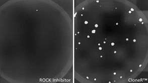 新闻STEMCELL Technologies to Launch CloneR™ to Facilitate Genome Editing of Human Pluripotent Stem Cells
 沪公网安备31010102008431号