Azari H et al. (JAN 2011)
Journal of visualized experiments : JoVE 49
Neural-colony forming cell assay: an assay to discriminate bona fide neural stem cells from neural progenitor cells.
The neurosphere assay (NSA) is one of the most frequently used methods to isolate,expand and also calculate the frequency of neural stem cells (NSCs). Furthermore,this serum-free culture system has also been employed to expand stem cells and determine their frequency from a variety of tumors and normal tissues. It has been shown recently that a one-to-one relationship does not exist between neurosphere formation and NSCs. This suggests that the NSA as currently applied,overestimates the frequency of NSCs in a mixed population of neural precursor cells isolated from both the embryonic and adult mammalian brain. This video practically demonstrates a novel collagen based semi- solid assay,the neural-colony forming cell assay (N-CFCA),which has the ability to discriminate stem from progenitor cells based on their long-term proliferative potential,and thus provides a method to enumerate NSC frequency. In the N-CFCA,colonies ≥2 mm in diameter are derived from cells that meet all the functional criteria of a NSC,while colonies textless 2mm are derived from progenitors. The N-CFCA procedure can be used for cells prepared from different sources including primary and cultured adult or embryonic mouse CNS cells. Here we use cells prepared from passage one neurospheres generated from embryonic day 14 mice brain to perform N-CFCA. The cultures are replenished with proliferation medium every seven days for three weeks to allow the plated cells to exhibit their full proliferative potential and then the frequency of neural progenitor and bona fide neural stem cells is calculated respectively by counting the number of colonies that are textless 2mm and the ones that are ≥2mm in reference to the number of cells that were initially plated.
View Publication
产品类型:
产品号#:
05740
产品名:
Kim E-K et al. (APR 2012)
Journal of cellular physiology 227 4 1680--7
Human mesenchymal stem cell differentiation to the osteogenic or adipogenic lineage is regulated by AMP-activated protein kinase.
AMP-activated protein kinase (AMPK) is an energy-sensing kinase that has recently been shown to regulate the differentiation of preadipocytes and osteoblasts. However,the role of AMPK in stem cell differentiation is largely unknown. Using in vitro culture models,the present study demonstrates that AMPK is a critical regulatory factor for osteogenic differentiation. We observed that expression and phosphorylation of AMPK were increased during osteogenesis in human adipose tissue-derived mesenchymal stem cells (hAMSC). To elucidate the role of AMPK in osteogenic differentiation,we investigated the effect of AMPK inhibition or knockdown on mineralization of hAMSC. Compound C,an AMPK inhibitor,reduced mineralized matrix deposition and suppressed the expression of osteoblast-specific genes,including alkaline phosphatase (ALP),runt-related transcription factor 2 (RUNX2),and osteocalcin (OCN). Knockdown of AMPK by shRNA-lentivirus infection also reduced osteogenesis. In addition,inhibition or knockdown of AMPK during osteogenesis inhibited ERK phosphorylation,which is required for osteogenesis. Interestingly,inhibition of AMPK induced adipogenic differentiation of hAMSC,even in osteogenic induction medium (OIM). These results provide a potential mechanism involving AMPK activation in osteogenic differentiation of hAMSC and suggest that commitment of hAMSC to osteogenic or adipogenic lineage is governed by activation or inhibition of AMPK,respectively.
View Publication
产品类型:
产品号#:
72102
100-0246
产品名:
Dorsomorphin
白消安(Busulfan)
Orr ME et al. (JUN 2012)
PLoS ONE 7 6 e39328
Genotype-Specific Differences between Mouse CNS Stem Cell Lines Expressing Frontotemporal Dementia Mutant or Wild Type Human Tau
Stem cell (SC) lines that capture the genetics of disease susceptibility provide new research tools. To assess the utility of mouse central nervous system (CNS) SC-containing neurosphere cultures for studying heritable neurodegenerative disease,we compared neurosphere cultures from transgenic mice that express human tau with the P301L familial frontotemporal dementia (FTD) mutation,rTg(tau(P301L))4510,with those expressing comparable levels of wild type human tau,rTg(tau(wt))21221. rTg(tau(P301L))4510 mice express the human tau(P301L) variant in their forebrains and display cellular,histological,biochemical and behavioral abnormalities similar to those in human FTD,including age-dependent differences in tau phosphorylation that distinguish them from rTg(tau(wt))21221 mice. We compared FTD-hallmark tau phosphorylation in neurospheres from rTg(tau(P301L))4510 mice and from rTg(tau(wt))21221 mice. The tau genotype-specific phosphorylation patterns in neurospheres mimicked those seen in mice,validating use of neurosphere cultures as models for studying tau phosphorylation. Genotype-specific tau phosphorylation was observed in 35 independent cell lines from individual fetuses; tau in rTg(tau(P301L))4510 cultures was hypophosphorylated in comparison with rTg(tau(wt))21221 as was seen in young adult mice. In addition,there were fewer human tau-expressing cells in rTg(tau(P301L))4510 than in rTg(tau(wt))21221 cultures. Following differentiation,neuronal filopodia-spine density was slightly greater in rTg(tau(P301L))4510 than rTg(tau(wt))21221 and control cultures. Together with the recapitulation of genotype-specific phosphorylation patterns,the observation that neurosphere lines maintained their cell line-specific-differences and retained SC characteristics over several passages supports the utility of SC cultures as surrogates for analysis of cellular disease mechanisms.
View Publication
产品类型:
产品号#:
05700
05701
05702
产品名:
NeuroCult™ 基础培养基(小鼠和大鼠)
NeuroCult™ 扩增添加物(小鼠和大鼠)
NeuroCult™扩增试剂盒(小鼠和大鼠)
Huat T et al. (JUL 2014)
BMC Neuroscience 15 1 91
IGF-1 enhances cell proliferation and survival during early differentiation of mesenchymal stem cells to neural progenitor-like cells
BACKGROUND There has been increasing interest recently in the plasticity of mesenchymal stem cells (MSCs) and their potential to differentiate into neural lineages. To unravel the roles and effects of different growth factors in the differentiation of MSCs into neural lineages,we have differentiated MSCs into neural lineages using different combinations of growth factors. Based on previous studies of the roles of insulin-like growth factor 1 (IGF-1) in neural stem cell isolation in the laboratory,we hypothesized that IGF-1 can enhance proliferation and reduce apoptosis in neural progenitor-like cells (NPCs) during differentiation of MSCs into NCPs.We induced MSCs differentiation under four different combinations of growth factors: (A) EGF%+%bFGF,(B) EGF%+%bFGF%+%IGF-1,(C) EGF%+%bFGF%+%LIF,(D) EGF%+%bFGF%+%BDNF,and (E) without growth factors,as a negative control. The neurospheres formed were characterized by immunofluorescence staining against nestin,and the expression was measured by flow cytometry. Cell proliferation and apoptosis were also studied by MTS and Annexin V assay,respectively,at three different time intervals (24 hr,3 days,and 5 days). The neurospheres formed in the four groups were then terminally differentiated into neuron and glial cells. RESULTS The four derived NPCs showed a significantly higher expression of nestin than was shown by the negative control. Among the groups treated with growth factors,NPCs treated with IGF-1 showed the highest expression of nestin. Furthermore,NPCs derived using IGF-1 exhibited the highest cell proliferation and cell survival among the treated groups. The NPCs derived from IGF-1 treatment also resulted in a better yield after the terminal differentiation into neurons and glial cells than that of the other treated groups. CONCLUSIONS Our results suggested that IGF-1 has a crucial role in the differentiation of MSCs into neuronal lineage by enhancing the proliferation and reducing the apoptosis in the NPCs. This information will be beneficial in the long run for improving both cell-based and cell-free therapy for neurodegenerative diseases.
View Publication
产品类型:
产品号#:
05771
产品名:
Fornara O et al. (FEB 2016)
Cell death and differentiation 23 2 261--9
Cytomegalovirus infection induces a stem cell phenotype in human primary glioblastoma cells: prognostic significance and biological impact.
Glioblastoma (GBM) is associated with poor prognosis despite aggressive surgical resection,chemotherapy,and radiation therapy. Unfortunately,this standard therapy does not target glioma cancer stem cells (GCSCs),a subpopulation of GBM cells that can give rise to recurrent tumors. GBMs express human cytomegalovirus (HCMV) proteins,and previously we found that the level of expression of HCMV immediate-early (IE) protein in GBMs is a prognostic factor for poor patient survival. In this study,we investigated the relation between HCMV infection of GBM cells and the presence of GCSCs. Primary GBMs were characterized by their expression of HCMV-IE and GCSCs marker CD133 and by patient survival. The extent to which HCMV infection of primary GBM cells induced a GCSC phenotype was evaluated in vitro. In primary GBMs,a large fraction of CD133-positive cells expressed HCMV-IE,and higher co-expression of these two proteins predicted poor patient survival. Infection of GBM cells with HCMV led to upregulation of CD133 and other GSCS markers (Notch1,Sox2,Oct4,Nestin). HCMV infection also promoted the growth of GBM cells as neurospheres,a behavior typically displayed by GCSCs,and this phenotype was prevented by either chemical inhibition of the Notch1 pathway or by treatment with the anti-viral drug ganciclovir. GBM cells that maintained expression of HCMV-IE failed to differentiate into neuronal or astrocytic phenotypes. Our findings imply that HCMV infection induces phenotypic plasticity of GBM cells to promote GCSC features and may thereby increase the aggressiveness of this tumor.
View Publication
产品类型:
产品号#:
05752
产品名:
NeuroCult™ NS-A 分化试剂盒(人)
M. Astorkia et al. (Jul 2024)
Heliyon 10 14
Molecular and network disruptions in neurodevelopment uncovered by single cell transcriptomics analysis of CHD8 heterozygous cerebral organoids
More than 100 genes have been associated with significantly increased risks of autism spectrum disorders (ASD) with an estimate of ∼1000 genes that may contribute. The new challenge is to investigate the molecular and cellular functions of these genes during neural and brain development,and then even more challenging,to link the altered molecular and cellular phenotypes to the ASD clinical manifestations. In this study,we used single-cell RNA-seq analysis to study one of the top risk genes,CHD8,in cerebral organoids,which models early neural development. We identified 21 cell clusters in the organoid samples,representing non-neuronal cells,neural progenitors,and early differentiating neurons at the start of neural cell fate commitment. Comparisons of the cells with one copy of a CHD8 knockout allele,generated by CRISPR/Cas9 editing,and their isogenic controls uncovered thousands of differentially expressed genes,which were enriched with functions related to neural and brain development,cilium organization,and extracellular matrix organization. The affected genes were also enriched with genes and pathways previously implicated in ASD,but surprisingly not for schizophrenia and intellectual disability risk genes. The comparisons also uncovered cell composition changes,indicating potentially altered neural differential trajectories upon CHD8 reduction. Moreover,we found that cell-cell communications were affected in the CHD8 knockout organoids,including the interactions between neural and glial cells. Taken together,our results provide new data and information for understanding CHD8 functions in the early stages of neural lineage development and interaction.
View Publication
产品类型:
产品号#:
08570
08571
产品名:
STEMdiff™ 脑类器官试剂盒
STEMdiff™ 脑类器官成熟试剂盒
A. W. Oehm et al. (Mar 2025)
Scientific Reports 15
Establishment and validation of red fox ( Vulpes vulpes ) airway epithelial cell cultures at the air-liquid-interface
The airway epithelium represents a central barrier against pathogens and toxins while playing a crucial role in modulating the immune response within the upper respiratory tract. Understanding these mechanisms is particularly relevant for red foxes ( Vulpes vulpes ),which serve as reservoirs for various zoonotic pathogens like rabies or the fox tapeworm ( Echinococcus multilocularis ). The study aimed to develop,establish,and validate an air-liquid interface (ALI) organoid model of the fox respiratory tract using primary airway epithelial cells isolated from the tracheas and main bronchi of hunted red foxes. The resulting ALI cultures exhibited a structurally differentiated,pseudostratified epithelium,characterised by ciliated cells,mucus secretion,and tight junctions,as confirmed through histological and immunohistochemical analysis. Functional assessments using a paracellular permeability assay and measurement of transepithelial electrical resistance,demonstrated a tight epithelial barrier. The potential of model’s utility for studying innate immune responses to respiratory infections was validated by exposing the cultures to lipopolysaccharide,phorbol-12-myristate-13-acetate and ionomycin,and nematode somatic antigens. Quantitative PCR revealed notable changes in the expression of pro-inflammatory cytokines TNF and IL-33. This in vitro model represents a significant advancement in respiratory research for non-classical species that may act as important wildlife reservoirs for a range of zoonotic pathogens.
View Publication
产品类型:
产品号#:
05001
05021
05022
产品名:
PneumaCult™-ALI 培养基
PneumaCult™-ALI 培养基含12 mm Transwell®插件
PneumaCult™-ALI 培养基含6.5 mm Transwell®插件
M. Koning et al. (Jul 2025)
NPJ Regenerative Medicine 10
Single cell transcriptomics of human kidney organoid endothelium reveals vessel growth processes and arterial maturation upon transplantation
Kidney organoids derived from human induced pluripotent stem cells lack a proper vasculature,hampering their applicability. Transplantation prevents the loss of organoid endothelial cells (ECs) observed in vitro,and promotes vascularization. In this study,we transplanted kidney organoids in chicken embryos and deployed single-cell RNA sequencing of ~12,000 organoid ECs to delineate their molecular landscape and identify key changes associated with transplantation. Transplantation significantly altered EC phenotypic composition. Consistent with angiogenesis,proliferating EC populations expanded 8 days after transplantation. Importantly,ECs underwent a major vein-to-arterial phenotypic shift. One of the transplantation-specific arterial EC populations,characterized by laminar shear stress response and Notch signalling,showed a similar transcriptome as human fetal kidney arterial/afferent arteriolar ECs. Consistently,transplantation-induced transcriptional changes involved proangiogenic and arteriogenic SOX7 transcription factor upregulation and regulon enrichment. These findings point to blood flow and candidate transcription factors such as SOX7 as possible targets to enhance kidney organoid vascularization. Subject terms: Nephrons,Transcriptomics,Angiogenesis,Angiogenesis,Stem cells,Stem-cell differentiation
View Publication


 EasySep™小鼠TIL(CD45)正选试剂盒





 重组小鼠干扰素γ 干扰素-γ
 抗人HLA-DR抗体,clone LN3 抗人、恒河猴HLA-DR的小鼠Monoclonal IgG2b抗体
 抗小鼠CD48(SLAMF2)抗体,clone HM48-1 仓鼠(亚美尼亚)抗小鼠 CD48(SLAMF2)Monoclonal IgG1 抗体
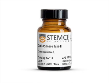 II型胶原酶 用于消化天然胶原纤维
 沪公网安备31010102008431号