M. Liu et al. (nov 2019)
Leukemia research 86 106225
Treatment of human T-cell acute lymphoblastic leukemia cells with CFTR inhibitor CFTRinh-172.
Our previous studies have demonstrated that a previously unrecognized role of CFTR in hematopoiesis and acute leukemia. Here,we show that CFTR inhibitor CFTR-inh172 possesses ability to inhibit human T-cell acute lymphoblastic leukemia cells. In detail,CFTR-inh172 inhibited cell proliferation,promoted apoptosis and arrested the cell cycle in human T-cell acute lymphoblastic leukemia cell CCRF-CEM,JURKAT and MOLT-4. Furthermore,transcriptome analysis reveals that CFTR-inh172 induces significant alteration of gene expression related to apoptosis and proliferation. These findings demonstrate the potential of CFTR inhibitor CFTR-inh172 in human T-cell acute lymphoblastic leukemia treatment.
View Publication
产品类型:
产品号#:
06005
产品名:
IntestiCult™ 类器官生长培养基 (小鼠)
(Aug 2024)
Scientific Reports 14
Rapid retinoic acid-induced trophoblast cell model from human induced pluripotent stem cells
A limited number of accessible and representative models of human trophoblast cells currently exist for the study of placentation. Current stem cell models involve either a transition through a naïve stem cell state or precise dynamic control of multiple growth factors and small-molecule cues. Here,we demonstrated that a simple five-day treatment of human induced pluripotent stem cells with two small molecules,retinoic acid (RA) and Wnt agonist CHIR 99021 (CHIR),resulted in rapid,synergistic upregulation of CDX2. Transcriptomic analysis of RA + CHIR-treated cells showed high similarity to primary trophectoderm cells. Multipotency was verified via further differentiation towards cells with syncytiotrophoblast or extravillous trophoblast features. RA + CHIR-treated cells were also assessed for the established criteria defining a trophoblast cell model,and they possess all the features necessary to be considered valid. Collectively,our data demonstrate a facile,scalable method for generating functional trophoblast-like cells in vitro to better understand the placenta.
View Publication
产品类型:
产品号#:
05854
05855
100-0483
100-0484
100-0276
100-1130
05990
产品名:
mFreSR™
mFreSR™
Hausser Scientificᵀᴹ 明线血球计数板
ReLeSR™
mTeSR™ Plus
mTeSR™ Plus
用于hESC/hiPSC维持培养的TeSR™-E8™
L. Bonneau et al. (Dec 2025)
Biology of the Cell 117 12
Generation of Intestinal and Colonic Organoids Derived From Human Pluripotent Stem Cells
Over the past decade,significant advancements have been made in understanding the developmental mechanisms involved in human gastrointestinal formation,with organoids emerging as key experimental models. These three‐dimensional in vitro cellular structures mimic the organization and functions of various gut regions,providing a powerful tool for research. By replicating critical stages of gut development,we can now direct the differentiation of cells into specific gastrointestinal tissues. In this protocol,we outline how to generate two types of organoids derived from human pluripotent stem cells (hPSCs): human intestinal organoids (HIOs) and human colonic organoids (HCOs). First,we induce definitive endoderm formation to produce these organoids and specify midgut/hindgut tissues. Three‐dimensional spheroids form spontaneously,can be collected,embedded in an extracellular matrix,and cultured over time. During this phase,the organoid epithelium develops,supported by a mesenchymal layer that promotes maturation and differentiation. After a month of culture,HIOs and HCOs reach a developmental and maturation stage comparable to that of the human fetal intestine. These organoids can be used to study human gastrointestinal development,model diseases,and test therapeutic agents. Human pluripotent stem cells can be guided through a stepwise differentiation process to produce self‐organizing intestinal and colonic organoids. The resulting organoids recapitulate fetal‐stage epithelial and mesenchymal organization,offering a powerful model to explore human gastrointestinal development and its disorders.
View Publication
Human blood IgM memory" B cells are circulating splenic marginal zone B cells harboring a prediversified immunoglobulin repertoire."
The human peripheral B-cell compartment displays a large population of immunoglobulin M-positive,immunoglobulin D-positive CD27(+) (IgM(+)IgD(+)CD27(+)) memory" B cells carrying a mutated immunoglobulin receptor. By means of phenotypic analysis�
View Publication
产品类型:
产品号#:
15024
15064
产品名:
RosetteSep™人B细胞富集抗体混合物
RosetteSep™人B细胞富集抗体混合物
Qiu C et al. (FEB 2008)
Blood 111 4 2400--8
Globin switches in yolk sac-like primitive and fetal-like definitive red blood cells produced from human embryonic stem cells.
We have previously shown that coculture of human embryonic stem cells (hESCs) for 14 days with immortalized fetal hepatocytes yields CD34(+) cells that can be expanded in serum-free liquid culture into large numbers of megaloblastic nucleated erythroblasts resembling yolk sac-derived cells. We show here that these primitive erythroblasts undergo a switch in hemoglobin (Hb) composition during late terminal erythroid maturation with the basophilic erythroblasts expressing predominantly Hb Gower I (zeta(2)epsilon(2)) and the orthochromatic erythroblasts hemoglobin Gower II (alpha(2)epsilon(2)). This suggests that the switch from Hb Gower I to Hb Gower II,the first hemoglobin switch in humans is a maturation switch not a lineage switch. We also show that extending the coculture of the hESCs with immortalized fetal hepatocytes to 35 days yields CD34(+) cells that differentiate into more developmentally mature,fetal liver-like erythroblasts,that are smaller,express mostly fetal hemoglobin,and can enucleate. We conclude that hESC-derived erythropoiesis closely mimics early human development because the first 2 human hemoglobin switches are recapitulated,and because yolk sac-like and fetal liver-like cells are sequentially produced. Development of a method that yields erythroid cells with an adult phenotype remains necessary,because the most mature cells that can be produced with current systems express less than 2% adult beta-globin mRNA.
View Publication
产品类型:
产品号#:
09600
09650
18056
18056RF
产品名:
StemSpan™ SFEM
StemSpan™ SFEM
Ko J-Y et al. (AUG 2014)
Stem cells and development 23 15 1788--1797
Osteogenesis from human induced pluripotent stem cells: an in vitro and in vivo comparison with mesenchymal stem cells.
The purpose of this study was to examine the in vitro and in vivo osteogenic potential of human induced pluripotent stem cells (hiPSCs) against that of human bone marrow mesenchymal stem cells (hBMMSCs). Embryoid bodies (EBs),which were formed from undifferentiated hiPSCs,were dissociated into single cells and underwent osteogenic differentiation using the same medium as hBMMSCs for 14 days. Osteoinduced hiPSCs were implanted on the critical-size calvarial defects and long bone segmental defects in rats. The healing of defects was evaluated after 8 weeks and 12 weeks of implantation,respectively. Osteoinduced hiPSCs showed relatively lower and delayed in vitro expressions of the osteogenic marker COL1A1 and bone sialoprotein,as well as a weaker osteogenic differentiation through alkaline phosphatase staining and mineralization through Alizarin red staining compared with hBMMSCs. Calvarial defects treated with osteoinduced hiPSCs had comparable quality of new bone formation,including full restoration of bone width and robust formation of trabeculae,to those treated with hBMMSCs. Both osteoinduced hiPSCs and hBMMSCs persisted in regenerated bone after 8 weeks of implantation. In critical-size long bone segmental defects,osteoinduced hiPSC treatment also led to healing of segmental defects comparable to osteoinduced hBMMSC treatment after 12 weeks. In conclusion,despite delayed in vitro osteogenesis,hiPSCs have an in vivo osteogenic potential as good as hBMMSCs.
View Publication
产品类型:
产品号#:
05850
05857
05870
05875
85850
85857
85870
85875
产品名:
mTeSR™1
mTeSR™1
Rezania A et al. (NOV 2013)
STEM CELLS 31 11 2432--2442
Enrichment of human embryonic stem cell-derived NKX6.1-expressing pancreatic progenitor cells accelerates the maturation of insulin-secreting cells in vivo
Human embryonic stem cells (hESCs) are considered a potential alternative to cadaveric islets as a source of transplantable cells for treating patients with diabetes. We previously described a differentiation protocol to generate pancreatic progenitor cells from hESCs,composed of mainly pancreatic endoderm (PDX1/NKX6.1-positive),endocrine precursors (NKX2.2/synaptophysin-positive,hormone/NKX6.1-negative),and polyhormonal cells (insulin/glucagon-positive,NKX6.1-negative). However,the relative contributions of NKX6.1-negative versus NKX6.1-positive cell fractions to the maturation of functional β-cells remained unclear. To address this question,we generated two distinct pancreatic progenitor cell populations using modified differentiation protocols. Prior to transplant,both populations contained a high proportion of PDX1-expressing cells (˜85%-90%) but were distinguished by their relatively high (˜80%) or low (˜25%) expression of NKX6.1. NKX6.1-high and NKX6.1-low progenitor populations were transplanted subcutaneously within macroencapsulation devices into diabetic mice. Mice transplanted with NKX6.1-low cells remained hyperglycemic throughout the 5-month post-transplant period whereas diabetes was reversed in NKX6.1-high recipients within 3 months. Fasting human C-peptide levels were similar between groups throughout the study,but only NKX6.1-high grafts displayed robust meal-,glucose- and arginine-responsive insulin secretion as early as 3 months post-transplant. NKX6.1-low recipients displayed elevated fasting glucagon levels. Theracyte devices from both groups contained almost exclusively pancreatic endocrine tissue,but NKX6.1-high grafts contained a greater proportion of insulin-positive and somatostatin-positive cells,whereas NKX6.1-low grafts contained mainly glucagon-expressing cells. Insulin-positive cells in NKX6.1-high,but not NKX6.1-low grafts expressed nuclear MAFA. Collectively,this study demonstrates that a pancreatic endoderm-enriched population can mature into highly functional β-cells with only a minor contribution from the endocrine subpopulation.
View Publication
Feeder-free differentiation of human iPSCs into natural killer cells with cytotoxic potential against malignant brain rhabdoid tumor cells
Natural killer (NK) cells are cytotoxic immune cells that can eliminate target cells without prior stimulation. Human induced pluripotent stem cells (iPSCs) provide a robust source of NK cells for safe and effective cell-based immunotherapy against aggressive cancers. In this in vitro study,a feeder-free iPSC differentiation was performed to obtain iPSC-NK cells,and distinct maturational stages of iPSC-NK were characterized. Mature cells of CD56bright CD16bright phenotype showed upregulation of CD56,CD16,and NK cell activation markers NKG2D and NKp46 upon IL-15 exposure,while exposure to aggressive atypical teratoid/rhabdoid tumor (ATRT) cell lines enhanced NKG2D and NKp46 expression. Malignant cell exposure also increased CD107a degranulation markers and stimulated IFN-? secretion in activated NK cells. CD56bright CD16bright iPSC-NK cells showed a ratio-dependent killing of ATRT cells,and the percentage lysis of CHLA-05-ATRT was higher than that of CHLA-02-ATRT. The iPSC-NK cells were also cytotoxic against other brain,kidney,and lung cancer cell lines. Further NK maturation yielded CD56?ve CD16bright cells,which lacked activation markers even after exposure to interleukins or ATRT cells - indicating diminished cytotoxicity. Generation and characterization of different NK phenotypes from iPSCs,coupled with their promising anti-tumor activity against ATRT in vitro,offer valuable insights into potential immunotherapeutic strategies for brain tumors. Graphical abstractImage 1 Highlights•Natural killer (NK) cells were derived from human induced pluripotent stem cells (iPSCs) in the absence of feeder cells.•Various maturational subtypes of iPSC-NK cells were characterized,and the phenotypic and functional properties were studied.•iPSC-NK cells of CD56bright CD16bright phenotype expressed activation markers in response to interleukin stimuli.•iPSC-NK cells were cytotoxic toward human atypical teratoid and rhabdoid tumor (ATRT) cells and other human cancer cells.•The cytotoxicity of iPSC-NK cells against various cancer cells in vitro might be translated into an in vivo immunotherapy.
View Publication
产品类型:
产品号#:
09600
09605
09650
09655
09915
09950
09960
100-0276
100-1130
产品名:
StemSpan™ SFEM
StemSpan™ SFEM II
StemSpan™ SFEM
StemSpan™ SFEM II
StemSpan™淋系祖细胞扩增添加物(10X)
StemSpan™ NK细胞分化添加物(100X)
StemSpan™ NK细胞生成试剂盒
mTeSR™ Plus
mTeSR™ Plus
Chu et al. (Jul 2025)
International Journal of Molecular Sciences 26 13
Limited Myelination Capacity in Human Schwann Cells in Experimental Models in Comparison to Rodent and Porcine Schwann Cells
Schwann cells (SCs) play a crucial role in peripheral nerve repair by supporting axonal regeneration and remyelination. While extensive research has been conducted using rodent SCs,increasing attention is being directed toward human SCs due to species-specific differences in phenotypical and functional properties,and accessibility of human SCs derived from diverse sources. A major challenge in translating SC-based therapies for nerve repair lies in the inability to replicate human SC myelination in vitro,posing a significant obstacle to drug discovery and preclinical research. In this study,we compared the myelination capacity of human,rodent,and porcine SCs in various co-culture conditions,including species-matched and cross-species neuronal environments in a serum-free medium. Our results confirmed that rodent and porcine SCs readily myelinate neurites under standard culture conditions after treatment with ascorbic acid for two weeks,whereas human SCs,at least within the four-week observation period,failed to show myelin staining in all co-cultures. Furthermore,we investigated whether cell culture manipulation impairs human SC myelination by transplanting freshly harvested and predegenerated human nerve segments into NOD-SCID mice for four weeks. Despite supporting host axonal regeneration into the grafts,human SCs exhibited very limited myelination,suggesting an intrinsic species-specific restriction rather than a cell culture-induced defect. These observations suggest fundamental differences between human and rodent SCs and highlight the need for human-specific models and protocols to advance our understanding of SC myelination.
View Publication

 EasySep™小鼠TIL(CD45)正选试剂盒


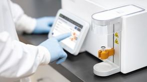 实验方案Gene Editing Human Pluripotent Stem Cells (hPSCs) Using the CellPore™ Transfection System
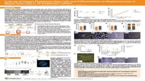 科学海报Scale-up of Human Pluripotent Stem Cells and Differentiation to Megakaryocytes or Neural Crest Cells in 3D Suspension Culture

 沪公网安备31010102008431号