
 EasySep™小鼠TIL(CD45)正选试剂盒


搜索结果: 'methocult media formulations for human hematopoietic cells serum containing'
-

产品类型:
产品号#:
05850
05857
05870
05875
85850
85857
85870
85875
产品名:
mTeSR™1
mTeSR™1
-
产品类型:
产品号#:
73252
73254
产品名:
二甲双胍 (Hydrochloride)
二甲双胍 (Hydrochloride)
-
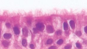 新闻NEW PneumaCult™-ALI Medium for Air-Liquid Interface Culture of Bronchial Epithelial Cells
产品类型:
产品号#:
产品名:
发布日期: 06/05/2012 -
产品类型:
产品号#:
09600
09650
18056
18056RF
60012
60012FI
60012FI.1
100-1574
产品名:
StemSpan™ SFEM
StemSpan™ SFEM
抗人CD32抗体,克隆IV.3
抗人CD32抗体,clone IV.3,FITC
抗人CD32抗体,克隆IV.3,FITC
-
产品类型:
产品号#:
07930
07931
07940
07955
07956
07959
07954
100-1061
07952
产品名:
CryoStor® CS10
CryoStor® CS10
CryoStor® CS10
CryoStor® CS10
CryoStor® CS10
CryoStor® CS10
CryoStor® CS10
-
产品类型:
产品号#:
15022
15062
产品名:
RosetteSep™人CD4+ T细胞富集抗体混合物
RosetteSep™人CD4+ T细胞富集抗体混合物
-
产品类型:
产品号#:
05850
05857
05870
05875
85850
85857
85870
85875
产品名:
mTeSR™1
mTeSR™1
-
产品类型:
产品号#:
04435
04445
产品名:
MethoCult™ H4435 Enriched
MethoCult™ H4435 Enriched
-
产品类型:
产品号#:
03134
产品名:
MethoCult™ M3134
-
产品类型:
产品号#:
05501
05502
产品名:
-
产品类型:
产品号#:
07801
17936
18060
18061
07861
07811
17936RF
产品名:
EasySep™人祖细胞富集试剂盒II
Lymphoprep™
Lymphoprep™
Lymphoprep™
Lymphoprep™
RoboSep™ 人祖细胞富集试剂盒II

 沪公网安备31010102008431号