Hughes CS et al. (FEB 2011)
Proteomics 11 4 675--90
Proteomics of human embryonic stem cells.
Human embryonic stem cells (hESCs) offer exciting potential in regenerative medicine for the treatment of a host of diseases including cancer,Alzheimer's and Parkinson's disease. They also provide insight into human development and disease and can be used as models for drug discovery and toxicity analyses. The key properties of hESCs that make them so promising for medical use are that they have the ability to self-renew indefinitely in culture and they are pluripotent,which means that they can differentiate into any of more than 200 human cell types. Since proteins are the effectors of cellular processes,it is important to investigate hESC expression at the protein level as well as at the transcript level. In addition,post-translational modifications,such as phosphorylation,may influence the activity of pivotal proteins in hESCs,and this information can only be determined by studying the proteome. In this review,we summarize the results obtained from several proteomics analyses of hESCs that have been reported in the last few years.
View Publication
产品类型:
产品号#:
05850
05857
05870
05875
85850
85857
85870
85875
产品名:
mTeSR™1
mTeSR™1
Maciejewski JP et al. (SEP 1996)
Blood 88 6 1983--91
A severe and consistent deficit in marrow and circulating primitive hematopoietic cells (long-term culture-initiating cells) in acquired aplastic anemia.
We examined the stem cell compartment of patients with acquired aplastic anemia (AA) using the long-term culture-initiating cell assay (LTC-IC),in parallel with measurements of CD34+ cells and mature hematopoietic progenitors. Secondary colonies from cells surviving 5 weeks of long-term bone marrow culture (LTBMC) were determined for the peripheral blood (PB) of 68 AA patients and 13 normal controls and for BM of 49 AA patients and 14 controls; because of low cell numbers,formal limiting dilution analysis could only be performed in 10 patients. The relationship of cell input in LTBMC and the output of secondary colonies was linear,allowing quantification of LTC-IC number from bulk cultures. Secondary colony formation was markedly abnormal in severe AA. In contrast to 7.8 colony-forming cells (CFC)/10(5) mononuclear cells in normal BM and 0.14 CFC/10(5) normal PB mononuclear cells,patients with severe disease showed 0.024 CFC/10(5) in BM and 0.0068 CFC/10(5) in PB. Under limiting dilution conditions,patients' cells also showed markedly lower colony-forming ability. In contrast to 4.3 +/- 1 colonies/normal LTC-IC,we obtained only 1.27 +/- 0.09 and 2.0 +/- 0.35 colonies from BM of acute and recovered cases,respectively. These values were used to extrapolate LTC-IC numbers from secondary colony formation in suspension cultures. In PB,calculated LTC-IC were decreased 7.4-fold in new and relapsed severe AA and 2.8-fold in recovered AA. In BM,LTC-IC were decreased 10-fold in new and relapsed AA and sixfold in recovered cases. Compared with measurements obtained on presentation,LTC-IC were lower in post-treatment samples from patients who had failed to recover after intensive immunosuppression and relatively higher in cases at relapse. In recovered patients,LTC-IC number increased but remained below the normal range in 20 of 25. In patients studied serially for 3 to 12 months after treatment,LTC-IC numbers remained stable but low. LTC-IC number correlated with concurrently determined CD34+ cell number and primary hematopoietic colony formation. These results indicate that stem cell numbers,as quantitated by the LTC-IC assay,are markedly diminished in number in all severe AA. Additionally,the function of the stem cell or the stem cell compartment in AA is also abnormal,as inferred from the low clonogenic potential in secondary colony assays. Early hematologic improvement in some patients occurs without increasing numbers of LTC-IC,and a minority of recovered cases show apparent repopulation of the LTC-IC compartment years after treatment.
View Publication
产品类型:
产品号#:
05150
05350
产品名:
MyeloCult™ H5100
Deng Y et al. (JAN 2014)
Carbohydrate Polymers 101 1 36--39
Peptide-decorated polyvinyl alcohol/hyaluronan nanofibers for human induced pluripotent stem cell culture
Realization of the full potential of human induced pluripotent stem cells (hiPSCs) in clinical applications requires development of well-defined conditions for their growth and differentiation. A novel fully defined polyvinyl alcohol/hyaluronan (PVA/HA) polysaccharide nanofiber was developed for hiPSCs culture in commercially available xeno-free,chemically defined medium. Vitronectin peptide (VP) was immobilized to PVA/HA nanofibers through NHS/EDC chemistry. The hiPSCs successfully grew and proliferated on the VP-decorated PVA/HA nanofibers,similar to those on MatrigelTM. Such well-defined,xeno-free and safe nanofiber substrate that supports culture of hiPSCs will not only help to accelerate the translational perspectives of hiPSCs,but also provide a platform to investigate the cell-nanofiber interaction mechanisms that regulate stem cell proliferation and differentiation. ?? 2013 Elsevier Ltd. All rights reserved.
View Publication
A TALEN genome-editing system for generating human stem cell-based disease models.
Transcription activator-like effector nucleases (TALENs) are a new class of engineered nucleases that are easier to design to cleave at desired sites in a genome than previous types of nucleases. We report here the use of TALENs to rapidly and efficiently generate mutant alleles of 15 genes in cultured somatic cells or human pluripotent stem cells,the latter for which we differentiated both the targeted lines and isogenic control lines into various metabolic cell types. We demonstrate cell-autonomous phenotypes directly linked to disease - dyslipidemia,insulin resistance,hypoglycemia,lipodystrophy,motor-neuron death,and hepatitis C infection. We found little evidence of TALEN off-target effects,but each clonal line nevertheless harbors a significant number of unique mutations. Given the speed and ease with which we were able to derive and characterize these cell lines,we anticipate TALEN-mediated genome editing of human cells becoming a mainstay for the investigation of human biology and disease. textcopyright 2013 Elsevier Inc.
View Publication
产品类型:
产品号#:
05850
05857
05870
05875
85850
85857
85870
85875
产品名:
mTeSR™1
mTeSR™1
Nagata S et al. ( )
Nature 319 6052 415--8
Molecular cloning and expression of cDNA for human granulocyte colony-stimulating factor.
Granulocyte colony-stimulating factor (G-CSF) is a member of the CSF family of hormone-like glycoproteins that regulate haematopoietic cell proliferation and differentiation,and G-CSF almost exclusively stimulates the colony formation of granulocytes from committed precursor cells in semi-solid agar culture. Recently,Nomura et al. have established a human squamous carcinoma cell line (designated CHU-2) from a human oral cavity tumour which produces large quantities of CSF constitutively,and the CSF produced by CHU-2 cells has been purified to homogeneity from the conditioned medium. We have now determined the partial amino-acid sequence of the purified G-CSF protein,and by using oligonucleotides as probes,have isolated several clones containing G-CSF complementary DNA from the cDNA library prepared with messenger RNA from CHU-2 cells. The complete nucleotide sequences of two of these cDNAs were determined and the expression of the cDNA in monkey COS cells gave rise to a protein showing authentic G-CSF activity. Furthermore,Southern hybridization analysis of DNA from normal leukocytes and CHU-2 cells suggests that the human genome contains only one gene for G-CSF and that some rearrangement has occurred within one of the alleles of the G-CSF gene in CHU-2 cells.
View Publication
产品类型:
产品号#:
02615
02855
产品名:
(Mar 2025)
Bio-protocol 15 5
Protocol for Screening Host-Targeting Antivirals (HTAs) Using Human PBMCs and pDCs
This protocol offers an ex vivo method for screening host-targeting antivirals (HTAs) using human peripheral blood mononuclear cells (PBMCs) or plasmacytoid dendritic cells (pDCs). Unlike virus-targeting antivirals (VTAs),HTAs provide advantages in overcoming drug resistance and offering broad-spectrum protection,especially against rapidly mutating or newly emerging viruses. By focusing on PBMCs or pDCs,known for their high production of humoral factors such as Type I interferons (IFNs),the protocol enables the screening of antivirals that modulate immune responses against viruses. Targeting host pathways,especially innate immunity,allows for species-independent antiviral activity,reducing the likelihood of viral escape mutations. Additionally,the protocol's versatility makes it a powerful tool for testing potential antivirals against various viral pathogens,including emerging viruses,positioning it as an essential resource in both pandemic preparedness and broad-spectrum antiviral research. This approach differentiates itself from existing protocols by focusing on host immune modulation through pDCs,offering a novel avenue for HTA discovery. Key features • Optimized protocol for screening HTAs against dengue virus (DENV),chikungunya virus (CHIKV),and Zika virus (ZIKV). • This protocol is ideal for screening soluble or intravenous-formulated compounds for evaluating their efficacy in experimental settings. • This protocol builds upon the method developed by Tsuji et al. [1] and extends its application to PBMCs and testing against DENV,CHIKV,and ZIKV.
View Publication
产品类型:
产品号#:
20144
17977
17977RF
产品名:
EasySep™缓冲液
EasySep™人浆细胞样DC分选试剂盒
RoboSep™ 人浆细胞样DC分选试剂盒
P. S. Martinez et al. (May 2026)
Journal of Histochemistry and Cytochemistry 23 12
Human iPSC-Derived Blood Vessel Organoids for Studying Chronic Hypoxia-Induced Microvascular Dysfunction
Microvascular dysfunction due to hypoxia is a key contributor in the pathogenesis of many disorders including cancer and retinal and cardiovascular diseases,but relevant human models are missing. Here,we present a robust 3D in vitro method with the use of human induced pluripotent stem cell–derived blood vessel organoids to analyze in vitro microvascular remodeling. We present a detailed practical pipeline combining optical tissue clearing,high-resolution immunofluorescence,and surface marker analysis to quantitatively assess hypoxia-driven changes in endothelial cells,pericytes,and the basal lamina. Exposure of these blood vessel organoids to chronic hypoxia (1% O2) for 1 week recapitulated key pathological features,including structural remodeling and a dysregulated secretome with altered vascular endothelial growth factor signaling. This approach establishes a versatile and human-relevant platform to study microvascular remodeling induced by chronic hypoxia and other pathological stimuli and their contribution to microvascular-related diseases.
View Publication
SummaryInterleukin-33 (IL-33) is an immunoregulatory cytokine that moderately suppresses experimental autoimmune encephalomyelitis (EAE),a murine model of multiple sclerosis (MS). However,poor pharmacokinetics and toxicity hinder its clinical translation. To address these limitations,we develop an activity-attenuated IL-33 by recombinant fusion to serum albumin (SA). SA-IL-33 exhibits reduced toxicity and prolonged residence in the secondary lymphoid organs (SLOs),sites of T cell priming in autoimmunity,compared to wild-type (WT) IL-33. Prophylactic SA-IL-33 administration prevents EAE with superior efficacy to WT IL-33 and comparable efficacy to fingolimod (FTY720),a Food and Drug Administration (FDA)-approved MS drug. Therapeutic SA-IL-33 treatment also reduces disease severity in both chronic and relapsing-remitting EAE. SA-IL-33 modulates immunity in EAE by suppressing CD45+ cell infiltration (including myelin-reactive T helper 17 [TH17] cells) in the spinal cord,while expanding type 2 immune cells (including type 2 innate lymphoid cells [ILC2s],ST2+ regulatory T cells [Tregs],T helper 2 [TH2] cells,and M2-polarized macrophages) in the SLOs. These findings suggest that SA-IL-33 is a promising therapeutic for neuroinflammatory diseases. Graphical abstract Highlights•Fusion of serum albumin (SA) to interleukin-33 (IL-33) attenuates its activity and toxicity•Engineered SA-IL-33 exhibits prolonged residence in the secondary lymphoid organs (SLOs)•SA-IL-33 treatment both prevents the onset of and reduces established neuroinflammation in mice•Cytokine therapy suppresses TH17 cells in the CNS and promotes immunoregulation in the SLOs The clinical utility of interleukin-33 is hindered by poor pharmacokinetics and toxicity. Budina et al. develop a fusion of serum albumin and interleukin-33 (SA-IL-33) with reduced toxicity and prolonged lymph node residence. SA-IL-33 prevents the onset of and suppresses established inflammation-mediated paralysis in mice,demonstrating promise as a therapeutic for neuroinflammatory diseases.
View Publication
Petersen OW and Polyak K (MAY 2010)
Cold Spring Harbor perspectives in biology 2 5 a003160
Stem cells in the human breast.
The origins of the epithelial cells participating in the development,tissue homeostasis,and cancer of the human breast are poorly understood. However,emerging evidence suggests a role for adult tissue-specific stem cells in these processes. In a hierarchical manner,these generate the two main mammary cell lineages,producing an increasing number of cells with distinct properties. Understanding the biological characteristics of human breast stem cells and their progeny is crucial in attempts to compare the features of normal stem cells and cancer precursor cells and distinguish these from nonprecursor cells and cells from the bulk of a tumor. A historical overview of research on human breast stem cells in primary tissue and in culture reveals the progress that has been made in this area,whereas a focus on the cell-of-origin and reprogramming that occurs during neoplastic conversion provides insight into the enigmatic way in which human breast cancers are skewed toward the luminal epithelial lineage.
View Publication
产品类型:
产品号#:
01700
01705
01702
产品名:
ALDEFLUOR™ 试剂盒
ALDEFLUOR™ DEAB试剂, 1.5 mM, 1 mL
ALDEFLUOR™检测缓冲液
Zhu H et al. (MAR 2015)
Stem Cells International 2015 621057
Development of a xeno-free substrate for human embryonic stem cell growth
Traditionally,human embryonic stem cells (hESCs) are cultured on inactivated live feeder cells. For clinical application using hESCs,there is a requirement to minimize the risk of contamination with animal components. Extracellular matrix (ECM) derived from feeder cells is the most natural way to provide xeno-free substrates for hESC growth. In this study,we optimized the step-by-step procedure for ECM processing to develop a xeno-free ECM that supports the growth of undifferentiated hESCs. In addition,this newly developed xeno-free substrate can be stored at 4°C and is ready to use upon request,which serves as an easier way to amplify hESCs for clinical applications.
View Publication
产品类型:
产品号#:
05850
05857
05870
05875
05940
07923
07174
85850
85857
85870
85875
100-0485
100-1077
产品名:
Dispase (1 U/mL)
mTeSR™1
mTeSR™1
温和细胞解离试剂
ReLeSR™
Reshkin SJ et al. ( 2003)
Clinical cancer research : an official journal of the American Association for Cancer Research 9 6 2366--2373
Paclitaxel induces apoptosis via protein kinase A- and p38 mitogen-activated protein-dependent inhibition of the Na+/H+ exchanger (NHE) NHE isoform 1 in human breast cancer cells.
PURPOSE: The molecular signal components essential to paclitaxel-dependent apoptosis in breast cancers are potential targets for combined therapy. However,the signal mechanisms underlying paclitaxel action still need to be better defined. EXPERIMENTAL DESIGN: In a breast cancer cell line,pharmacological agents and transient transfection with dominant interfering and constitutive active mutants were used to identify the signal transduction module involved in the regulation of paclitaxel-induced apoptosis and to evaluate its potential as a therapeutic target. RESULTS: In MDA-MB-435 cells,paclitaxel treatment stimulated the activity of both protein kinase A and p38,and inhibited the activity of the Na(+)/H(+) exchanger isoform 1 (NHE1) with similar IC(50) concentrations as for its activation of apoptosis. Activation and inhibition experiments demonstrated that protein kinase A and p38 participate sequentially upstream of the NHE1 in regulating the paclitaxel-induced apoptotic pathway. Importantly,concurrent specific inhibition of the NHE1 with paclitaxel treatment resulted in a synergistic induction of apoptosis and a reduction in the paclitaxel IC(50) for apoptosis. This sensitization of paclitaxel apoptotic action by specific inhibition of NHE1 was verified in breast cancer cell lines with different paclitaxel sensitivity. CONCLUSIONS: We have,for the first time,identified NHE1 as an essential component of paclitaxel-induced apoptosis in breast cancer cells and,importantly,identified that simultaneous inhibition of the NHE1 results in a synergistic potentiation of low-dose paclitaxel apoptotic action. As specific NHE1 inhibitors have finished Phase II/Phase III clinical trials for myocardial protection,there is the possibility for a rapid biological translation of this novel therapeutic strategy to a clinical setting.
View Publication

 EasySep™小鼠TIL(CD45)正选试剂盒


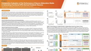 科学海报Comparative Evaluation of the Performance of Neuron Maturation Media in Generating Functional hPSC-Derived Forebrain Neurons
 沪公网安备31010102008431号