Fibromodulin reprogrammed cells: A novel cell source for bone regeneration.
Pluripotent or multipotent cell-based therapeutics are vital for skeletal reconstruction in non-healing critical-sized defects since the local endogenous progenitor cells are not often adequate to restore tissue continuity or function. However,currently available cell-based regenerative strategies are hindered by numerous obstacles including inadequate cell availability,painful and invasive cell-harvesting procedures,and tumorigenesis. Previously,we established a novel platform technology for inducing a quiescent stem cell-like stage using only a single extracellular proteoglycan,fibromodulin (FMOD),circumventing gene transduction. In this study,we further purified and significantly increased the reprogramming rate of the yield multipotent FMOD reprogrammed (FReP) cells. We also exposed the 'molecular blueprint' of FReP cell osteogenic differentiation by gene profiling. Radiographic analysis showed that implantation of FReP cells into a critical-sized SCID mouse calvarial defect,contributed to the robust osteogenic capability of FReP cells in a challenging clinically relevant traumatic scenario in vivo. The persistence,engraftment,and osteogenesis of transplanted FReP cells without tumorigenesis in vivo were confirmed by histological and immunohistochemical staining. Taken together,we have provided an extended potency,safety,and molecular profile of FReP cell-based bone regeneration. Therefore,FReP cells present a high potential for cellular and gene therapy products for bone regeneration.
View Publication
产品类型:
产品号#:
05850
05857
05870
05875
05872
05873
05893
85850
85857
85870
85875
27845
27945
27840
27865
27940
27965
100-0483
100-0484
产品名:
AggreWell™ EB形成培养基
mTeSR™1
mTeSR™1
Hausser Scientificᵀᴹ 明线血球计数板
ReLeSR™
D. Merrick et al. ( 2019)
Science (New York,N.Y.) 364 6438
Identification of a mesenchymal progenitor cell hierarchy in adipose tissue.
Metabolic health depends on the capacity of adipose tissue progenitor cells to undergo de novo adipogenesis. The cellular hierarchy and mechanisms governing adipocyte progenitor differentiation are incompletely understood. Through single-cell RNA sequence analyses,we show that the lineage hierarchy of adipocyte progenitors consists of distinct mesenchymal cell types that are present in both mouse and human adipose tissues. Cells marked by dipeptidyl peptidase-4 (DPP4)/CD26 expression are highly proliferative,multipotent progenitors. During the development of subcutaneous adipose tissue in mice,these progenitor cells give rise to intercellular adhesion molecule-1 (ICAM1)/CD54-expressing (CD54+) committed preadipocytes and a related adipogenic cell population marked by Clec11a and F3/CD142 expression. Transforming growth factor-beta maintains DPP4+ cell identity and inhibits adipogenic commitment of DPP4+ and CD142+ cells. Notably,DPP4+ progenitors reside in the reticular interstitium,a recently appreciated fluid-filled space within and between tissues,including adipose depots.
View Publication
Loss of symmetric cell division of apical neural progenitors drives
Developmental and epileptic encephalopathies (DEEs) feature altered brain development,developmental delay and seizures,with seizures exacerbating developmental delay. Here we identify a cohort with biallelic variants in DENND5A,encoding a membrane trafficking protein,and develop animal models with phenotypes like the human syndrome. We demonstrate that DENND5A interacts with Pals1/MUPP1,components of the Crumbs apical polarity complex required for symmetrical division of neural progenitor cells. Human induced pluripotent stem cells lacking DENND5A fail to undergo symmetric cell division with an inherent propensity to differentiate into neurons. These phenotypes result from misalignment of the mitotic spindle in apical neural progenitors. Cells lacking DENND5A orient away from the proliferative apical domain surrounding the ventricles,biasing daughter cells towards a more fate-committed state,ultimately shortening the period of neurogenesis. This study provides a mechanism for DENND5A-related DEE that may be generalizable to other developmental conditions and provides variant-specific clinical information for physicians and families. Developmental and epileptic encephalopathies are devastating neurological disorders. Here,the authors establish a cohort of patients with variants in the gene DENND5A and use human stem cells to discover a disease mechanism involving altered cell division.
View Publication
产品类型:
产品号#:
05833
08581
08582
100-0483
100-0484
05990
85850
85857
产品名:
STEMdiff™神经前体细胞培养基
STEMdiff™SMADi神经诱导试剂盒
STEMdiff™SMADi神经诱导试剂盒,2套
Hausser Scientificᵀᴹ 明线血球计数板
ReLeSR™
用于hESC/hiPSC维持培养的TeSR™-E8™
mTeSR™1
mTeSR™1
L. M. Weiss et al. (Sep 2024)
Communications Biology 7
RUNX1 interacts with lncRNA SMANTIS to regulate monocytic cell functions
Monocytes,the circulating macrophage precursors,contribute to diseases like atherosclerosis and asthma. Long non-coding RNAs (lncRNAs) have been shown to modulate the phenotype and inflammatory capacity of monocytes. We previously discovered the lncRNA SMANTIS,which contributes to cellular phenotype expression by controlling BRG1 in mesenchymal cells. Here,we report that SMANTIS is particularly highly expressed in monocytes and lost during differentiation into macrophages. Moreover,different types of myeloid leukemia presented specific SMANTIS expression patterns. Interaction studies revealed that SMANTIS binds RUNX1,a transcription factor frequently mutated in AML,primarily through its Alu-element on the RUNT domain. RNA-seq after CRISPR/Cas9-mediated deletion of SMANTIS or RUNX1 revealed an association with cell adhesion and both limited the monocyte adhesion to endothelial cells. Mechanistically,SMANTIS KO reduced RUNX1 genomic binding and altered the interaction of RUNX1 with EP300 and CBFB. Collectively,SMANTIS interacts with RUNX1 and attenuates monocyte adhesion,which might limit monocyte vascular egress. Subject terms: Long non-coding RNAs,Transcription
View Publication
W.-X. Ding et al. (feb 2007)
The Journal of biological chemistry 282 7 4702--10
Differential effects of endoplasmic reticulum stress-induced autophagy on cell survival.
Autophagy is a cellular response to adverse environment and stress,but its significance in cell survival is not always clear. Here we show that autophagy could be induced in the mammalian cells by chemicals,such as A23187,tunicamycin,thapsigargin,and brefeldin A,that cause endoplasmic reticulum stress. Endoplasmic reticulum stress-induced autophagy is important for clearing polyubiquitinated protein aggregates and for reducing cellular vacuolization in HCT116 colon cancer cells and DU145 prostate cancer cells,thus mitigating endoplasmic reticulum stress and protecting against cell death. In contrast,autophagy induced by the same chemicals does not confer protection in a normal human colon cell line and in the non-transformed murine embryonic fibroblasts but rather contributes to cell death. Thus the impact of autophagy on cell survival during endoplasmic reticulum stress is likely contingent on the status of cells,which could be explored for tumor-specific therapy.
View Publication
Alkylator-Induced and Patient-Derived Xenograft Mouse Models of Therapy-Related Myeloid Neoplasms Model Clinical Disease and Suggest the Presence of Multiple Cell Subpopulations with Leukemia Stem Cell Activity.
Acute myeloid leukemia (AML) is a heterogeneous group of aggressive bone marrow cancers arising from transformed hematopoietic stem and progenitor cells (HSPC). Therapy-related AML and MDS (t-AML/MDS) comprise a subset of AML cases occurring after exposure to alkylating chemotherapy and/or radiation and are associated with a very poor prognosis. Less is known about the pathogenesis and disease-initiating/leukemia stem cell (LSC) subpopulations of t-AML/MDS compared to their de novo counterparts. Here,we report the development of mouse models of t-AML/MDS. First,we modeled alkylator-induced t-AML/MDS by exposing wild type adult mice to N-ethyl-N-nitrosurea (ENU),resulting in several models of AML and MDS that have clinical and pathologic characteristics consistent with human t-AML/MDS including cytopenia,myelodysplasia,and shortened overall survival. These models were limited by their inability to transplant clinically aggressive disease. Second,we established three patient-derived xenograft models of human t-AML. These models led to rapidly fatal disease in recipient immunodeficient xenografted mice. LSC activity was identified in multiple HSPC subpopulations suggesting there is no canonical LSC immunophenotype in human t-AML. Overall,we report several new t-AML/MDS mouse models that could potentially be used to further define disease pathogenesis and test novel therapeutics.
View Publication
产品类型:
产品号#:
21000
20119
20155
产品名:
RoboSep™- S
RoboSep™ 吸头组件抛光剂
RoboSep™分选管套装(9个塑料管)
Chiew MY et al. (MAY 2016)
Leukemia & lymphoma 1--9
Generation of a MLL-AF9-specific stem cell model of acute monocytic leukemia.
Acute monocytic leukemia (AML-M5),a subtype of acute myeloid leukemia (AML),affects mostly young children and has poor prognosis. The mechanisms of treatment failure of AML-M5 are still unclear. In this study,we generated iPSC from THP-1 cells from a patient with AML-M5,using retroviruses encoding the pluripotency-associated genes (OCT3/4,SOX2,KLF4 and c-MYC). These AML-M5-derived iPSC showed features similar with those of human embryonic stem cells in terms of the morphology,gene expression,protein/antigen expression and differentiation capability. Parental-specific markers were down-regulated in these AML-M5-derived iPSCs. Expression of MLL-AF9 fusion gene (previously identified to be associated with pathogenesis of AML-M5) was observed in all iPSC clones as well as parental cells. We conclude that AML-M5-specific iPSC clones have been successfully developed. This disease model may provide a novel approach for future study of pathogenesis and therapeutic intervention of AML-M5.
View Publication
产品类型:
产品号#:
05850
05857
05870
05875
85850
85857
85870
85875
产品名:
mTeSR™1
mTeSR™1
La Spada A et al. (DEC 2016)
The journal of histochemistry and cytochemistry : official journal of the Histochemistry Society 64 12 739--751
Cell Line Macroarray: An Alternative High-Throughput Platform to Analyze hiPSC Lines.
In the past decade,tissue microarray (TMA) technology has evolved as an innovative tool for high-throughput proteomics analysis and mainly for biomarker validation. Similarly,enormous amount of data can be obtained from the cell line macroarray (CLMA) technology,which developed from the TMA using formalin-fixed,paraffin-embedded cell pellets. Here,we applied CLMA technology in stem cell research and in particular to identify bona fide neogenerated human induced pluripotent stem cell (hiPSC) clones suitable for down the line differentiation. All hiPSC protocols generate tens of clones,which need to be tested to determine genetically stable cell lines suitable for differentiation. Screening methods generally rely on fluorescence-activated cell sorting isolation and coverslip cell growth followed by immunofluorescence; these techniques could be cumbersome. Here,we show the application of CLMA to identify neogenerated pluripotent cell colonies and neuronal differentiated cell products. We also propose the use of the automated image analyzer,TissueQuest,as a reliable tool to quickly select the best clones,based upon the level of expression of multiple pluripotent biomarkers.
View Publication


 EasySep™小鼠TIL(CD45)正选试剂盒





 产品手册Highway1™: Fast, Gentle, and Automated Cell Sorting for Every Lab
 实验方案How to Collect Plasma from Whole Blood Before Cell Isolation
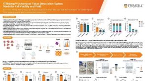 科学海报STEMprep™ Automated Tissue Dissociation System: Maximize Cell Viability and Yield

 沪公网安备31010102008431号