Lannutti BJ et al. (FEB 2009)
Blood 113 8 1778--85
Incomplete restoration of Mpl expression in the mpl-/- mouse produces partial correction of the stem cell-repopulating defect and paradoxical thrombocytosis.
Expression of Mpl is restricted to hematopoietic cells in the megakaryocyte lineage and to undifferentiated progenitors,where it initiates critical cell survival and proliferation signals after stimulation by its ligand,thrombopoietin (TPO). As a result,a deficiency in Mpl function in patients with congenital amegakaryocytic thrombocytopenia (CAMT) and in mpl(-/-) mice produces profound thrombocytopenia and a severe stem cell-repopulating defect. Gene therapy has the potential to correct the hematopoietic defects of CAMT by ectopic gene expression that restores normal Mpl receptor activity. We rescued the mpl(-/-) mouse with a transgenic vector expressing mpl from the promoter elements of the 2-kb region of DNA just proximal to the natural gene start site. Transgene rescued mice exhibit thrombocytosis but only partial correction of the stem cell defect. Furthermore,they show very low-level expression of Mpl on platelets and megakaryocytes,and the transgene-rescued megakaryocytes exhibit diminished TPO-dependent kinase phosphorylation and reduced platelet production in bone marrow chimeras. Thrombocytosis is an unexpected consequence of reduced Mpl expression and activity. However,impaired TPO homeostasis in the transgene-rescued mice produces elevated plasma TPO levels,which serves as an unchecked stimulus to drive the observed excessive megakaryocytopoiesis.
View Publication
Megakaryoblastic leukemia 1 (MKL1),identified as part of the t(1;22) translocation specific to acute megakaryoblastic leukemia,is highly expressed in differentiated muscle cells and promotes muscle differentiation by activating serum response factor (SRF). Here we show that Mkl1 expression is up-regulated during murine megakaryocytic differentiation and that enforced overexpression of MKL1 enhances megakaryocytic differentiation. When the human erythroleukemia (HEL) cell line is induced to differentiate with 12-O-tetradecanoylphorbol 13-acetate,overexpression of MKL1 results in an increased number of megakaryocytes with a concurrent increase in ploidy. MKL1 overexpression also promotes megakaryocytic differentiation of primary human CD34(+) cells cultured in the presence of thrombopoietin. The effect of MKL1 is abrogated when SRF is knocked down,suggesting that MKL1 acts through SRF. Consistent with these findings in human cells,knockout of Mkl1 in mice leads to reduced platelet counts in peripheral blood,and reduced ploidy in bone marrow megakaryocytes. In conclusion,MKL1 promotes physiologic maturation of human and murine megakaryocytes.
View Publication
产品类型:
产品号#:
09500
09600
09650
04960
04902
04900
04963
04962
04970
04971
04901
产品名:
BIT 9500血清替代物
StemSpan™ SFEM
StemSpan™ SFEM
MegaCult™-C胶原和无细胞因子培养基
胶原蛋白溶液
MegaCult™-C无细胞因子培养基
双室载玻片套件
MegaCult™-C CFU-Mk染色试剂盒
MegaCult™-C无细胞因子全套试剂盒
MegaCult™-C含细胞因子全套试剂盒
MegaCult™-C含细胞因子培养基
Halene S et al. (SEP 2010)
Blood 116 11 1942--50
Serum response factor is an essential transcription factor in megakaryocytic maturation.
Serum response factor (Srf) is a MADS-box transcription factor that is critical for muscle differentiation. Its function in hematopoiesis has not yet been revealed. Mkl1,a cofactor of Srf,is part of the t(1;22) translocation in acute megakaryoblastic leukemia,and plays a critical role in megakaryopoiesis. To test the role of Srf in megakaryocyte development,we crossed Pf4-Cre mice,which express Cre recombinase in cells committed to the megakaryocytic lineage,to Srf(F/F) mice in which functional Srf is no longer expressed after Cre-mediated excision. Pf4-Cre/Srf(F/F) knockout (KO) mice are born with normal Mendelian frequency,but have significant macrothrombocytopenia with approximately 50% reduction in platelet count. In contrast,the BM has increased number and percentage of CD41(+) megakaryocytes (WT: 0.41% ± 0.06%; KO: 1.92% ± 0.12%) with significantly reduced ploidy. KO mice show significantly increased megakaryocyte progenitors in the BM by FACS analysis and CFU-Mk. Megakaryocytes lacking Srf have abnormal stress fiber and demarcation membrane formation,and platelets lacking Srf have abnormal actin distribution. In vitro and in vivo assays reveal platelet function defects in KO mice. Critical actin cytoskeletal genes are down-regulated in KO megakaryocytes. Thus,Srf is required for normal megakaryocyte maturation and platelet production partly because of regulation of cytoskeletal genes.
View Publication
产品类型:
产品号#:
09500
09600
09650
04971
04902
04901
04963
04962
产品名:
BIT 9500血清替代物
StemSpan™ SFEM
StemSpan™ SFEM
MegaCult™-C含细胞因子全套试剂盒
胶原蛋白溶液
MegaCult™-C含细胞因子培养基
双室载玻片套件
MegaCult™-C CFU-Mk染色试剂盒
Migliaccio AR et al. (FEB 2003)
The Journal of experimental medicine 197 3 281--96
GATA-1 as a regulator of mast cell differentiation revealed by the phenotype of the GATA-1low mouse mutant.
Here it is shown that the phenotype of adult mice lacking the first enhancer (DNA hypersensitive site I) and the distal promoter of the GATA-1 gene (neo Delta HS or GATA-1(low) mutants) reveals defects in mast cell development. These include the presence of morphologically abnormal alcian blue(+) mast cells and apoptotic metachromatic(-) mast cell precursors in connective tissues and peritoneal lavage and numerous (60-70% of all the progenitors) unique" trilineage cells committed to erythroid�
View Publication
产品类型:
产品号#:
04960
04902
04900
04961
04901
04963
04962
04970
04971
产品名:
MegaCult™-C胶原和无细胞因子培养基
胶原蛋白溶液
MegaCult™-C无细胞因子培养基
MegaCult™-C胶原和含细胞因子培养基
MegaCult™-C含细胞因子培养基
双室载玻片套件
MegaCult™-C CFU-Mk染色试剂盒
MegaCult™-C无细胞因子全套试剂盒
MegaCult™-C含细胞因子全套试剂盒
Wang Q et al. (FEB 2004)
Blood 103 4 1278--85
BUBR1 deficiency results in abnormal megakaryopoiesis.
The physiologic function of BUBR1,a key component of the spindle checkpoint,was examined by generating BUBR1-mutant mice. BUBR1(-/-) embryos failed to survive beyond day 8.5 in utero as a result of extensive apoptosis. Whereas BUBR1(+/-) blastocysts grew relatively normally in vitro,BUBR1(-/-) blastocysts exhibited impaired proliferation and atrophied. Adult BUBR1(+/-) mice manifested splenomegaly and abnormal megakaryopoiesis. BUBR1 haploinsufficiency resulted in an increase in the number of splenic megakaryocytes,which was correlated with an increase in megakaryocytic,but a decrease in erythroid,progenitors in bone marrow cells. RNA interference-mediated down-regulation of BUBR1 also caused an increase in polyploidy formation in murine embryonic fibroblast cells and enhanced megakaryopoiesis in bone marrow progenitor cells. However,enhanced megakaryopoiesis in BUBR1(+/-) mice was not correlated with a significant increase in platelets in peripheral blood,which was at least partly due to a defect in the formation of proplatelet-producing megakaryocytes. Together,these results indicate that BUBR1 is essential for early embryonic development and normal hematopoiesis.
View Publication
产品类型:
产品号#:
09600
09650
04960
04902
04900
04961
04901
04963
04962
04970
04971
产品名:
StemSpan™ SFEM
StemSpan™ SFEM
MegaCult™-C胶原和无细胞因子培养基
胶原蛋白溶液
MegaCult™-C无细胞因子培养基
MegaCult™-C胶原和含细胞因子培养基
MegaCult™-C含细胞因子培养基
双室载玻片套件
MegaCult™-C CFU-Mk染色试剂盒
MegaCult™-C无细胞因子全套试剂盒
MegaCult™-C含细胞因子全套试剂盒
Drayer AL et al. (JAN 2006)
Stem cells (Dayton,Ohio) 24 1 105--14
Mammalian target of rapamycin is required for thrombopoietin-induced proliferation of megakaryocyte progenitors.
Thrombopoietin (TPO) is a potent regulator of megakaryopoiesis and stimulates megakaryocyte (MK) progenitor expansion and MK differentiation. In this study,we show that TPO induces activation of the mammalian target of rapamycin (mTOR) signaling pathway,which plays a central role in translational regulation and is required for proliferation of MO7e cells and primary human MK progenitors. Treatment of MO7e cells,human CD34+,and primary MK cells with the mTOR inhibitor rapamycin inhibits TPO-induced cell cycling by reducing cells in S phase and blocking cells in G0/G1. Rapamycin markedly inhibits the clonogenic growth of MK progenitors with high proliferative capacity but does not reduce the formation of small MK colonies. Addition of rapamycin to MK suspension cultures reduces the number of MK cells,but inhibition of mTOR does not significantly affect expression of glycoproteins IIb/IIIa (CD41) and glycoprotein Ib (CD42),nuclear polyploidization levels,cell size,or cell survival. The downstream effectors of mTOR,p70 S6 kinase (S6K) and 4E-binding protein 1 (4E-BP1),are phosphorylated by TPO in a rapamycin- and LY294002-sensitive manner. Part of the effect of the phosphatidyl inositol 3-kinase pathway in regulating megakaryopoiesis may be mediated by the mTOR/S6K/4E-BP1 pathway. In conclusion,these data demonstrate that the mTOR pathway is activated by TPO and plays a critical role in regulating proliferation of MK progenitors,without affecting differentiation or cell survival.
View Publication
产品类型:
产品号#:
04961
04902
04901
04971
04963
04962
产品名:
MegaCult™-C胶原和含细胞因子培养基
胶原蛋白溶液
MegaCult™-C含细胞因子培养基
MegaCult™-C含细胞因子全套试剂盒
双室载玻片套件
MegaCult™-C CFU-Mk染色试剂盒
Matsumura-Takeda K et al. (APR 2007)
Stem cells (Dayton,Ohio) 25 4 862--70
CD41+/CD45+ cells without acetylcholinesterase activity are immature and a major megakaryocytic population in murine bone marrow.
Murine megakaryocytes (MKs) are defined by CD41/CD61 expression and acetylcholinesterase (AChE) activity; however,their stages of differentiation in bone marrow (BM) have not been fully elucidated. In murine lineage-negative (Lin(-))/CD45(+) BM cells,we found CD41(+) MKs without AChE activity (AChE(-)) except for CD41(++) MKs with AChE activity (AChE(+)),in which CD61 expression was similar to their CD41 level. Lin(-)/CD41(+)/CD45(+)/AChE(-) MKs could differentiate into AChE(+),with an accompanying increase in CD41/CD61 during in vitro culture. Both proplatelet formation (PPF) and platelet (PLT) production for Lin(-)/CD41(+)/CD45(+)/AChE(-) MKs were observed later than for Lin(-)/CD41(++)/CD45(+)/AChE(+) MKs,whereas MK progenitors were scarcely detected in both subpopulations. GeneChip and semiquantitative polymerase chain reaction analyses revealed that the Lin(-)/CD41(+)/CD45(+)/AChE(-) MKs are assigned at the stage between the progenitor and PPF preparation phases in respect to the many MK/PLT-specific gene expressions,including beta1-tubulin. In normal mice,the number of Lin(-)/CD41(+)/CD45(+)/AChE(-) MKs was 100 times higher than that of AChE(+) MKs in BM. When MK destruction and consequent thrombocytopenia were caused by an antitumor agent,mitomycin-C,Lin(-)/CD41(+)/CD45(+)/AChE(-) MKs led to an increase in AChE(+) MKs and subsequent PLT recovery with interleukin-11 administration. It was concluded that MKs in murine BM at least in part consist of immature Lin(-)/CD41(+)/CD45(+)/AChE(-) MKs and more differentiated Lin(-)/CD41(++)/CD45(+)/AChE(+) MKs. Immature Lin(-)/CD41(+)/CD45(+)/AChE(-) MKs are a major MK population compared with AChE(+) MKs in BM and play an important role in rapid PLT recovery in vivo.
View Publication
产品类型:
产品号#:
03231
04960
04902
04900
04961
04901
04963
04962
04970
04971
产品名:
MethoCult™ M3231
MegaCult™-C胶原和无细胞因子培养基
胶原蛋白溶液
MegaCult™-C无细胞因子培养基
MegaCult™-C胶原和含细胞因子培养基
MegaCult™-C含细胞因子培养基
双室载玻片套件
MegaCult™-C CFU-Mk染色试剂盒
MegaCult™-C无细胞因子全套试剂盒
MegaCult™-C含细胞因子全套试剂盒
Levay K and Slepak VZ (SEP 2007)
The Journal of clinical investigation 117 9 2672--83
Tescalcin is an essential factor in megakaryocytic differentiation associated with Ets family gene expression.
We show here that the process of megakaryocytic differentiation requires the presence of the recently discovered protein tescalcin. Tescalcin is dramatically upregulated during the differentiation and maturation of primary megakaryocytes or upon PMA-induced differentiation of K562 cells. This upregulation requires sustained signaling through the ERK pathway. Overexpression of tescalcin in K562 cells initiates events of spontaneous megakaryocytic differentiation,such as expression of specific cell surface antigens,inhibition of cell proliferation,and polyploidization. Conversely,knockdown of this protein in primary CD34+ hematopoietic progenitors and cell lines by RNA interference suppresses megakaryocytic differentiation. In cells lacking tescalcin,the expression of Fli-1,Ets-1,and Ets-2 transcription factors,but not GATA-1 or MafB,is blocked. Thus,tescalcin is essential for the coupling of ERK cascade activation with the expression of Ets family genes in megakaryocytic differentiation.
View Publication
产品类型:
产品号#:
04960
04902
04900
04961
04901
04963
04962
04970
04971
产品名:
MegaCult™-C胶原和无细胞因子培养基
胶原蛋白溶液
MegaCult™-C无细胞因子培养基
MegaCult™-C胶原和含细胞因子培养基
MegaCult™-C含细胞因子培养基
双室载玻片套件
MegaCult™-C CFU-Mk染色试剂盒
MegaCult™-C无细胞因子全套试剂盒
MegaCult™-C含细胞因子全套试剂盒
V. Cesarini et al. (aug 2019)
Scientific reports 9 1 12206
Regulation of PDE5 expression in human aorta and thoracic aortic aneurysms.
Aneurysms and dissections affecting thoracic aorta are associated with smooth muscle cell (SMC) dysfunction. NO/cGMP signaling pathway in smooth muscle cells has been shown to be affected in sporadic thoracic aortic aneurysms. We analyzed the mRNA levels of PDE5,a cGMP-hydrolyzing enzyme highly expressed in aortic SMCs,that regulates arterious vascular tone by lowering cGMP levels. We found that aortic tissue obtained from Marfan,tricuspid and bicuspid thoracic aneurysms expressed lower levels of PDE5 mRNA compared to control aortas. In particular,we found that affected aortas showed lower levels of all the PDE5A isoforms,compared to control aortas. Transfection of vascular SMCs (VSMCs) with NOTCH3 activated domain (NICD3) induced the expression of PDE5A1 and A3 protein isoforms,but not that of the corresponding mRNAs. VSMC stimulation with GSNO,a nitric oxide analogue or with 8-br-cGMP,but not with 8-br-cAMP,up-regulated PDE5 and NOTCH-3 protein levels,indicating a negative feedback loop to protect the arterial wall from excessive relaxation. Finally,we found that PDE5 is expressed early during human aorta development,suggesting that if loss of function mutations of PDE5 occur,they might potentially affect aortic wall development.
View Publication
产品类型:
产品号#:
04961
04965
产品名:
MegaCult™-C胶原和含细胞因子培养基
M. S. Tavangar et al. (may 2020)
Clinical and experimental dental research
Differential expression of drug resistance genes in CD146 positive dental pulp derived stem cells and CD146 negative fibroblasts.
INTRODUCTION The stem cell portion of the dental pulp derived cultures (DPSCs) showed a higher resistance to cytotoxic effect of restorative dental materials compared to pulpal fibroblasts (DPFs). Here,we aimed to compare the expression of some drug resistant genes between these cells. METHODS AND MATERIALS To separate DPSCs from DPFs,we used magnetic cell sorting technique based on CD146 expression. To assess the stem cell properties,the positive and negative portions underwent colony forming assays and were induced to be differentiated into the adipocytes,osteoblasts,hepatocytes,and neural cells. Cell surface antigen panels were checked using immune fluorescence and flow-cytometry techniques. The mRNA expression of 14 ABC transporters including ABCA2,ABCB1,ABCB11,ABCC1,ABCC2,ABCC3,ABCC4,ABCC5-2,ABCC5-4,ABCC5-13,ABCC6,ABCC10,ABCC11,and ABCG2 genes was assessed,using quantitative RT-PCR technique. RESULTS Only the CD146 positive portion could be differentiated into the desired fates,and they formed higher colonies (16.7 ± 3.32 vs. 1.7 ± 1.67,p {\textless} .001). The cell surface antigen panels were the same,except for CD146 and STRO-1 markers which were expressed only in the positive portion. Among the ABC transporter genes studied,the positive portion showed a higher expression (approximately two-fold) of ABCA2,ABCC5-13,and ABCC5-2 genes. CONCLUSION Dental pulp stem cells which can be separated from dental pulp fibroblasts based on CD146 expression,express higher levels of some drug resistance genes which probably accounts for their features of more resistance to cytotoxic effects of some dental materials. This needs to be more validated in future.
View Publication

 EasySep™小鼠TIL(CD45)正选试剂盒


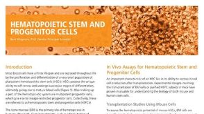 研究综述Hematopoietic Stem and Progenitor Cells (HSPCs): Isolation, Culture, and Assays
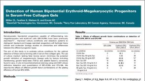 科学海报Detection of Human Bipotential Erythroid-Megakaryocytic Progenitors in Serum-Free Collagen Gels
 沪公网安备31010102008431号