Sengupta A et al. (JUN 2011)
Proceedings of the National Academy of Sciences of the United States of America 108 24 9957--62
Atypical protein kinase C (aPKCzeta and aPKClambda) is dispensable for mammalian hematopoietic stem cell activity and blood formation.
The stem-cell pool is considered to be maintained by a balance between symmetric and asymmetric division of stem cells. The cell polarity model proposes that the facultative use of symmetric and asymmetric cell division is orchestrated by a polarity complex consisting of partitioning-defective proteins Par3 and Par6,and atypical protein kinase C (aPKCζ and aPKCλ),which regulates planar symmetry of dividing stem cells with respect to the signaling microenvironment. However,the role of the polarity complex is unexplored in mammalian adult stem-cell functions. Here we report that,in contrast to accepted paradigms,polarization and activity of adult hematopoietic stem cell (HSC) do not depend on either aPKCζ or aPKCλ or both in vivo. Mice,having constitutive and hematopoietic-specific (Vav1-Cre) deletion of aPKCζ and aPKCλ,respectively,have normal hematopoiesis,including normal HSC self-renewal,engraftment,differentiation,and interaction with the bone marrow microenvironment. Furthermore,inducible complete deletion of aPKCλ (Mx1-Cre) in aPKCζ(-/-) HSC does not affect HSC polarization,self-renewal,engraftment,or lineage repopulation. In addition,aPKCζ- and aPKCλ-deficient HSCs elicited a normal pattern of hematopoietic recovery secondary to myeloablative stress. Taken together,the expression of aPKCζ,aPKCλ,or both are dispensable for primitive and adult HSC fate determination in steady-state and stress hematopoiesis,contrary to the hypothesis of a unique,evolutionary conserved aPKCζ/λ-directed cell polarity signaling mechanism in mammalian HSC fate determination.
View Publication
产品类型:
产品号#:
03434
03444
产品名:
MethoCult™ GF M3434
MethoCult™ GF M3434
Ellis BW et al. (MAR 2017)
Biomicrofluidics 11 2 024105
Human iPSC-derived myocardium-on-chip with capillary-like flow for personalized medicine.
The heart wall tissue,or the myocardium,is one of the main targets in cardiovascular disease prevention and treatment. Animal models have not been sufficient in mimicking the human myocardium as evident by the very low clinical translation rates of cardiovascular drugs. Additionally,current in vitro models of the human myocardium possess several shortcomings such as lack of physiologically relevant co-culture of myocardial cells,lack of a 3D biomimetic environment,and the use of non-human cells. In this study,we address these shortcomings through the design and manufacture of a myocardium-on-chip (MOC) using 3D cell-laden hydrogel constructs and human induced pluripotent stem cell (hiPSC) derived myocardial cells. The MOC utilizes 3D spatially controlled co-culture of hiPSC derived cardiomyocytes (iCMs) and hiPSC derived endothelial cells (iECs) integrated among iCMs as well as in capillary-like side channels,to better mimic the microvasculature seen in native myocardium. We first fully characterized iCMs using immunostaining,genetic,and electrochemical analysis and iECs through immunostaining and alignment analysis to ensure their functionality,and then seeded these cells sequentially into the MOC device. We showed that iECs could be cultured within the microfluidic device without losing their phenotypic lineage commitment,and align with the flow upon physiological level shear stresses. We were able to incorporate iCMs within the device in a spatially controlled manner with the help of photocrosslinkable polymers. The iCMs were shown to be viable and functional within the device up to 7 days,and were integrated with the iECs. The iCMs and iECs in this study were derived from the same hiPSC cell line,essentially mimicking the myocardium of an individual human patient. Such devices are essential for personalized medicine studies where the individual drug response of patients with different genetic backgrounds can be tested in a physiologically relevant manner.
View Publication
产品类型:
产品号#:
05850
05857
05870
05875
85850
85857
85870
85875
产品名:
mTeSR™1
mTeSR™1
Bielawski KS et al. (SEP 2016)
Tissue engineering. Part C,Methods
Real-Time Force and Frequency Analysis of Engineered Human Heart Tissue Derived from Induced Pluripotent Stem Cells Using Magnetic Sensing.
Engineered heart tissues made from human pluripotent stem cell-derived cardiomyocytes have been used for modeling cardiac pathologies,screening new therapeutics,and providing replacement cardiac tissue. Current methods measure the functional performance of engineered heart tissue by their twitch force and beating frequency,typically obtained by optical measurements. In this article,we describe a novel method for assessing twitch force and beating frequency of engineered heart tissue using magnetic field sensing,which enables multiple tissues to be measured simultaneously. The tissues are formed as thin structures suspended between two silicone posts,where one post is rigid and another is flexible and contains an embedded magnet. When the tissue contracts it causes the flexible post to bend in proportion to its twitch force. We measured the bending of the post using giant magnetoresistive (GMR) sensors located underneath a 24-well plate containing the tissues. We validated the accuracy of the readings from the GMR sensors against optical measurements. We demonstrated the utility and sensitivity of our approach by testing the effects of three concentrations of isoproterenol and verapamil on twitch force and beating frequency in real-time,parallel experiments. This system should be scalable beyond the 24-well format,enabling greater automation in assessing the contractile function of cardiomyocytes in a tissue-engineered environment.
View Publication
产品类型:
产品号#:
05850
05857
05870
05875
85850
85857
85870
85875
产品名:
mTeSR™1
mTeSR™1
Kang S et al. (APR 2009)
Molecular and cellular biology 29 8 2105--17
Fibroblast growth factor receptor 3 associates with and tyrosine phosphorylates p90 RSK2, leading to RSK2 activation that mediates hematopoietic transformation.
Dysregulation of the receptor tyrosine kinase fibroblast growth factor receptor 3 (FGFR3) plays a pathogenic role in a number of human hematopoietic malignancies and solid tumors. These include t(4;14) multiple myeloma associated with ectopic expression of FGFR3 and t(4;12)(p16;p13) acute myeloid leukemia associated with expression of a constitutively activated fusion tyrosine kinase,TEL-FGFR3. We recently reported that FGFR3 directly tyrosine phosphorylates RSK2 at Y529,which consequently regulates RSK2 activation. Here we identified Y707 as an additional tyrosine in RSK2 that is phosphorylated by FGFR3. Phosphorylation at Y707 contributes to RSK2 activation,through a putative disruption of the autoinhibitory alphaL-helix on the C terminus of RSK2,unlike Y529 phosphorylation,which facilitates ERK binding. Moreover,we found that FGFR3 interacts with RSK2 through residue W332 in the linker region of RSK2 and that this association is required for FGFR3-dependent phosphorylation of RSK2 at Y529 and Y707,as well as the subsequent RSK2 activation. Furthermore,in a murine bone marrow transplant assay,genetic deficiency in RSK2 resulted in a significantly delayed and attenuated myeloproliferative syndrome induced by TEL-FGFR3 as compared with wild-type cells,suggesting a critical role of RSK2 in FGFR3-induced hematopoietic transformation. Our current and previous findings represent a paradigm for tyrosine phosphorylation-dependent regulation of serine-threonine kinases.
View Publication
Optimization of seeding density of OP9 cells to improve hematopoietic differentiation efficiency
BackgroundOP9 mouse stromal cell line has been widely used to induce differentiation of human embryonic stem cells (hESCs) into hematopoietic stem/progenitor cells (HSPCs). However,the whole co-culture procedure usually needs 14–18 days,including preparing OP9 cells at least 4 days. Therefore,the inefficient differentiation system is not appreciated. We aimed to optimize the culture conditions to improve differentiation efficiency.MethodsIn the experimental group,we set six different densities of OP9 cells and just cultured them for 24 h before co-culture,and in the control group,OP9 cells were cultured for 4 days to reach an overgrown state before co-culture. Then we compared the hematopoietic differentiation efficiency among them.ResultsOP9 cells were randomly assigned into two groups. In the experimental group,six different plated numbers of OP9 cells were cultured for 1 day before co-culture with hESCs. In contrast,in the control group,OP9 cells were cultured for 4 days at a total number of 3.1 × 10⁴ cells/cm2 in a 6-well plate to reach an overgrown state before co-culture. Hematopoietic differentiation was evaluated with CD34 immunostaining,and compared between these two groups. We could not influence the differentiation efficiency of OP9 cells with a total number of 10.4 × 10⁴ cells/cm2 in a 6-well plate which was cultured just for 1 day,followed by co-culture with hESCs. It reached the same differentiation efficiency 5 days earlier than the control group.ConclusionThe peak of CD34 + cells appeared 2 days earlier compared to the control group. A total number of 1.0 × 10⁶ cells in a 6-well plate for OP9 cells was appropriate to have high differentiation efficiency.
View Publication
产品类型:
产品号#:
85850
85857
产品名:
mTeSR™1
mTeSR™1
M. C. Czarnog\'orski et al. (nov 2022)
Immunity & ageing : I & A 19 1 51
Ageing-resembling phenotype of long-term allogeneic hematopoietic cells recipients compared to their donors.
BACKGROUND Ageing is a complex phenomenon that leads to decreased proliferative activity,loss of function of the cells,and cellular senescence. Senescence of the immune system exacerbates individual's immune response,both humoral and cellular but increases the frequency of infections. We hypothesized that physiological ageing of adaptive immune system occurs in recipients of allogeneic hematopoietic cells transplant (allo-HCT) at faster rate when compared to their respective donors since the small number of donor cells undergo immense proliferative stress restoring recipients hematopoiesis. We compared molecular characterizations of ageing between recipients and donors of allo-HCT: telomeric length and immunophenotypic changes in main lymphocyte subsets - CD4+,CD8+,CD19+,CD56+. RESULTS Median telomeric length (TL) of CD8+ lymphocytes was significantly longer in donors compared to recipients (on average 2,1 kb and 1,7 kb respectively,p??=??0,02). Similar trends were observed for CD4+ and CD19+ although the results did not reach statistical significance. We have also found trends in the immunophenotype between recipients and donors in the subpopulations of CD4+ (na{\{i}}ve and effector memory) CD8+ Eomes+ and B-lymphocytes (B1 and B2). Lower infection risk recipients had also a significantly greater percentage of NK cells (22 3%) than high-risk patients (9 3%) p??=??0 04. CONCLUSION Our data do not support the initial hypothesis of accelerated aging in the long term all-HCT recipients with the exception of the recipients lymphocytes (mainly CD8+) which present some molecular features characteristic for physiological ageing (telomeric shortening immunophenotype) when compared to their respective donors. However a history of lower infection numbers in HCT recipients seems to be associated with increased percentage of NK cells. The history of GVHD seems not to affect the rate of ageing. Therefore it is safe to conclude that the observed subtle differences between recipients' and donors' cells result mainly from the proliferative stress in the early period after allo-HCT and the difference between hosts' and recipients' microenvironments."
View Publication
产品类型:
产品号#:
19655
19655RF
产品名:
EasySep™ Direct人总淋巴细胞分选试剂盒
RoboSep™ Direct人总淋巴细胞分选试剂盒
D. M. Gravano et al. (DEC 2016)
Journal of autoimmunity 75 58--67
CD8+ T cells drive autoimmune hematopoietic stem cell dysfunction and bone marrow failure.
Bone marrow (BM) failure syndrome encompasses a group of disorders characterized by BM stem cell dysfunction,resulting in varying degrees of hypoplasia and blood pancytopenia,and in many patients is autoimmune and inflammatory in nature. The important role of T helper 1 (Th1) polarized CD4+ T cells in driving BM failure has been clearly established in several models. However,animal model data demonstrating a functional role for CD8+ T cells in BM dysfunction is largely lacking and our objective was to test the hypothesis that CD8+ T cells play a non-redundant role in driving BM failure. Clinical evidence implicates a detrimental role for CD8+ T cells in BM failure and a beneficial role for Foxp3+ regulatory T cells (Tregs) in maintaining immune tolerance in the BM. We demonstrate that IL-2-deficient mice,which have a deficit in functional Tregs,develop spontaneous BM failure. Furthermore,we demonstrate a critical role for CD8+ T cells in the development of BM failure,which is dependent on the cytokine,IFNgamma$. CD8+ T cells promote hematopoietic stem cell dysfunction and depletion of myeloid lineage progenitor cells,resulting in anemia. Adoptive transfer experiments demonstrate that CD8+ T cells dramatically expedite disease progression and promote CD4+ T cell accumulation in the BM. Thus,BM dysregulation in IL-2-deficient mice is mediated by a Th1 and IFNgamma$-producing CD8+ T cell (Tc1) response.
View Publication
产品类型:
产品号#:
18556
18556RF
产品名:
Christ O et al. (SEP 2007)
Haematologica 92 9 1165--72
Improved purification of hematopoietic stem cells based on their elevated aldehyde dehydrogenase activity.
BACKGROUND AND OBJECTIVES: Primitive human hematopoietic cells contain higher levels of aldehyde dehydrogenase (ALDH) activity than their terminally differentiating progeny but the particular stages when ALDH levels change have not been well defined. The objective of this study was to compare ALDH levels among the earliest stages of hematopoietic cell differentiation and to determine whether these could be exploited to obtain improved purity of human cord blood cells with long-term lympho-myeloid repopulating activity in vivo. DESIGN AND METHODS: ALDEFLUOR-stained human cord blood cells displaying different levels of ALDH activity were first analyzed for co-expression of various surface markers. Subsets of these cells were then isolated by multi-parameter flow cytometry and assessed for short-and long-term repopulating activity in sublethally irradiated immunodeficient mice. RESULTS: Most short-term myeloid repopulating cells (STRC-M) and all long-term lympho-myeloid repopulating cells (LTRC-ML) stained selectively as ALDH+. Limiting dilution analysis of the frequencies of both STRC-M and LTRC-ML showed that they were similarly and most highly enriched in the 10% top ALDH+ cells. Removal of cells expressing CD2,CD3,CD7,CD14,CD16,CD24,CD36,CD38,CD56,CD66b,or glycophorin A from the ALDH+ low-density fraction of human cord blood cells with low light side-scattering properties yielded a population containing LTRC-ML at a frequency of 1/360. INTERPRETATION AND CONCLUSION: Elevated ALDH activity is a broadly inclusive property of primitive human cord blood cells that,in combination with other markers,allows easy isolation of the stem cell fraction at unprecedented purities.
View Publication
产品类型:
产品号#:
01700
01705
01702
产品名:
ALDEFLUOR™ 试剂盒
ALDEFLUOR™ DEAB试剂, 1.5 mM, 1 mL
ALDEFLUOR™检测缓冲液
Li Y et al. (MAR 2009)
Blood 113 10 2342--51
Mesenchymal stem/progenitor cells promote the reconstitution of exogenous hematopoietic stem cells in Fancg-/- mice in vivo.
Fanconi anemia (FA) is a heterogeneous genetic disorder characterized by bone marrow failure and complex congenital anomalies. Although mutations in FA genes result in a characteristic phenotype in the hematopoietic stem/progenitor cells (HSPCs),little is known about the consequences of a nonfunctional FA pathway in other stem/progenitor cell compartments. Given the intense functional interactions between HSPCs and the mesenchymal microenvironment,we investigated the FA pathway on the cellular functions of murine mesenchymal stem/progenitor cells (MSPCs) and their interactions with HSPCs in vitro and in vivo. Here,we show that loss of the murine homologue of FANCG (Fancg) results in a defect in MSPC proliferation and in their ability to support the adhesion and engraftment of murine syngeneic HSPCs in vitro or in vivo. Transplantation of wild-type (WT) but not Fancg(-/-) MSPCs into the tibiae of Fancg(-/-) recipient mice enhances the HSPC engraftment kinetics,the BM cellularity,and the number of progenitors per tibia of WT HSPCs injected into lethally irradiated Fancg(-/-) recipients. Collectively,these data show that FA proteins are required in the BM microenvironment to maintain normal hematopoiesis and provide genetic and quantitative evidence that adoptive transfer of WT MSPCs enhances hematopoietic stem cell engraftment.
View Publication
Monoclonal antibodies specific for human monocytes, granulocytes and endothelium.
Four monoclonal antibodies against antigens of human myeloid cells have been produced and thoroughly characterized in terms of their reactions with peripheral blood cells,cell lines,nine lymphoid and non-lymphoid tissues and the polypeptides with which they react. UCHM1 and SmO identify antigens present on the majority of blood monocytes and a variable,but lower,proportion of tissue macrophages. From their morphology and location in tissues,these cells appear to be recirculating monocytes. SMO antigen is also present on platelets. In addition,both antibodies stained endothelial cells,SMO in all tissues examined and UCHM1 variably. Biochemical investigation indicated that the UCHM1 antigen is a protein of 52,000 MW while the SMO antigen could not be indentified. The antibodies TG1 and 28 identify antigens mainly present on granulocytes. While mAb 28 reacted with neutrophils,TG1 also stained eosinophils and stained strongly a proportion of monocytes. TG1 also reacted variably with some non-haemopoietic cell lines. Both antibodies reacted predominantly with granulocytes in tissue sections. MAb TG1 precipitated a single polypeptide of 156,000 MW from monocytes and granulocytes,while mAb 28 precipitated non-convalently associated polypeptides of 83,000 and 155,000 MW from granulocytes but only a single molecule from monocytes,corresponding to the lower MW chain of 83,000. The epitope with which mAb 28 reacts appears not to be exposed on the surface of intact monocytes. This suggests that a similar or identical 83,000 MW molecule is made by both neutrophils and monocytes,but that its expression differs according to cell type.
View Publication

 EasySep™小鼠TIL(CD45)正选试剂盒


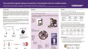 科学海报Fully Automated Magnetic Labeling and Separation of Hematopoietic Cells from Multiple Samples
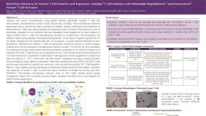 科学海报Workflow Solutions for Human T Cell Isolation and Expansion
 沪公网安备31010102008431号