Phondeechareon T et al. (OCT 2016)
Annals of hematology 95 10 1617--1625
Generation of induced pluripotent stem cells as a potential source of hematopoietic stem cells for transplant in PNH patients.
Paroxysmal nocturnal hemoglobinuria (PNH) is an acquired hemolytic anemia caused by lack of CD55 and CD59 on blood cell membrane leading to increased sensitivity of blood cells to complement. Hematopoietic stem cell transplantation (HSCT) is the only curative therapy for PNH,however,lack of HLA-matched donors and post-transplant complications are major concerns. Induced pluripotent stem cells (iPSCs) derived from patients are an attractive source for generating autologous HSCs to avoid adverse effects resulting from allogeneic HSCT. The disease involves only HSCs and their progeny; therefore,other tissues are not affected by the mutation and may be used to produce disease-free autologous HSCs. This study aimed to derive PNH patient-specific iPSCs from human dermal fibroblasts (HDFs),characterize and differentiate to hematopoietic cells using a feeder-free protocol. Analysis of CD55 and CD59 expression was performed before and after reprogramming,and hematopoietic differentiation. Patients' dermal fibroblasts expressed CD55 and CD59 at normal levels and the normal expression remained after reprogramming. The iPSCs derived from PNH patients had typical pluripotent properties and differentiation capacities with normal karyotype. After hematopoietic differentiation,the differentiated cells expressed early hematopoietic markers (CD34 and CD43) with normal CD59 expression. The iPSCs derived from HDFs of PNH patients have normal levels of CD55 and CD59 expression and hold promise as a potential source of HSCs for autologous transplantation to cure PNH patients.
View Publication
产品类型:
产品号#:
07923
07920
04435
04445
85850
85857
产品名:
Dispase (1 U/mL)
ACCUTASE™
MethoCult™H4435富集
MethoCult™H4435富集
mTeSR™1
mTeSR™1
文献
Zhang CC et al. (FEB 2006)
Proceedings of the National Academy of Sciences of the United States of America 103 7 2184--9
Prion protein is expressed on long-term repopulating hematopoietic stem cells and is important for their self-renewal.
Although the wild-type prion protein (PrP) is abundant and widely expressed in various types of tissues and cells,its physiological function(s) remain unknown,and PrP knockout mice do not exhibit overt and undisputed phenotypes. Here we showed that PrP is expressed on the surface of several bone marrow cell populations successively enriched in long-term (LT) hematopoietic stem cells (HSCs) using flow cytometry analysis. Affinity purification of the PrP-positive and -negative fractions from these populations,followed by competitive bone marrow reconstitution assays,shows that all LT HSCs express PrP. HSCs from PrP-null bone marrow exhibited impaired self-renewal in serial transplantation of lethally irradiated mouse recipients both in the presence and absence of competitors. When treated with a cell cycle-specific myelotoxic agent,the animals reconstituted with PrP-null HSCs exhibit increased sensitivity to hematopoietic cell depletion. Ectopic expression of PrP in PrP-null bone marrow cells by retroviral infection rescued the defective hematopoietic engraftment during serial transplantation. Therefore,PrP is a marker for HSCs and supports their self-renewal.
View Publication
J. Qiu et al. (dec 2022)
STAR protocols 3 4 101828
Protocol to identify and analyze mouse and human quiescent hematopoietic stem cells using flow cytometry combined with confocal imaging.
Mitochondrial membrane potential (MMP) segregates functionally distinct subsets within highly purified hematopoietic stem cells (HSCs). Here,we detail a protocol for FACS isolation of MMP sub-fractions of phenotypically defined mouse and human HSCs. These steps are followed by high-/super-resolution immunofluorescence microscopy of HSCs' lysosomes. While the protocol describes the isolation of quiescent HSCs,which are the most potent subsets,it could also be applied to other HSC subsets. This protocol overcomes some experimental challenges associated with low HSC numbers. For complete details on the use and execution of this protocol,please refer to Liang et al. (2020) and Qiu et al. (2021).
View Publication
Dobo I et al. (JAN 2001)
The hematology journal : the official journal of the European Haematology Association / EHA 2 6 396--403
Comparison of four serum-free, cytokine-free media for analysis of endogenous erythroid colony growth in polycythemia vera and essential thrombocythemia.
INTRODUCTION: The assay of endogenous erythroid colony formation (EEC),a characteristic of polycythemia vera and essential thrombocythemia,is not standardized. In this multicentric study,we tested four semisolid,serum-free,cytokine-free media based on either methylcellulose (M1,M2) or collagen (C1,C2) commercialized for the EEC assay. MATERIALS AND METHODS: Bone marrow mononuclear cells (BMMC) from 73 individuals (62 patients with either polycythemia vera (26),essential thrombocythemia (19),secondary polyglobuly (17) or chronic myeloid leukemia (2) and 11 healthy donors) were grown in parallel in the four media without,or with 0.01 U/ml erythropoietin (EPo). RESULTS: In all four media EEC formation was specific,as it was not observed in cultures of patients with secondary polyglobuly or chronic myeloid leukemia,nor of healthy donors. Analysis of fresh or MGG-stained collagen gel cultures allowed detection of EEC formation significantly more frequently than methylcellulose-based media; addition of 0.01 U/ml of EPo had little or no effect on EEC formation. Collagen-based medium C1 gave better results than the other media tested: the 'C1' EEC assay was positive for 68.2% of polycythemia vera cultures with significantly higher median EEC numbers (6.5/10(5) BMMC for patients with one major criteria of polycythemia vera and 19 and 21/10(5) BMMC for patients with two or three major criteria,respectively). Medium C1 was also better for essential thrombocythemia cultures with 47.4% of positive results but with a low median EEC number (6.7/10(5) BMMC). When associated with the ELISA dosage of serum EPo,the 'C1' EEC assay allowed confirmation or elimination of the diagnosis of polycythemia vera for 91% (20/22) of polyglobulic patients. CONCLUSION: We propose that serum-free collagen-based culture systems be considered to standardize the EEC assay,now part of the new criteria of polycythemia vera.
View Publication
产品类型:
产品号#:
04961
04962
04850
04974
04902
04960
04900
04901
04963
04970
04971
产品名:
MegaCult™-C胶原蛋白和细胞因子培养基
MegaCult™-C cfu染色试剂盒
MegaCult™-C含脂培养基
MegaCult™-C胶原蛋白和脂质培养基
胶原蛋白溶液
MegaCult™-C胶原蛋白和不含细胞因子的培养基
MegaCult™-C培养基无细胞因子
MegaCult™-C细胞因子培养基
双室载玻片试剂盒
MegaCult™-C不含细胞因子完整试剂盒
MegaCult™-C细胞因子完整试剂盒
文献
Bruserud &O et al. (MAY 2003)
Leukemia research 27 5 455--64
In vitro culture of human acute lymphoblastic leukemia (ALL) cells in serum-free media; a comparison of native ALL blasts, ALL cell lines and virus-transformed B cell lines.
The aim of this study was to standardize in vitro culture conditions for human acute lymphoblastic leukemia (ALL) cells. The cells were cultured in medium containing 10% fetal calf serum (FCS) and in the four serum-free media X-vivo 10,X-vivo 15,X-vivo 20 and Stem Span. Native ALL blasts could proliferate in all four serum-free media,but the strongest responses were usually observed with Stem Span. Native leukemia blasts were also cultured in the presence of various single cytokines or cytokine combinations. The highest proliferation was usually observed in the presence of Flt3-Ligand (Flt3-L) when single cytokines were examined,and these responses could be further increased especially by combining Flt3-L with interleukin 3 (IL3),IL7 or stem cell factor (SCF). Proliferation could also be increased when ALL blasts were cultured in the presence of two commercially available fibroblast cell lines (Hs27 and HFL1). Based on these results we suggest that in vitro culture conditions for native human ALL blasts can be standardized by using serum-free culture media supplemented with exogenous Flt3-L+IL3+SCF,and the use of accessory cells can also be standardized by using well-characterized fibroblast cell lines. Detectable ALL blast proliferation can then be observed for most patients. Our experimental model can thereby be used for in vitro evaluation of possible antileukemic treatment strategies,and it will then allow comparison of experimental results between different studies.
View Publication
产品类型:
产品号#:
产品名:
文献
Ma YD et al. (NOV 2009)
Blood 114 20 4402--10
Defects in osteoblast function but no changes in long-term repopulating potential of hematopoietic stem cells in a mouse chronic inflammatory arthritis model.
Recent studies support the notion that there is an intricate relationship between hematopoiesis and bone homeostasis in normal steady states. Using mice undergoing chronic inflammatory arthritis,we investigated the relationship between hematopoiesis and bone homeostasis in pathologic conditions. We demonstrate that mice undergoing chronic inflammatory arthritis displayed osteoporosis resulting from a severe defect in osteoblast function. Despite the defective osteoblast function,however,the hematopoietic stem cells from these mice exhibited normal properties in either long-term repopulation or cell cycling. Therefore,the bone-forming capacity of osteoblasts is distinct from their ability to maintain hematopoietic stem cells in chronic inflammatory conditions.
View Publication
beta-Catenin expression in the bone marrow microenvironment is required for long-term maintenance of primitive hematopoietic cells.
Hematopoiesis is dependent upon the bone marrow microenvironment,which is comprised of multiple mesenchymal cell types,including fibroblasts,endothelial cells,osteoblasts,and stroma progenitors. The canonical Wnt signaling pathway,which relies on the beta-catenin protein to mediate its signal,is necessary for the normal development of mesenchymal tissue. We hypothesized that canonical Wnt signaling regulates the cellular composition and function of the bone marrow microenvironment. We observed that a beta-catenin-deficient bone marrow microenvironment maintained hematopoietic stem cells but exhibited a decreased capacity to support primitive hematopoietic cells. These results correlated with decreased numbers of osteoblasts and with decreased production of basic fibroblast growth factor,stem cell factor,and vascular cell adhesion molecule-1. From these data,we propose a model in which beta-catenin in the microenvironment is required noncell autonomously for long-term maintenance of hematopoietic progenitors.
View Publication

 EasySep™小鼠TIL(CD45)正选试剂盒


 文献
文献 产品手册UM171 & UM729: Novel Small Molecules for the Ex Vivo Expansion of Human Hematopoietic Stem Cells
 产品手册Neural Stem Cells: Standardized Media and Reagents
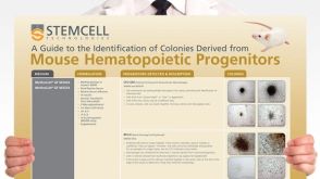 挂图Identification of Colonies Derived from Mouse Hematopoietic Progenitors Representative colony images and tips for identifying progenitor subtypes in CFU assays
