Liu C-G et al. (JUN 2011)
Annals of surgery 253 6 1165--71
Clinical implications of stem cell gene Oct-4 expression in breast cancer.
PURPOSE: To explore the expression of stem cell genes in breast cancer and the relationship between stem cell gene expression and clinical and pathological characteristics and prognosis of breast cancer. BACKGROUND: By now,stem cell differentiation-related genes and the relationship between the genes and clinic-pathological characteristics and prognosis of breast cancer are still unclear. MATERIALS AND METHODS: CD44+/CD24- tumor cells were selected by Flow cytometry. The differential expression of genes between CD44+/CD24- tumor cells and non-CD44+/CD24- tumor cells were detected by RT(2) Profiler™ PCR Array. The expression of stem cell gene Octamer-4 (Oct-4) was analyzed by immunohistochemistry staining and the relationship between Oct-4 and clinicopathological parameters of breast cancer was determined. RESULTS: Seven different genes including stem cell differentiation-related factors (CD44,Oct-4,and nestin),cell cycle regulators (APC and CDC2),and growth factors (HGF and TGF) were detected as significantly differently expressed between CD44+/CD24- tumor cells and non-CD44+/CD24- tumor cells. Oct-4 protein expressed significantly higher in cancerous tissues than adjacent-tumor tissues (P = 0.001). Moreover,we observed that the expression of Oct-4 protein was related to histological type,lymph node status and molecular type of breast cancer (P = 0.001,0.006,and 0.001,respectively). After survival analysis,the cases with highly expressed Oct-4 protein attained a significantly poorer postoperative disease-specific survival than those with none/low expressed Oct-4 protein (P = 0.001). In the Cox regression test,tumor size,histological type,disease stage,lymph node metastasis,Her-2 and Oct-4 were detected as the independent prognostic factors (P = 0.031,0.012,0.001,0.002,0.030,and 0.003,respectively). CONCLUSIONS: Oct-4 was highly expressed in CD44+/CD24- tumor cells,and may be a potential biomarker for the initiation,progression,and differentiation of breast cancer.
View Publication
产品类型:
产品号#:
05620
产品名:
MammoCult™ 人源培养基套装
Liebmann JE et al. ( 1993)
British journal of cancer 68 6 1104--1109
Cytotoxic studies of paclitaxel (Taxol) in human tumour cell lines.
The cytotoxicity of paclitaxel against eight human tumour cell lines has been studied with in vitro clonogenic assays. The fraction of surviving cells fell sharply after exposure for 24 h to paclitaxel concentrations ranging from 2 to 20 nM; the paclitaxel IC50 was found to range between 2.5 and 7.5 nM. Increasing the paclitaxel concentration above 50 nM,however,resulted in no additional cytotoxicity after a 24 h drug exposure. Cells incubated in very high concentrations of paclitaxel (10,000 nM) had an increase in survival compared with cells treated with lower concentrations of the drug. Prolonging the time of exposure of cells to paclitaxel from 24 to 72 h increased cytotoxicity from 5 to 200 fold in different cell lines. Exponentially growing cells were more sensitive to paclitaxel than were cells in the plateau phase of growth. Cremophor EL,the diluent in which the clinical preparation of paclitaxel is formulated,antagonised paclitaxel at concentrations of 0.135% (v/v). These data suggest that paclitaxel will be most effective clinically when there is prolonged exposure of tumour to the drug. Further,it appears that modest concentrations (i.e.,50 nM) should be as effective as higher concentrations of paclitaxel. Finally,we have noted that Cremophor EL is a biologically active diluent and,at high concentrations (0.135% v/v),can antagonise paclitaxel cytotoxicity.
View Publication
Shackleton M et al. (JAN 2006)
Nature 439 7072 84--8
Generation of a functional mammary gland from a single stem cell.
The existence of mammary stem cells (MaSCs) has been postulated from evidence that the mammary gland can be regenerated by transplantation of epithelial fragments in mice. Interest in MaSCs has been further stimulated by their potential role in breast tumorigenesis. However,the identity and purification of MaSCs has proved elusive owing to the lack of defined markers. We isolated discrete populations of mouse mammary cells on the basis of cell-surface markers and identified a subpopulation (Lin-CD29hiCD24+) that is highly enriched for MaSCs by transplantation. Here we show that a single cell,marked with a LacZ transgene,can reconstitute a complete mammary gland in vivo. The transplanted cell contributed to both the luminal and myoepithelial lineages and generated functional lobuloalveolar units during pregnancy. The self-renewing capacity of these cells was demonstrated by serial transplantation of clonal outgrowths. In support of a potential role for MaSCs in breast cancer,the stem-cell-enriched subpopulation was expanded in premalignant mammary tissue from MMTV-wnt-1 mice and contained a higher number of MaSCs. Our data establish that single cells within the Lin-CD29hiCD24+ population are multipotent and self-renewing,properties that define them as MaSCs.
View Publication
产品类型:
产品号#:
01700
01705
05601
05610
05620
01702
产品名:
ALDEFLUOR™ 试剂盒
ALDEFLUOR™ DEAB试剂, 1.5 mM, 1 mL
EpiCult™-B 人培养基
EpiCult™-B 小鼠培养基
MammoCult™ 人源培养基套装
ALDEFLUOR™检测缓冲液
Wittman VP et al. (SEP 2006)
The Journal of Immunology 177 6 4187--95
Antibody targeting to acClass I MHC-peptide epitope promotes tumor cell death
Therapeutic mAbs that target tumor-associated Ags on the surface of malignant cells have proven to be an effective and specific option for the treatment of certain cancers. However,many of these protein markers of carcinogenesis are not expressed on the cells' surface. Instead these tumor-associated Ags are processed into peptides that are presented at the cell surface,in the context of MHC class I molecules,where they become targets for T cells. To tap this vast source of tumor Ags,we generated a murine IgG2a mAb,3.2G1,endowed with TCR-like binding specificity for peptide-HLA-A*0201 (HLA-A2) complex and designated this class of Ab as TCR mimics (TCRm). The 3.2G1 TCRm recognizes the GVL peptide (GVLPALPQV) from human chorionic gonadotropin beta presented by the peptide-HLA-A*0201 complex. When used in immunofluorescent staining reactions using GVL peptide-loaded T2 cells,the 3.2G1 TCRm specifically stained the cells in a peptide and Ab concentration-dependent manner. Staining intensity correlated with the extent of cell lysis by complement-dependent cytotoxicity (CDC),and a peptide concentration-dependent threshold level existed for the CDC reaction. Staining of human tumor lines demonstrated that 3.2G1 TCRm was able to recognize endogenously processed peptide and that the breast cancer cell line MDA-MB-231 highly expressed the target epitope. The 3.2G1 TCRm-mediated CDC and Ab-dependent cellular cytotoxicity of a human breast carcinoma line in vitro and inhibited in vivo tumor implantation and growth in nude mice. These results provide validation for the development of novel TCRm therapeutic reagents that specifically target and kill tumors via recognition and binding to MHC-peptide epitopes.
View Publication
产品类型:
产品号#:
03800
03801
03802
03803
03804
03805
03806
03831
产品名:
ClonaCell™-HY杂交瘤试剂盒
ClonaCell™-HY培养基A
ClonaCell™-HY 培养基 B
ClonaCell™-HY 培养基 C
ClonaCell™-HY 培养基 D
ClonaCell™-HY 培养基 E
ClonaCell™-HY PEG
ClonaCell™-HY 液体 HAT 筛选培养基
Korkaya H et al. (OCT 2008)
Oncogene 27 47 6120--30
HER2 regulates the mammary stem/progenitor cell population driving tumorigenesis and invasion.
The cancer stem cell hypothesis proposes that cancers arise in stem/progenitor cells through disregulation of self-renewal pathways generating tumors,which are driven by a component of 'tumor-initiating cells' retaining stem cell properties. The HER2 gene is amplified in 20-30% of human breast cancers and has been implicated in mammary tumorigenesis as well as in mediating aggressive tumor growth and metastasis. We demonstrate that HER2 overexpression drives mammary carcinogenesis,tumor growth and invasion through its effects on normal and malignant mammary stem cells. HER2 overexpression in normal mammary epithelial cells (NMEC) increases the proportion of stem/progenitor cells as demonstrated by in vitro mammosphere assays and the expression of stem cell marker aldehyde dehydrogenase (ALDH) as well as by generation of hyperplastic lesions in humanized fat pads of NOD (nucleotide-binding oligomerization domain)/SCID (severe combined immunodeficient) mice. Overexpression of HER2 in a series of breast carcinoma cell lines increases the ALDH-expressing 'cancer stem cell' population which displays increased expression of stem cell regulatory genes,increased invasion in vitro and increased tumorigenesis in NOD/SCID mice. The effects of HER2 overexpression on breast cancer stem cells are blocked by trastuzumab in sensitive,but not resistant,cell lines,an effect mediated by the PI3-kinase/Akt pathway. These studies provide support for the cancer stem cell hypothesis by suggesting that the effects of HER2 amplification on carcinogenesis,tumorigenesis and invasion may be due to its effects on normal and malignant mammary stem/progenitor cells. Furthermore,the clinical efficacy of trastuzumab may relate to its ability to target the cancer stem cell population in HER2-amplified tumors.
View Publication
产品类型:
产品号#:
01700
01705
01702
产品名:
ALDEFLUOR™ 试剂盒
ALDEFLUOR™ DEAB试剂, 1.5 mM, 1 mL
ALDEFLUOR™检测缓冲液
Mortensen M et al. (MAR 2011)
The Journal of experimental medicine 208 3 455--67
The autophagy protein Atg7 is essential for hematopoietic stem cell maintenance.
The role of autophagy,a lysosomal degradation pathway which prevents cellular damage,in the maintenance of adult mouse hematopoietic stem cells (HSCs) remains unknown. Although normal HSCs sustain life-long hematopoiesis,malignant transformation of HSCs leads to leukemia. Therefore,mechanisms protecting HSCs from cellular damage are essential to prevent hematopoietic malignancies. In this study,we crippled autophagy in HSCs by conditionally deleting the essential autophagy gene Atg7 in the hematopoietic system. This resulted in the loss of normal HSC functions,a severe myeloproliferation,and death of the mice within weeks. The hematopoietic stem and progenitor cell compartment displayed an accumulation of mitochondria and reactive oxygen species,as well as increased proliferation and DNA damage. HSCs within the Lin(-)Sca-1(+)c-Kit(+) (LSK) compartment were significantly reduced. Although the overall LSK compartment was expanded,Atg7-deficient LSK cells failed to reconstitute the hematopoietic system of lethally irradiated mice. Consistent with loss of HSC functions,the production of both lymphoid and myeloid progenitors was impaired in the absence of Atg7. Collectively,these data show that Atg7 is an essential regulator of adult HSC maintenance.
View Publication
产品类型:
产品号#:
03434
03444
产品名:
MethoCult™ GF M3434
MethoCult™ GF M3434
Dalley AJ et al. (JAN 2013)
Journal of oral pathology & medicine : official publication of the International Association of Oral Pathologists and the American Academy of Oral Pathology 42 1 37--46
Organotypic culture of normal, dysplastic and squamous cell carcinoma-derived oral cell lines reveals loss of spatial regulation of CD44 and p75 NTR in malignancy.
Oral squamous cell carcinomas (OSCC) often arise from dysplastic lesions. The role of cancer stem cells in tumour initiation is widely accepted,yet the potential existence of pre-cancerous stem cells in dysplastic tissue has received little attention. Cell lines from oral diseases ranging in severity from dysplasia to malignancy provide opportunity to investigate the involvement of stem cells in malignant progression from dysplasia. Stem cells are functionally defined by their ability to generate hierarchical tissue structures in consortium with spatial regulation. Organotypic cultures readily display tissue hierarchy in vitro; hence,in this study,we compared hierarchical expression of stem cell-associated markers in dermis-based organotypic cultures of oral epithelial cells from normal tissue (OKF6-TERT2),mild dysplasia (DOK),severe dysplasia (POE-9n) and OSCC (PE/CA P J15). Expression of CD44,p75(NTR),CD24 and ALDH was studied in monolayers by flow cytometry and in organotypic cultures by immunohistochemistry. Spatial regulation of CD44 and p75(NTR) was evident for organotypic cultures of normal (OKF6-TERT2) and dysplasia (DOK and POE-9n) but was lacking for OSCC (PE/CA PJ15)-derived cells. Spatial regulation of CD24 was not evident. All monolayer cultures exhibited CD44,p75(NTR),CD24 antigens and ALDH activity (ALDEFLUOR(®) assay),with a trend towards loss of population heterogeneity that mirrored disease severity. In monolayer,increased FOXA1 and decreased FOXA2 expression correlated with disease severity,but OCT3/4,Sox2 and NANOG did not. We conclude that dermis-based organotypic cultures give opportunity to investigate the mechanisms that underlie loss of spatial regulation of stem cell markers seen with OSCC-derived cells.
View Publication


 EasySep™小鼠TIL(CD45)正选试剂盒





 产品手册Highway1™: Fast, Gentle, and Automated Cell Sorting for Every Lab
 实验方案How to Collect Plasma from Whole Blood Before Cell Isolation
 产品手册Highway1™: Fast, Gentle, and Automated Cell Sorting for Every Lab
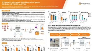 科学海报STEMprep™ Automated Tissue Dissociation System: Maximize Cell Viability and Yield

 沪公网安备31010102008431号