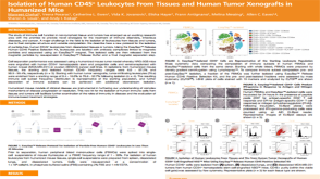
Eric Song
Priming the Immune System Against Brain Tumors
产品类型:
研究方向:
免疫学,癌症研究
产品号#:
产品名:
发布日期: 04/09/2020
Biswas S et al. (OCT 2009)
Journal of immunology (Baltimore,Md. : 1950) 183 8 5050--8
Elevated levels of select gangliosides in T cells from renal cell carcinoma patients is associated with T cell dysfunction.
Increased expression of gangliosides by different tumor types including renal cell carcinoma (RCC) is thought to contribute to the immune suppression observed in cancer patients. In this study,we report an increase in apoptotic T cells from RCC patients compared with T cells from normal donors that coincided with the detection of T cells staining positive for GM2 and that the apoptosis was predominantly observed in the GM2(+) but not the GM2(-) T cell population. Ganglioside shedding from tumor rather than endogenous production accounts for GM2(+) T cells since there was no detectable level of mRNA for GM2 synthase in RCC patient T cells and in T cells from normal healthy donors after incubation with either purified GM2 or supernatant from RCC cell lines despite their staining positive for GM2. Moreover,reactive oxygen species as well as activated caspase 3,8,and 9 were predominantly elevated in GM2(+) but not GM2(-) T cells. Similarly,increased staining for GD2 and GD3 but not GD1a was detected with patient T cells with elevated levels of apoptosis in the GD2(+) and GD3(+) cells. These findings suggest that GM2,GD2,and GD3 play a significant role in immune dysfunction observed in RCC patient T cells.
View Publication
产品类型:
产品号#:
19051
19051RF
产品名:
EasySep™人T细胞富集试剂盒
RoboSep™ 人T细胞富集试剂盒含滤芯吸头
Y. Zhang et al. ( 2015)
The Journal of Immunology 194 5937-5947
Genetic Vaccines To Potentiate the Effective CD103+ Dendritic Cell-Mediated Cross-Priming of Antitumor Immunity
The development of effective cancer vaccines remains an urgent,but as yet unmet,clinical need. This deficiency is in part due to an incomplete understanding of how to best invoke dendritic cells (DC) that are crucial for the induction of tumor-specific CD8(+) T cells capable of mediating durable protective immunity. In this regard,elevated expression of the transcription factor X box-binding protein 1 (XBP1) in DC appears to play a decisive role in promoting the ability of DC to cross-present Ags to CD8(+) T cells in the therapeutic setting. Delivery of DNA vaccines encoding XBP1 and tumor Ag to skin DC resulted in increased IFN-? production by plasmacytoid DC (pDC) from skin/tumor draining lymph nodes and the cross-priming of Ag-specific CD8(+) T cell responses associated with therapeutic benefit. Antitumor protection was dependent on cross-presenting Batf3(+) DC,pDC,and CD8(+) T cells. CD103(+) DC from the skin/tumor draining lymph nodes of the immunized mice appeared responsible for activation of Ag-specific naive CD8(+) T cells,but were dependent on pDC for optimal effectiveness. Similarly,human XBP1 improved the capacity of human blood- and skin-derived DC to activate human T cells. These data support an important intrinsic role for XBP1 in DC for effective cross-priming and orchestration of Batf3(+) DC-pDC interactions,thereby enabling effective vaccine induction of protective antitumor immunity.
View Publication
Zhang M et al. (SEP 2014)
International journal of cancer 135 5 1132--41
Anti-β₂M monoclonal antibodies kill myeloma cells via cell- and complement-mediated cytotoxicity.
Our previous studies showed that anti-β2M monoclonal antibodies (mAbs) at high doses have direct apoptotic effects on myeloma cells,suggesting that anti-β2M mAbs might be developed as a novel therapeutic agent. In this study,we investigated the ability of the mAbs at much lower concentrations to indirectly kill myeloma cells by utilizing immune effector cells or molecules. Our results showed that anti-β2M mAbs effectively lysed MM cells via antibody-dependent cell-mediated cytotoxicity (ADCC) and complement-dependent cytotoxicity (CDC),which were correlated with and dependent on the surface expression of β2M on MM cells. The presence of MM bone marrow stromal cells or addition of IL-6 did not attenuate anti-β2M mAb-induced ADCC and CDC activities against MM cells. Furthermore,anti-β2M mAbs only showed limited cytotoxicity toward normal B cells and nontumorous mesenchymal stem cells,indicating that the ADCC and CDC activities of the anti-β2M mAbs were more prone to the tumor cells. Lenalidomide potentiated in vitro ADCC activity against MM cells and in vivo tumor inhibition capacity induced by the anti-β2M mAbs by enhancing the activity of NK cells. These results support clinical development of anti-β2M mAbs,both as a monotherapy and in combination with lenalidomide,to improve MM patient outcome.
View Publication
产品类型:
产品号#:
18387
18387RF
产品名:
Cammenga J et al. (JAN 2007)
Cancer research 67 2 537--45
Mutations in the RUNX1 gene are found at high frequencies in minimally differentiated acute myelogenous leukemia. In addition to null mutations,many of the mutations generate Runx1 DNA-binding (RDB) mutants. To determine if these mutants antagonize wild-type protein activity,cDNAs were transduced into murine bone marrow or human cord blood cells using retroviral vectors. Significantly,the RDB mutants did not act in a transdominant fashion in vivo to disrupt Runx1 activity in either T-cell or platelet development,which are highly sensitive to Runx1 dosage. However,RDB mutant expression impaired expansion and differentiation of the erythroid compartment in which Runx1 expression is normally down-regulated,showing that a RDB-independent function is incompatible with erythroid differentiation. Significantly,both bone marrow progenitors expressing RDB mutants or deficient for Runx1 showed increased replating efficiencies in vitro,accompanied by the accumulation of myeloblasts and dysplastic progenitors,but the effect was more pronounced in RDB cultures. Disruption of the interface that binds CBFbeta,an important cofactor of Runx1,did not impair RDB mutant replating activity,arguing against inactivation of Runx1 function by CBFbeta sequestration. We propose that RDB mutants antagonize Runx1 function in early progenitors by disrupting a critical balance between DNA-binding-independent and DNA-binding-dependent signaling.
View Publication
产品类型:
产品号#:
03434
03444
09500
09600
09650
18096
18096RF
84434
84444
产品名:
MethoCult™ GF M3434
MethoCult™ GF M3434
BIT 9500血清替代物
StemSpan™ SFEM
StemSpan™ SFEM
Timm MM et al. (OCT 2006)
Leukemia 20 10 1863--9
Thymoglobulin targets multiple plasma cell antigens and has in vitro and in vivo activity in multiple myeloma.
Multiple myeloma is characterized by the proliferation of clonal plasma cells that have a heterogeneous expression of various cell surface markers,precluding successful use of monoclonal antibodies for therapeutic targeting of the tumor cell. Thymoglobulin (rabbit-derived polyclonal anti-thymocyte globulin),by virtue of its method of preparation,contains antibodies against several B-cell and plasma cell antigens and offers an attractive option for immunotherapy of myeloma. Here,we demonstrate potent anti-myeloma activity of the rabbit anti-thymocyte globulin preparation Thymoglobulin in vitro and in vivo in an animal model of myeloma. Thymoglobulin was able to induce dose- and time-dependent apoptosis of several myeloma cell lines,including those resistant to conventional anti-myeloma agents. Importantly,the anti-myeloma activity was preserved even when myeloma cells were grown with different cytokines demonstrating the ability to overcome microenvironment-mediated resistance. Thymoglobulin induced apoptosis of freshly isolated primary myeloma cells from patients. Using a competitive flow cytometric analysis,we were able to identify the potential antigen targets for Thymoglobulin preparation. Finally,in a plasmacytoma mouse model of myeloma,Thymoglobulin delayed the tumor growth in a dose-dependent manner providing convincing evidence for continued evaluation of this agent in the clinic in patients with myeloma,either alone or in combination with other agents.
View Publication
产品类型:
产品号#:
18357
18357RF
21000
20119
20155
产品名:
RoboSep™- S
RoboSep™ 吸头组件抛光剂
RoboSep™分选管套装(9个塑料管)
Akatsuka A et al. (SEP 2010)
International immunology 22 9 783--90
Tumor cells of non-hematopoietic and hematopoietic origins express activation-induced C-type lectin, the ligand for killer cell lectin-like receptor F1.
Killer cell lectin-like receptor F1 (KLRF1) is an activating C-type lectin-like receptor expressed on human NK cells and subsets of T cells. In this study,we show that activation-induced C-type lectin (AICL) is a unique KLRF1 ligand expressed on tumor cell lines of hematopoietic and non-hematopoietic origins. We screened a panel of human tumor cell lines using the KLRF1 reporter cells and found that several tumor lines expressed KLRF1 ligands. We characterized a putative KLRF1 ligand expressed on the U937 cell line. The molecular mass for the deglycosylated ligand was 28 kDa under non-reducing condition and 17 kDa under reducing condition,suggesting that the KLRF1 ligand is a homodimer. By expression cloning from a U937 cDNA library,we identified AICL as a KLRF1 ligand. We generated mAbs against AICL to identify the KLRF1 ligands on non-hematopoietic tumor lines. The anti-AICL mAbs stained the tumor lines that express the KLRF1 ligands and importantly the interaction of KLRF1 with the KLRF1 ligand on non-hematopoietic tumors was completely blocked by the two anti-AICL mAbs. Moreover,NK cell degranulation triggered by AICL-expressing targets was partially inhibited by the anti-AICL mAb. Finally,we demonstrate that AICL is expressed in human primary liver cancers. These results suggest that AICL is expressed on tumor cells of non-hematopoietic origins and raise the possibility that AICL may contribute to NK cell surveillance of tumor cells.
View Publication
产品类型:
产品号#:
18554
18554RF
18564
18564RF
产品名:
Wang X et al. (OCT 2009)
Cancer research 69 19 7612--8
Correction of the abnormal trafficking of primary myelofibrosis CD34+ cells by treatment with chromatin-modifying agents.
The abnormal trafficking of CD34+ cells is a unique characteristic of primary myelofibrosis (PMF). We have further studied the behavior of PMF CD34+ cells by examining their homing to the marrow and the spleens of nonobese diabetic/severe combined immunodeficient (NOD/SCID) mice. Following the infusion of PMF and normal granulocyte colony-stimulating factor-mobilized peripheral blood (mPB) CD34+ cells into NOD/SCID mice,reduced numbers of PMF CD34+ cells and granulocyte-macrophage colony-forming unit (CFU-GM) compared with mPB were detected in the marrow of these mice,whereas similar numbers of PMF and mPB CD34+ cells and CFU-GM homed to their spleens. The abnormal homing of PMF CD34+ cells was associated with reduced expression of CXCR4,but was not related to the presence of JAK2V617F. The sequential treatment of PMF CD34+ cells with the chromatin-modifying agents 5-aza-2'-deoxycytidine (5azaD) and trichostatin A (TSA),but not treatment with small molecule inhibitors of JAK2,resulted in the generation of increased numbers of CD34+CXCR4+ cells,which was accompanied by enhanced homing of PMF CD34+ cells to the marrow but not the spleens of NOD/SCID mice. Following 5azaD/TSA treatment,JAK2V617F-negative PMF hematopoietic progenitor cells preferentially homed to the marrow but not the spleens of recipient mice. Our data suggest that PMF CD34+ cells are characterized by a reduced ability to home to the marrow but not the spleens of NOD/SCID mice and that this homing defect can be corrected by sequential treatment with chromatin-modifying agents.
View Publication
产品类型:
产品号#:
18056
18056RF
产品名:
Popovic R et al. (APR 2009)
Blood 113 14 3314--22
Regulation of mir-196b by MLL and its overexpression by MLL fusions contributes to immortalization.
Chromosomal translocations involving the Mixed Lineage Leukemia (MLL) gene produce chimeric proteins that cause abnormal expression of a subset of HOX genes and leukemia development. Here,we show that MLL normally regulates expression of mir-196b,a hematopoietic microRNA located within the HoxA cluster,in a pattern similar to that of the surrounding 5' Hox genes,Hoxa9 and Hoxa10,during embryonic stem (ES) cell differentiation. Within the hematopoietic lineage,mir-196b is most abundant in short-term hematopoietic stem cells and is down-regulated in more differentiated hematopoietic cells. Leukemogenic MLL fusion proteins cause overexpression of mir-196b,while treatment of MLL-AF9 transformed bone marrow cells with mir-196-specific antagomir abrogates their replating potential in methylcellulose. This demonstrates that mir-196b function is necessary for MLL fusion-mediated immortalization. Furthermore,overexpression of mir-196b was found specifically in patients with MLL associated leukemias as determined from analysis of 55 primary leukemia samples. Overexpression of mir-196b in bone marrow progenitor cells leads to increased proliferative capacity and survival,as well as a partial block in differentiation. Our results suggest a mechanism whereby increased expression of mir-196b by MLL fusion proteins significantly contributes to leukemia development.
View Publication

 EasySep™小鼠TIL(CD45)正选试剂盒


 科学海报Isolation of Human CD45+ Leukocytes From Tissues and Human Tumor Xenografts in Humanized Mice
 专家访谈Eric Song Priming the Immune System Against Brain Tumors


 沪公网安备31010102008431号