Effector T Cells Abrogate Stroma-Mediated Chemoresistance in Ovarian Cancer.
Effector T cells and fibroblasts are major components in the tumor microenvironment. The means through which these cellular interactions affect chemoresistance is unclear. Here,we show that fibroblasts diminish nuclear accumulation of platinum in ovarian cancer cells,resulting in resistance to platinum-based chemotherapy. We demonstrate that glutathione and cysteine released by fibroblasts contribute to this resistance. CD8(+) T cells abolish the resistance by altering glutathione and cystine metabolism in fibroblasts. CD8(+) T-cell-derived interferon (IFN)γ controls fibroblast glutathione and cysteine through upregulation of gamma-glutamyltransferases and transcriptional repression of system xc(-) cystine and glutamate antiporter via the JAK/STAT1 pathway. The presence of stromal fibroblasts and CD8(+) T cells is negatively and positively associated with ovarian cancer patient survival,respectively. Thus,our work uncovers a mode of action for effector T cells: they abrogate stromal-mediated chemoresistance. Capitalizing upon the interplay between chemotherapy and immunotherapy holds high potential for cancer treatment.
View Publication
产品类型:
产品号#:
17953
17953RF
15022
15062
100-0710
产品名:
EasySep™人CD8+ T细胞分选试剂盒
RoboSep™ 人CD8+ T细胞分选试剂盒
RosetteSep™人CD4+ T细胞富集抗体混合物
RosetteSep™人CD4+ T细胞富集抗体混合物
EasySep™人CD8+ T细胞分选试剂盒
Brooks SE et al. ( 2015)
PloS one 10 10 e0140483
Application of the pMHC Array to Characterise Tumour Antigen Specific T Cell Populations in Leukaemia Patients at Disease Diagnosis.
Immunotherapy treatments for cancer are becoming increasingly successful,however to further improve our understanding of the T-cell recognition involved in effective responses and to encourage moves towards the development of personalised treatments for leukaemia immunotherapy,precise antigenic targets in individual patients have been identified. Cellular arrays using peptide-MHC (pMHC) tetramers allow the simultaneous detection of different antigen specific T-cell populations naturally circulating in patients and normal donors. We have developed the pMHC array to detect CD8+ T-cell populations in leukaemia patients that recognise epitopes within viral antigens (cytomegalovirus (CMV) and influenza (Flu)) and leukaemia antigens (including Per Arnt Sim domain 1 (PASD1),MelanA,Wilms' Tumour (WT1) and tyrosinase). We show that the pMHC array is at least as sensitive as flow cytometry and has the potential to rapidly identify more than 40 specific T-cell populations in a small sample of T-cells (0.8-1.4 x 10(6)). Fourteen of the twenty-six acute myeloid leukaemia (AML) patients analysed had T cells that recognised tumour antigen epitopes,and eight of these recognised PASD1 epitopes. Other tumour epitopes recognised were MelanA (n = 3),tyrosinase (n = 3) and WT1(126-134) (n = 1). One of the seven acute lymphocytic leukaemia (ALL) patients analysed had T cells that recognised the MUC1(950-958) epitope. In the future the pMHC array may be used provide point of care T-cell analyses,predict patient response to conventional therapy and direct personalised immunotherapy for patients.
View Publication
产品类型:
产品号#:
19053
19053RF
产品名:
EasySep™人CD8+ T细胞富集试剂盒
RoboSep™ 人CD8+ T细胞富集试剂盒含滤芯吸头
Ortega V et al. (MAR 2016)
Cancer genetics 209 3 82--6
Optimal strategy for obtaining routine chromosome analysis by using negative fractions of CD138 enriched plasma cells.
Fluorescence in situ hybridization (FISH) is superior to routine chromosome analysis (RCA) in detecting important prognostic genetic abnormalities in plasma cell dyscrasia (PCD); however,its sensitivity is hampered due to paucity of plasma cells (PC) in whole bone marrow (BM). Studies showed that the abnormality detection rate in enriched plasma cells (EPC) is greater than unselected plasma cells (UPC),but purification techniques are limiting to only FISH when sample volumes are inadequate. Not performing RCA may compromise patient care since RCA is equally important for detecting non-PC related abnormalities when the diagnosis is undefined. To resolve this critical issue,we designed a study where an immuno-magnetic CD138 enriched positive selection was used for FISH while the negative fraction (NF) was used to retrieve other myeloid elements for RCA. Parallel FISH studies were performed using UPC and CD138 EPC,while karyotyping was achieved using whole BM and discarded myeloid elements from the NF. Results showed that the abnormality rate of EPC was doubled compared to UPC for FISH,and CA displayed 100% success rate using the NF. PCD related chromosome abnormalities were confined to whole BM while non-PCD related abnormalities were found in both whole BM and NF. Our results demonstrate the feasibility of using the NF for RCA.
View Publication
Maes C et al. (MAY 2006)
The Journal of clinical investigation 116 5 1230--42
Placental growth factor mediates mesenchymal cell development, cartilage turnover, and bone remodeling during fracture repair.
Current therapies for delayed- or nonunion bone fractures are still largely ineffective. Previous studies indicated that the VEGF homolog placental growth factor (PlGF) has a more significant role in disease than in health. Therefore we investigated the role of PlGF in a model of semi-stabilized bone fracture healing. Fracture repair in mice lacking PlGF was impaired and characterized by a massive accumulation of cartilage in the callus,reminiscent of delayed- or nonunion fractures. PlGF was required for the early recruitment of inflammatory cells and the vascularization of the fracture wound. Interestingly,however,PlGF also played a role in the subsequent stages of the repair process. Indeed in vivo and in vitro findings indicated that PlGF induced the proliferation and osteogenic differentiation of mesenchymal progenitors and stimulated cartilage turnover by particular MMPs. Later in the process,PlGF was required for the remodeling of the newly formed bone by stimulating osteoclast differentiation. As PlGF expression was increased throughout the process of bone repair and all the important cell types involved expressed its receptor VEGFR-1,the present data suggest that PlGF is required for mediating and coordinating the key aspects of fracture repair. Therefore PlGF may potentially offer therapeutic advantages for fracture repair.
View Publication
产品类型:
产品号#:
03534
03334
03434
03444
18753
18753RF
产品名:
MethoCult™ GF M3534
MethoCult™ M3334
MethoCult™ GF M3434
MethoCult™ GF M3434
Wunderlich M et al. (SEP 2006)
Blood 108 5 1690--7
Human CD34+ cells expressing the inv(16) fusion protein exhibit a myelomonocytic phenotype with greatly enhanced proliferative ability.
The t(16:16) and inv(16) are associated with FAB M4Eo myeloid leukemias and result in fusion of the CBFB gene to the MYH11 gene (encoding smooth muscle myosin heavy chain [SMMHC]). Knockout of CBFbeta causes embryonic lethality due to lack of definitive hematopoiesis. Although knock-in of CBFB-MYH11 is not sufficient to cause disease,expression increases the incidence of leukemia when combined with cooperating events. Although mouse models are valuable tools in the study of leukemogenesis,little is known about the contribution of CBFbeta-SMMHC to human hematopoietic stem and progenitor cell self-renewal. We introduced the CBFbeta-MYH11 cDNA into human CD34+ cells via retroviral transduction. Transduced cells displayed an initial repression of progenitor activity but eventually dominated the culture,resulting in the proliferation of clonal populations for up to 7 months. Long-term cultures displayed a myelomonocytic morphology while retaining multilineage progenitor activity and engraftment in NOD/SCID-B2M-/- mice. Progenitor cells from long-term cultures showed altered expression of genes defining inv(16) identified in microarray studies of human patient samples. This system will be useful in examining the effects of CBFbeta-SMMHC on gene expression in the human preleukemic cell,in characterizing the effect of this oncogene on human stem cell biology,and in defining its contribution to the development of leukemia.
View Publication
产品类型:
产品号#:
04100
18056
18056RF
产品名:
MethoCult™ H4100
Carlsten M et al. (FEB 2007)
Cancer research 67 3 1317--25
DNAX accessory molecule-1 mediated recognition of freshly isolated ovarian carcinoma by resting natural killer cells.
Although natural killer (NK) cells are well known for their ability to kill tumors,few studies have addressed the interactions between resting (nonactivated) NK cells and freshly isolated human tumors. Here,we show that human leukocyte antigen class I(low) tumor cells isolated directly from patients with advanced ovarian carcinoma trigger degranulation by resting allogeneic NK cells. This was paralleled by induction of granzyme B and caspase-6 activities in the tumor cells and significant tumor cell lysis. Ovarian carcinoma cells displayed ubiquitous expression of the DNAX accessory molecule-1 (DNAM-1) ligand PVR and sparse/heterogeneous expression of the NKG2D ligands MICA/MICB and ULBP1,ULBP2,and ULBP3. In line with the NK receptor ligand expression profiles,antibody-mediated blockade of activating receptor pathways revealed a dominant role for DNAM-1 and a complementary contribution of NKG2D signaling in tumor cell recognition. These results show that resting NK cells are capable of directly recognizing freshly isolated human tumor cells and identify ovarian carcinoma as a potential target for adoptive NK cell-based immunotherapy.
View Publication
产品类型:
产品号#:
18259
18259RF
产品名:
Pereira WdO et al. (OCT 2013)
BMC research notes 6 433
Development of plasma cell myeloma in a B-cell chronic lymphocytic leukemia patient with chromosome 12 trisomy.
BACKGROUND Cancer development results from the progressive accumulation of genomic abnormalities that culminate in the neoplastic phenotype. Cytogenetic alterations,mutations and rearrangements may be considered as molecular legacy which trace the clonal history of the disease. Concomitant tumors are reported and they may derive from a common or divergent founder clone. B-cell chronic lymphocytic leukemia (B-CLL) and plasma cell myeloma (PCM) are both mature B-cell neoplasms,and their concomitancy,albeit rare,is documented. CASE PRESENTATION Here,we described a patient with prior B-CLL with secondary development of PCM. Cytogenetic and multi parametric flow cytometry analyses were performed. The B-CLL population presented chromosome 12 trisomy,unlikely the arisen PCM population. CONCLUSION The close follow up of B-CLL patients is important for early intervention in case of development of other malignancy,such as myeloma. Our observation suggests these two diseases may have arisen from different clones. We understand that the investigation of clonal origin may provide important information regarding therapeutic decisions,and should be considered in concomitant neoplasm.
View Publication
产品类型:
产品号#:
18387
18387RF
产品名:
Beer PA et al. (JAN 2015)
Blood 125 3 504--15
Disruption of IKAROS activity in primitive chronic-phase CML cells mimics myeloid disease progression.
Without effective therapy,chronic-phase chronic myeloid leukemia (CP-CML) evolves into an acute leukemia (blast crisis [BC]) that displays either myeloid or B-lymphoid characteristics. This transition is often preceded by a clinically recognized,but biologically poorly characterized,accelerated phase (AP). Here,we report that IKAROS protein is absent or reduced in bone marrow blasts from most CML patients with advanced myeloid disease (AP or BC). This contrasts with primitive CP-CML cells and BCR-ABL1-negative acute myeloid leukemia blasts,which express readily detectable IKAROS. To investigate whether loss of IKAROS contributes to myeloid disease progression in CP-CML,we examined the effects of forced expression of a dominant-negative isoform of IKAROS (IK6) in CP-CML patients' CD34(+) cells. We confirmed that IK6 disrupts IKAROS activity in transduced CP-CML cells and showed that it confers on them features of AP-CML,including a prolonged increased output in vitro and in xenografted mice of primitive cells with an enhanced ability to differentiate into basophils. Expression of IK6 in CD34(+) CP-CML cells also led to activation of signal transducer and activator of transcription 5 and transcriptional repression of its negative regulators. These findings implicate loss of IKAROS as a frequent step and potential diagnostic harbinger of progressive myeloid disease in CML patients.
View Publication
产品类型:
产品号#:
18056
18056RF
产品名:
Lelaidier M et al. (OCT 2015)
Oncotarget 6 30 29440--55
TRAIL-mediated killing of acute lymphoblastic leukemia by plasmacytoid dendritic cell-activated natural killer cells.
Acute lymphoblastic leukemia (ALL) still frequently recurs after hematopoietic stem cell transplantation (HSCT),underscoring the need to improve the graft-versus-leukemia (GvL) effect. Natural killer (NK) cells reconstitute in the first months following HSCT when leukemia burden is at its lowest,but ALL cells have been shown to be resistant to NK cell-mediated killing. We show here that this resistance is overcome by NK cell stimulation with TLR-9-activated plasmacytoid dendritic cells (pDCs). NK cell priming with activated pDCs resulted in TRAIL and CD69 up-regulation on NK cells and IFN-γ production. NK cell activation was dependent on IFN-α produced by pDCs,but was not reproduced by IFN-α alone. ALL killing was further enhanced by inhibition of KIR engagement. We showed that ALL lysis was mainly mediated by TRAIL engagement,while the release of cytolytic granules was involved when ALL expressed NK cell activating receptor ligands. Finally,adoptive transfers of activated-pDCs in ALL-bearing humanized mice delayed the leukemia onset and cure 30% of mice. Our data therefore demonstrate that TLR-9 activated pDCs are a powerful tool to overcome ALL resistance to NK cell-mediated killing and to reinforce the GvL effect of HSCT. These results open new therapeutic avenues to prevent relapse in children with ALL.
View Publication

 EasySep™小鼠TIL(CD45)正选试剂盒


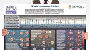 挂图Natural Killer Cells Overview of NK cell receptors, subsets, activation and function


 沪公网安备31010102008431号