Liyanage UK et al. (SEP 2002)
Journal of immunology (Baltimore,Md. : 1950) 169 5 2756--61
Prevalence of regulatory T cells is increased in peripheral blood and tumor microenvironment of patients with pancreas or breast adenocarcinoma.
Regulatory T cells (T(reg)) that prevent autoimmune diseases by suppression of self-reactive T cells may also suppress the immune response against cancer. In mice,depletion of T(reg) by Ab therapy leads to more efficient tumor rejection. T(reg)-mediated suppression of antitumor immune responses may partly explain the poor clinical response to vaccine-based immunotherapy for human cancer. In this study,we measured the prevalence of T(reg) that coexpress CD4 and CD25 in the PBLs,tumor-infiltrating lymphocytes,and regional lymph node lymphocytes from 65 patients with either pancreas or breast cancer. In breast cancer patients (n = 35),pancreas cancer patients (n = 30),and normal donors (n = 35),the prevalence of T(reg) were 16.6% (SE 1.22),13.2% (SE 1.13),and 8.6% (SE 0.71) of the total CD4(+) cells,respectively. The prevalence of T(reg) were significantly higher in breast cancer patients (p textless 0.01) and pancreas cancer patients (p textless 0.01) when compared with normal donors. In tumor-infiltrating lymphocytes and lymph node lymphocytes,the T(reg) prevalence were 20.2% (SE 3.93) and 20.1% (SE 4.3),respectively. T(reg) constitutively coexpressed CTLA-4 and CD45RO markers,and secreted TGF-beta and IL-10 but did not secrete IFN-gamma. When cocultured with activated CD8(+) cells or CD4(+)25(-) cells,T(reg) potently suppressed their proliferation and secretion of IFN-gamma. We conclude that the prevalence of T(reg) is increased in the peripheral blood as well as in the tumor microenvironment of patients with invasive breast or pancreas cancers. These T(reg) may mitigate the immune response against cancer,and may partly explain the poor immune response against tumor Ags.
View Publication
产品类型:
产品号#:
15022
15062
15023
15063
产品名:
RosetteSep™人CD4+ T细胞富集抗体混合物
RosetteSep™人CD4+ T细胞富集抗体混合物
RosetteSep™人CD8+ T细胞富集抗体混合物
RosetteSep™人CD8+ T细胞富集抗体混合物
Pospori C et al. (JUN 2011)
Blood 117 25 6813--24
Specificity for the tumor-associated self-antigen WT1 drives the development of fully functional memory T cells in the absence of vaccination.
Recently,vaccines against the Wilms Tumor antigen 1 (WT1) have been tested in cancer patients. However,it is currently not known whether physiologic levels of WT1 expression in stem and progenitor cells of normal tissue result in the deletion or tolerance induction of WT1-specific T cells. Here,we used an human leukocyte antigen-transgenic murine model to study the fate of human leukocyte antigen class-I restricted,WT1-specific T cells in the thymus and in the periphery. Thymocytes expressing a WT1-specific T-cell receptor derived from high avidity human CD8 T cells were positively selected into the single-positive CD8 population. In the periphery,T cells specific for the WT1 antigen differentiated into CD44-high memory phenotype cells,whereas T cells specific for a non-self-viral antigen retained a CD44(low) naive phenotype. Only the WT1-specific T cells,but not the virus-specific T cells,displayed rapid antigen-specific effector function without prior vaccination. Despite long-term persistence of WT1-specific memory T cells,the animals did not develop autoimmunity,and the function of hematopoietic stem and progenitor cells was unimpaired. This is the first demonstration that specificity for a tumor-associated self-antigen may drive differentiation of functionally competent memory T cells.
View Publication
Ex vivo induction of multiple myeloma-specific cytotoxic T lymphocytes.
Multiple myeloma (MM) is an incurable plasma cell malignancy characterized by immunosuppression. In this study,we identified factors in patients' bone marrow (BM) sera inhibiting autologous anti-MM immunity and developed an ex vivo strategy for inducing MM-specific cytotoxic T lymphocytes (CTLs). We found that sera from BM of MM patients inhibited induction of dendritic cells (DCs),evidenced by both phenotype and only weak stimulation of T-cell proliferation. Anti-vascular endothelial growth factor (anti-VEGF) and/or anti-interleukin 6 (anti-IL-6) antibodies neutralized this inhibitory effect,confirming that VEGF and IL-6,at least in part,mediate immunosuppression in MM patients. To induce MM-specific CTLs ex vivo,immature DCs were generated by culture of adherent mononuclear cells in medium containing granulocyte-macrophage colony-stimulating factor (GM-CSF) and IL-4 for 5 days and then cocultured with apoptotic MM bodies in the presence of tumor necrosis factor alpha (TNF-alpha) for 3 days to induce their maturation. Autologous BM or peripheral blood mononuclear cells were stimulated weekly with these DCs,and cytotoxicity was examined against the MM cells used to pulse DCs. DCs cultured with apoptotic bodies stimulated significantly greater T-cell proliferation (stimulation index [SI] = 23.2 at a T-DC ratio of 360:1) than T cells stimulated by MM cells only (SI = 5.6),DCs only (SI = 9.3),or MM lysate-pulsed DCs (SI = 13.5). These CTLs from MM patients demonstrated specific cytotoxicity (24.7% at the effector-target [E/T] ratio of 40:1) against autologous primary MM cells. These studies therefore show that CTLs from MM patients can recognize and lyse autologous tumor cells and provide the framework for novel immunotherapy to improve patient outcome in MM.
View Publication
Wu K et al. (JAN 2011)
The Journal of biological chemistry 286 3 2132--42
Cell fate determination factor Dachshund reprograms breast cancer stem cell function.
The cell fate determination factor Dachshund was cloned as a dominant inhibitor of the hyperactive epidermal growth factor receptor ellipse. The expression of Dachshund is lost in human breast cancer associated with poor prognosis. Breast tumor-initiating cells (TIC) may contribute to tumor progression and therapy resistance. Here,endogenous DACH1 was reduced in breast cancer cell lines with high expression of TIC markers and in patient samples of the basal breast cancer phenotype. Re-expression of DACH1 reduced new tumor formation in serial transplantations in vivo,reduced mammosphere formation,and reduced the proportion of CD44(high)/CD24(low) breast tumor cells. Conversely,lentiviral shRNA to DACH1 increased the breast (B)TIC population. Genome-wide expression studies of mammary tumors demonstrated DACH1 repressed a molecular signature associated with stem cells (SOX2,Nanog,and KLF4) and genome-wide ChIP-seq analysis identified DACH1 binding to the promoter of the Nanog,KLF4,and Lin28 genes. KLF4/c-Myc and Oct4/Sox2 antagonized DACH1 repression of BTIC. Mechanistic studies demonstrated DACH1 directly repressed the Nanog and Sox2 promoters via a conserved domain. Endogenous DACH1 regulates BTIC in vitro and in vivo.
View Publication
产品类型:
产品号#:
01700
01705
01702
产品名:
ALDEFLUOR™ 试剂盒
ALDEFLUOR™ DEAB试剂, 1.5 mM, 1 mL
ALDEFLUOR™检测缓冲液
Wang TH et al. ( 2000)
Cancer 88 11 2619--2628
Paclitaxel-induced cell death: where the cell cycle and apoptosis come together.
BACKGROUND: Compelling evidence indicates that paclitaxel kills cancer cells through the induction of apoptosis. Paclitaxel binds microtubules and causes kinetic suppression (stabilization) of microtubule dynamics. The consequent arrest of the cell cycle at mitotic phase has been considered to be the cause of paclitaxel-induced cytotoxicity. However,the biochemical events,downstream from paclitaxel's binding to microtubules,that lead to apoptosis are not well understood. METHODS: The authors examined recent scientific literature about the mechanisms by which paclitaxel exerts cytotoxicity. RESULTS: In addition to an arrest of the cell cycle at the mitotic phase in paclitaxel-treated cells,recent discoveries of activation of signaling molecules by paclitaxel and paclitaxel-induced transcriptional activation of various genes indicate that paclitaxel initiates apoptosis through multiple mechanisms. The checkpoint of mitotic spindle assembly,aberrant activation of cyclin-dependent kinases,and the c-Jun N-terminal kinase/stress-activated protein kinase (JNK/SAPK) are shown to be involved in paclitaxel-induced apoptosis. Consistent with observations that microtubules of different status (e.g.,cytoskeletal microtubules vs. mitotic spindles) have different sensitivity to paclitaxel,the concentration of paclitaxel appears to be the major determinant of its apoptogenic mechanisms. CONCLUSIONS: Advances in research of the cell cycle and apoptosis have extended our understanding of the mechanisms of paclitaxel-induced cell death. Further elucidation of resistance and enhancement of paclitaxel-induced apoptosis should expedite the development of better paclitaxel-based regimens for cancer therapy.
View Publication


 EasySep™小鼠TIL(CD45)正选试剂盒




 StemSpan™淋系祖细胞扩增添加物(10X) 用于人CD34+细胞扩增及分化为淋系祖细胞的添加物
 抗人CD25抗体,clone BC96 抗人、恒河猴、食蟹猴CD25的小鼠Monoclonal IgG1抗体
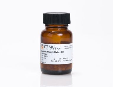 大豆胰蛋白酶抑制剂,ACF 用于抑制胰蛋白酶的活性
 抗人CD298抗体,clone LNH-94 小鼠Monoclonal IgG1抗体,抗人CD298
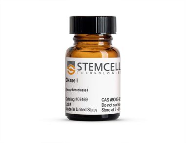 DNase I 用于消化DNA
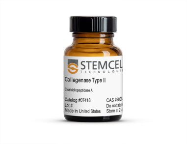 II型胶原酶 用于消化天然胶原纤维
 III型胶原酶 用于消化天然胶原纤维
 沪公网安备31010102008431号