miR-34a contributes to megakaryocytic differentiation of K562 cells independently of p53.
The role of miRNAs in regulating megakaryocyte differentiation was examined using bipotent K562 human leukemia cells. miR-34a is strongly up-regulated during phorbol ester-induced megakaryocyte differentiation,but not during hemin-induced erythrocyte differentiation. Enforced expression of miR-34a in K562 cells inhibits cell proliferation,induces cell-cycle arrest in G(1) phase,and promotes megakaryocyte differentiation as measured by CD41 induction. miR-34a expression is also up-regulated during thrombopoietin-induced differentiation of CD34(+) hematopoietic precursors,and its enforced expression in these cells significantly increases the number of megakaryocyte colonies. miR-34a directly regulates expression of MYB,facilitating megakaryocyte differentiation,and of CDK4 and CDK6,to inhibit the G(1)/S transition. However,these miR-34a target genes are down-regulated rapidly after inducing megakaryocyte differentiation before miR-34a is induced. This suggests that miR-34a is not responsible for the initial down-regulation but may contribute to maintaining their suppression later on. Previous studies have implicated miR-34a as a tumor suppressor gene whose transcription is activated by p53. However,in p53-null K562 cells,phorbol esters induce miR-34a expression independently of p53 by activating an alternative phorbol ester-responsive promoter to produce a longer pri-miR-34a transcript.
View Publication
Houtenbos I et al. (MAR 2006)
Haematologica 91 3 348--55
Leukemia-derived dendritic cells: towards clinical vaccination protocols in acute myeloid leukemia.
The ability of acute myeloid leukemic (AML) blasts to differentiate into leukemic dendritic cells (DC) thus acquiring the potential to present known and unknown leukemic antigens efficiently,holds promise as a possible new treatment for AML patients with minimal residual disease. Recent advances in culture methods have made the clinical use of leukemic DC feasible. However,additional measures appear to be essential in order to potentiate vaccines and to overcome the intrinsic tolerant state of the patients immune system. This review describes ways to improve AML-DC vaccines and discusses critical aspects concerning the development of clinical vaccination protocols.
View Publication
产品类型:
产品号#:
09600
09650
产品名:
StemSpan™ SFEM
StemSpan™ SFEM
Gallia GL et al. (FEB 2009)
Molecular cancer therapeutics 8 2 386--93
Inhibition of Akt inhibits growth of glioblastoma and glioblastoma stem-like cells.
A commonly activated signaling cascade in many human malignancies,including glioblastoma multiforme,is the Akt pathway. This pathway can be activated via numerous upstream alterations including genomic amplification of epidermal growth factor receptor,PTEN deletion,or PIK3CA mutations. In this study,we screened phosphatidylinositol 3-kinase/Akt small-molecule inhibitors in an isogenic cell culture system with an activated Akt pathway secondary to a PIK3CA mutation. One small molecule,A-443654,showed the greatest selective inhibition of cells with the mutant phenotype. Based on these findings,this inhibitor was screened in vitro against a panel of glioblastoma multiforme cell lines. All cell lines tested were sensitive to A-443654 with a mean IC(50) of approximately 150 nmol/L. An analogue of A-443654,methylated at a region that blocks Akt binding,was on average 36-fold less active. Caspase assays and dual flow cytometric analysis showed an apoptotic mechanism of cell death. A-443654 was further tested in a rat intracranial model of glioblastoma multiforme. Animals treated intracranially with polymers containing A-443654 had significantly extended survival compared with control animals; animals survived 79% and 43% longer than controls when A-443654-containing polymers were implanted simultaneously or in a delayed fashion,respectively. This small molecule also inhibited glioblastoma multiforme stem-like cells with similar efficacy compared with traditionally cultured glioblastoma multiforme cell lines. These results suggest that local delivery of an Akt small-molecule inhibitor is effective against experimental intracranial glioma,with no observed resistance to glioblastoma multiforme cells grown in stem cell conditions.
View Publication
产品类型:
产品号#:
05751
产品名:
NeuroCult™ NS-A 扩增试剂盒(人)
Rawat VPS et al. (SEP 2010)
Proceedings of the National Academy of Sciences of the United States of America 107 39 16946--51
The vent-like homeobox gene VENTX promotes human myeloid differentiation and is highly expressed in acute myeloid leukemia.
Recent data indicate that a variety of regulatory molecules active in embryonic development may also play a role in the regulation of early hematopoiesis. Here we report that the human Vent-like homeobox gene VENTX,a putative homolog of the Xenopus xvent2 gene,is a unique regulatory hematopoietic gene that is aberrantly expressed in CD34(+) leukemic stem-cell candidates in human acute myeloid leukemia (AML). Quantitative RT-PCR documented expression of the gene in lineage positive hematopoietic subpopulations,with the highest expression in CD33(+) myeloid cells. Notably,expression levels of VENTX were negligible in normal CD34(+)/CD38(-) or CD34(+) human progenitor cells. In contrast to this,leukemic CD34(+)/CD38(-) cells from AML patients with translocation t(8,21) and normal karyotype displayed aberrantly high expression of VENTX. Gene expression and pathway analysis demonstrated that in normal CD34(+) cells enforced expression of VENTX initiates genes associated with myeloid development and down-regulates genes involved in early lymphoid development. Functional analyses confirmed that aberrant expression of VENTX in normal CD34(+) human progenitor cells perturbs normal hematopoietic development,promoting generation of myeloid cells and impairing generation of lymphoid cells in vitro and in vivo. Stable knockdown of VENTX expression inhibited the proliferation of human AML cell lines. Taken together,these data extend our insights into the function of embryonic mesodermal factors in human postnatal hematopoiesis and indicate a role for VENTX in normal and malignant myelopoiesis.
View Publication
产品类型:
产品号#:
04434
04444
产品名:
MethoCult™ H4434 Classic
MethoCult™ H4434 Classic
Simõ et al. (AUG 2011)
Breast cancer research and treatment 129 1 23--35
Effects of estrogen on the proportion of stem cells in the breast.
There is increasing evidence that breast cancers contain tumor-initiating cells with stem cell properties. The importance of estrogen in the development of the mammary gland and in breast cancer is well known,but the influence of estrogen on the stem cell population has not been assessed. We show that estrogen reduces the proportion of stem cells in the normal human mammary gland and in breast cancer cells. The embryonic stem cell genes NANOG,OCT4,and SOX2 are expressed in normal breast stem cells and at higher levels in breast tumor cells and their expression decreases upon differentiation. Overexpression of each stem cell gene reduces estrogen receptor (ER) expression,and increases the number of stem cells and their capacity for invasion,properties associated with tumorigenesis and poor prognosis. These results indicate that estrogen reduces the size of the human breast stem cell pool and may provide an explanation for the better prognosis of ER-positive tumors.
View Publication
产品类型:
产品号#:
01700
01705
01702
产品名:
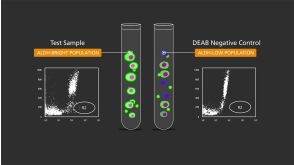
ALDEFLUOR™ 试剂盒
ALDEFLUOR™ DEAB试剂, 1.5 mM, 1 mL
ALDEFLUOR™检测缓冲液
Krishnamurthy S et al. (DEC 2010)
Cancer research 70 23 9969--78
Endothelial cell-initiated signaling promotes the survival and self-renewal of cancer stem cells.
Recent studies have demonstrated that cancer stem cells play an important role in the pathobiology of head and neck squamous cell carcinomas (HNSCC). However,little is known about functional interactions between head and neck cancer stem-like cells (CSC) and surrounding stromal cells. Here,we used aldehyde dehydrogenase activity and CD44 expression to sort putative stem cells from primary human HNSCC. Implantation of 1,000 CSC (ALDH+CD44+Lin-) led to tumors in 13 (out of 15) mice,whereas 10,000 noncancer stem cells (ALDH-CD44-Lin-) resulted in 2 tumors in 15 mice. These data demonstrated that ALDH and CD44 select a subpopulation of cells that are highly tumorigenic. The ability to self-renew was confirmed by the observation that ALDH+CD44+Lin- cells sorted from human HNSCC formed more spheroids (orospheres) in 3-D agarose matrices or ultra-low attachment plates than controls and were serially passaged in vivo. We observed that approximately 80% of the CSC were located in close proximity (within 100-μm radius) of blood vessels in human tumors,suggesting the existence of perivascular niches in HNSCC. In vitro studies demonstrated that endothelial cell-secreted factors promoted self-renewal of CSC,as demonstrated by the upregulation of Bmi-1 expression and the increase in the number of orospheres as compared with controls. Notably,selective ablation of tumor-associated endothelial cells stably transduced with a caspase-based artificial death switch (iCaspase-9) caused a marked reduction in the fraction of CSC in xenograft tumors. Collectively,these findings indicate that endothelial cell-initiated signaling can enhance the survival and self-renewal of head and neck CSC.
View Publication
产品类型:
产品号#:
01700
01705
01702
产品名:
ALDEFLUOR™ 试剂盒
ALDEFLUOR™ DEAB试剂, 1.5 mM, 1 mL
ALDEFLUOR™检测缓冲液
Bruserud &O et al. (APR 2004)
Haematologica 89 4 391--402
Osteoblasts increase proliferation and release of pro-angiogenic interleukin 8 by native human acute myelogenous leukemia blasts.
BACKGROUND AND OBJECTIVES: Interactions between acute myelogenous leukemia (AML) blasts and non-leukemic cells in the bone marrow seem to be important for both disease development and susceptibility to chemotherapy. Recent studies have focused on the endothelial cells,but other non-leukemic cells may also be involved. In the present study we investigated how osteoblasts affect native human AML blasts. DESIGN AND METHODS: AML cells were derived from a large group of consecutive patients. The AML blasts and osteoblastic sarcoma cell lines (Cal72,SJSA-1) were incubated together in different chambers separated by a semipermeable membrane. We investigated effects of co-culture on proliferation,apoptosis and cytokine release. RESULTS: The cross-talk between these two cell populations,achieved via release of soluble mediators,resulted in increased AML blast proliferation,including increased proliferation of clonogenic progenitors,but did not affect spontaneous in vitro apoptosis. Both interleukin (IL) 1-b and granulocyte-macrophage colony-stimulating factor were involved in this growth-enhancing cross-talk,and normal osteoblasts could also increase the AML blast proliferation. Furthermore,co-culture of AML blasts with osteoblastic sarcoma cells as well as normal osteoblasts increased the levels of the pro-angiogenic mediator IL8. INTERPRETATION AND CONCLUSIONS: Our in vitro results suggest that the release of soluble mediators by osteoblasts supports leukemic hematopoiesis through two major mechanisms: (i) direct enhancement of AML blast proliferation; and (ii) enhanced angiogenesis caused by increased IL8 levels.
View Publication
产品类型:
产品号#:
09600
09650
产品名:
StemSpan™ SFEM
StemSpan™ SFEM
Mahbub AA et al. (DEC 2013)
Anti-cancer agents in medicinal chemistry 13 10 1601--13
Differential effects of polyphenols on proliferation and apoptosis in human myeloid and lymphoid leukemia cell lines.
BACKGROUND Mortality rates for leukemia are high despite considerable improvements in treatment. Since polyphenols exert pro-apoptotic effects in solid tumors,our study investigated the effects of polyphenols in haematological malignancies. The effect of eight polyphenols (quercetin,chrysin,apigenin,emodin,aloe-emodin,rhein,cis-stilbene and trans-stilbene) were studied on cell proliferation,cell cycle and apoptosis in four lymphoid and four myeloid leukemic cells lines,together with normal haematopoietic control cells. METHODS Cellular proliferation was measured by CellTiter-Glo(®) luminescent assay; and cell cycle arrest was assessed using flow cytometry of propidium iodide stained cells. Apoptosis was investigated by caspase-3 activity assay using flow cytometry and apoptotic morphology was confirmed by Hoescht 33342 staining. RESULTS Emodin,quercetin,and cis-stilbene were the most effective polyphenols at decreasing cell viability (IC50 values of 5-22 μM,8-33 μM,and 25-85 μM respectively) and inducing apoptosis (AP50 values (the concentration which 50% of cells undergo apoptosis) of 2-27 μM,19-50 μM,and 8-50 μM respectively). Generally,lymphoid cell lines were more sensitive to polyphenol treatment compared to myeloid cell lines,however the most resistant myeloid (KG-1a and K562) cell lines were still found to respond to emodin and quercetin treatment at low micromolar levels. Non-tumor cells were less sensitive to all polyphenols compared to the leukemia cells. CONCLUSIONS These findings suggest that polyphenols have anti-tumor activity against leukemia cells with differential effects. Importantly,the differential sensitivity of emodin,quercetin,and cis-stilbene between leukemia and normal cells suggests that polyphenols are potential therapeutic agents for leukemia.
View Publication
产品类型:
产品号#:
70008
70008.1
70008.2
70008.3
70008.4
70008.5
70008.6
200-0002
200-0001
200-0000
产品名:
冻存的人脐带血CD34+细胞
冻存的人脐带血CD34+细胞
冻存的人脐带血CD34+细胞
冻存的人脐带血CD34+细胞
冻存的人脐带血CD34+细胞
冻存的人脐带血CD34+细胞
冻存的人脐带血CD34+细胞
冻存的人脐带血CD34+细胞
冻存的人脐带血CD34+细胞
Li S et al. ( 2013)
Oncology letters 5 2 717--721
The mTOR inhibitor AZD8055 inhibits proliferation and glycolysis in cervical cancer cells.
The aim of the present study was to determine the effect of AZD8055 on proliferation,apoptosis and glycolysis in the human cervical cancer cell line HeLa and to investigate the underlying mechanism(s) of action. HeLa human cervical cancer cells were treated with 10 nM AZD8055 for 24,48 or 72 h. MTT was used to determine cell proliferation. Annexin V/propidium iodide staining was used to determine cell apoptosis analyzed by fluorescence-activated cell sorting (FACS). Glycolytic activity was determined by measuring the activity of the key enzyme lactate dehydrogenase (LDH) and lactate production. RNA and protein expression were examined by qRT-PCR and western blotting,respectively. Treatment with AZD8055 inhibited proliferation and glycolysis,and induced apoptosis in HeLa cells in a time-dependent manner. During the prolonged treatment with AZD8055,the phosphorylation of mammalian target of rapamycin (mTOR) C1 substrates p70S6K and phosphorylation of the mTORC2 substrate Akt were deregulated,suggesting that the activity of mTOR was downregulated. Furthermore,our study showed that the expression of miR-143 was upregulated in a time-dependent manner in HeLa cells treated with AZD8055. In summary,the present study reveals a novel antitumor mechanism of AZD8055 in HeLa human cervical cancer cells.
View Publication
产品类型:
产品号#:
73002
73004
产品名:
AZD8055
AZD8055
A. Kuske et al. (DEC 2016)
Scientific reports 6 39736
Improved detection of circulating tumor cells in non-metastatic high-risk prostate cancer patients.
The relevance of blood-based assays to monitor minimal residual disease (MRD) in non-metastatic prostate cancer (PCa) remains unclear. Proving that clinically relevant circulating tumor cells (CTCs) can be detected with available technologies could address this. This study aimed to improve CTC detection in non-metastatic PCa patients by combining three independent CTC assays: the CellSearch system,an in vivo CellCollector and the EPISPOT. Peripheral blood samples from high-risk PCa patients were screened for CTCs before and three months after radical prostatectomy (RP). Combining the results of both time points,CTCs were detected in 37{\%},54.9{\%} and 58.7{\%} of patients using CellSearch,CellCollector and EPISPOT,respectively. The cumulative positivity rate of the three CTC assays was 81.3{\%} (87/107) with 21.5{\%} (23/107) of patients harboring ≥5 CTCs/7.5 ml blood. Matched pair analysis of 30 blood samples taken before and after surgery indicated a significant decrease in CTCs captured by the CellCollector from 66{\%} before RP to 34{\%} after therapy (p = 0.031). CTC detection by EPISPOT before RP significantly correlated with PSA serum values (p {\textless} 0.0001) and clinical tumor stage (p = 0.04),while the other assays showed no significant correlations. In conclusion,CTC-based liquid biopsies have the potential to monitor MRD in patients with non-metastatic prostate cancer.
View Publication

 EasySep™小鼠TIL(CD45)正选试剂盒


 沪公网安备31010102008431号