Hybridoma technology for the generation of rodent mAbs via classical fusion
Monoclonal antibodies (mAbs) have proven to be instrumental in the advancement of research,diagnostic,industrial vaccine,and therapeutic applications. The use of mAbs in laboratory protocols has been growing in an exponential fashion for the last four decades. Described herein are methods for the development of highly specific mAbs through traditional hybridoma fusion. For ultimate success,a series of simultaneously initiated protocols are to be undertaken with careful attention to cell health of both the myeloma fusion partner and immune splenocytes. Coordination and attention to detail will enable a researcher with basic tissue culture skills to generate mAbs from immunized rodents to a variety of antigens (including proteins,carbohydrates,DNA,and haptens) (see Note 1). Furthermore,in vivo and in vitro methods used for antigen sensitization of splenocytes prior to somatic fusion are described herein.
View Publication
Date Y et al. ( 2014)
Analytical Chemistry 86 6 2989--96
Label-free impedimetric immunoassay for trace levels of polychlorinated biphenyls in insulating oil
A rapid,ultrasensitive,and practical label-free impedimetric immunoassay for measuring trace levels of total polychlorinated biphenyls (PCBs) in insulating oil was developed. First,we developed a novel monoclonal antibody (RU6F9) for PCBs by using a designed immunogen and characterized its binding affinity for a commercial mixtures of PCBs and its main congeners. A micro comblike gold electrode was fabricated,and the antibody was covalently immobilized on the electrode through a self-assembled monolayer formed by dithiobis-N-succinimidyl propionate. The antigen-binding event on the surface of the functionalized electrode was determined as the change in charge transfer resistance by using electrochemical impedance spectroscopy. The resulting impedimetric immunoassay in aqueous solution achieved a wide determination range (0.01-10 μg/L) and a low detection limit (LOD) of 0.001 μg/L,which was 100-fold more sensitive than a conventional flow-based immunoassay for PCBs. By combining the impedimetric immunoassay with a cleanup procedure for insulating oil utilizing a multilayer cleanup column followed by DMSO partitioning,an LOD of 0.052 mg/kg-oil was achieved,which satisfied the Japanese regulation criterion of 0.5 mg/kg-oil. Finally,the immunoassay was employed to determine total PCB levels in actual used insulating oils (n = 33) sampled from a used transformer containing trace levels of PCBs,and the results agreed well with the Japanese official method (HRGC/HRMS).
View Publication
产品类型:
产品号#:
03800
产品名:
ClonaCell™-HY杂交瘤试剂盒
Lei IL et al. (JAN 2015)
Journal of visualized experiments : JoVE January 52047. doi: 10.3791/52047.
Derivation of cardiac progenitor cells from embryonic stem cells.
Cardiac progenitor cells (CPCs) have the capacity to differentiate into cardiomyocytes,smooth muscle cells (SMC),and endothelial cells and hold great promise in cell therapy against heart disease. Among various methods to isolate CPCs,differentiation of embryonic stem cell (ESC) into CPCs attracts great attention in the field since ESCs can provide unlimited cell source. As a result,numerous strategies have been developed to derive CPCs from ESCs. In this protocol,differentiation and purification of embryonic CPCs from both mouse and human ESCs is described. Due to the difficulty of using cell surface markers to isolate embryonic CPCs,ESCs are engineered with fluorescent reporters activated by CPC-specific cre recombinase expression. Thus,CPCs can be enriched by fluorescence-activated cell sorting (FACS). This protocol illustrates procedures to form embryoid bodies (EBs) from ESCs for CPC specification and enrichment. The isolated CPCs can be subsequently cultured for cardiac lineage differentiation and other biological assays. This protocol is optimized for robust and efficient derivation of CPCs from both mouse and human ESCs.
View Publication
Wen Y and Jin S (OCT 2014)
Journal of Biotechnology 188 122--129
Production of neural stem cells from human pluripotent stem cells
Despite significant advances in commercially available media and kits and the differentiation approaches for human neural stem cell (NSC) generation,NSC production from the differentiation of human pluripotent stem cell (hPSC) is complicated by its time-consuming procedure,complex medium composition,and purification step. In this study,we developed a convenient and simplified NSC production protocol to meet the demand of NSC production. We demonstrated that NSCs can be generated efficiently without requirement of specific small molecules or embryoid body formation stage. Our experimental results suggest that a short suspension culture period may facilitate ectoderm lineage specification rather than endoderm or mesoderm lineage specification from hPSCs. The method developed in this study shortens the turnaround time of NSC production from both human embryonic stem cells (hESCs) and induced pluripotent stem cells (iPSCs) differentiation. It provides a straightforward and useful strategy for generating NSCs that can benefit a wide range of research applications for human brain research.
View Publication
产品类型:
产品号#:
05832
05850
05857
05870
05875
07923
85850
85857
85870
85875
产品名:
STEMdiff™ 神经花环选择试剂
Dispase (1 U/mL)
mTeSR™1
mTeSR™1
Inagi R et al. (NOV 2007)
Nephrology,dialysis,transplantation : official publication of the European Dialysis and Transplant Association - European Renal Association 22 11 3311--7
Establishment of a sandwich ELISA for human megsin, a kidney-specific serine protease inhibitor.
BACKGROUND: We previously identified a novel serine protease inhibitor (serpin),megsin,which is predominantly expressed in the kidney. Megsin expression is up-regulated in human and experimental renal diseases associated with mesangial proliferation and expansion,suggesting that urinary megsin may be a novel diagnostic marker for some renal diseases. METHODS: We established a specific and sensitive sandwich enzyme-linked immunosorbent assay (ELISA) for megsin and measured urinary megsin of patients with various renal diseases. RESULTS: Megsin ELISA specifically detected megsin but not other serpins. The detection limit was 0.04 ng/ml,which allowed detection of urinary megsin in 3.6% of healthy individuals. The antigenic epitope in the urine detected by the ELISA was confirmed as megsin protein by time-of-flight mass spectrometry. Among patients with rapidly progressive glomerulonephritis (n = 18),55.6% were urinary megsin-positive,while 24.1% in IgA nephropathy (n = 112) and 15.1% in chronic non-IgA glomerulonephritis (n = 245) were urinary megsin-positive,respectively. Among patients with chronic renal failure due to unknown causes (n = 74),18.9% were positive for urinary megsin. In diabetic patients with or without nephropathy (n = 1073),12.3% were urinary megsin-positive,while positivity of urinary megsin in patients with non-renal diseases (n = 768) was equivalent (3.3%) to that of healthy individuals. Of note,when urinary megsin-positive patients with diabetic nephropathy (n = 71) were classified into four stages by their proteinuria and estimated glomerular filtration rate,urinary megsin excretion increased as the stage progressed up to stage 3A,suggesting correlation of that with mesangial expansion level. Urinary megsin decreased in the advanced stage,probably reflecting development of glomerulosclerosis. CONCLUSION: We established a high-sensitive megsin ELISA,which detects urinary megsin in some patients with renal diseases and in only a few healthy subjects. Megsin ELISA may be a novel diagnostic tool for renal diseases.
View Publication
产品类型:
产品号#:
03800
03801
03802
03803
03804
03805
03806
产品名:
ClonaCell™-HY杂交瘤试剂盒
ClonaCell™-HY培养基A
ClonaCell™-HY 培养基 B
ClonaCell™-HY 培养基 C
ClonaCell™-HY 培养基 D
ClonaCell™-HY 培养基 E
ClonaCell™-HY PEG
Katori S et al. (JUL 2009)
The Journal of neuroscience : the official journal of the Society for Neuroscience 29 29 9137--47
Protocadherin-alpha family is required for serotonergic projections to appropriately innervate target brain areas.
Serotonergic axons from the raphe nuclei in the brainstem project to every region of the brain,where they make connections through their extensive terminal arborizations. This serotonergic innervation contributes to various normal behaviors and psychiatric disorders. The protocadherin-alpha (Pcdha) family of clustered protocadherins consists of 14 cadherin-related molecules generated from a single gene cluster. We found that the Pcdhas were strongly expressed in the serotonergic neurons. To elucidate their roles,we examined serotonergic fibers in a mouse mutant (Pcdha(Delta CR/Delta CR)) lacking the Pcdha cytoplasmic region-encoding exons,which are common to the gene cluster. In the first week after birth,the distribution pattern of serotonergic fibers in Pcdha(Delta CR/Delta CR) mice was similar to wild-type,but by 3 weeks of age,when the serotonergic axonal termini complete their arborizations,the distribution of the projections was abnormal. In some target regions,notably the globus pallidus and substantia nigra,the normally even distribution of serotonin axonal terminals was,in the mutants,dense at the periphery of each region,but sparse in the center. In the stratum lacunosum-molecular of the hippocampus,the mutants showed denser serotonergic innervation than in wild-type,and in the dentate gyrus of the hippocampus and the caudate-putamen,the innervation was sparser. Together,the abnormalities suggested that Pcdha proteins are important in the late-stage maturation of serotonergic projections. Further examination of alternatively spliced exons encoding the cytoplasmic tail showed that the A-type (but not the B-type) cytoplasmic tail was essential for the normal development of serotonergic projections.
View Publication


 EasySep™小鼠TIL(CD45)正选试剂盒




 产品手册Highway1™: Fast, Gentle, and Automated Cell Sorting for Every Lab
 技术公告Protocol for Semi-Solid Hybridoma Cloning Using ClonaCell™-HY Medium in a 96-Well Plate
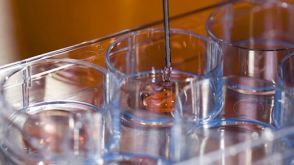 实验方案How to Prepare and Plate Semi-Solid Methylcellulose Medium for Cell Culture



 沪公网安备31010102008431号