Oxygen transport and stem cell aggregation in stirred-suspension bioreactor cultures
Stirred-suspension bioreactors are a promising modality for large-scale culture of 3D aggregates of pluripotent stem cells and their progeny. Yet,cells within these clusters experience limitations in the transfer of factors and particularly O2 which is characterized by low solubility in aqueous media. Cultured stem cells under different O2 levels may exhibit significantly different proliferation,viability and differentiation potential. Here,a transient diffusion-reaction model was built encompassing the size distribution and ultrastructural characteristics of embryonic stem cell (ESC) aggregates. The model was coupled to experimental data from bioreactor and static cultures for extracting the effective diffusivity and kinetics of consumption of O2 within mouse (mESC) and human ESC (hESC) clusters. Under agitation,mESC aggregates exhibited a higher maximum consumption rate than hESC aggregates. Moreover,the reaction-diffusion model was integrated with a population balance equation (PBE) for the temporal distribution of ESC clusters changing due to aggregation and cell proliferation. Hypoxia was found to be negligible for ESCs with a smaller radius than 100 µm but became appreciable for aggregates larger than 300 µm. The integrated model not only captured the O2 profile both in the bioreactor bulk and inside ESC aggregates but also led to the calculation of the duration that fractions of cells experience a certain range of O2 concentrations. The approach described in this study can be employed for gaining a deeper understanding of the effects of O2 on the physiology of stem cells organized in 3D structures. Such frameworks can be extended to encompass the spatial and temporal availability of nutrients and differentiation factors and facilitate the design and control of relevant bioprocesses for the production of stem cell therapeutics.
View Publication
产品类型:
产品号#:
05850
05857
05870
05875
85850
85857
85870
85875
产品名:
mTeSR™1
mTeSR™1
Wittman VP et al. (SEP 2006)
The Journal of Immunology 177 6 4187--95
Antibody targeting to acClass I MHC-peptide epitope promotes tumor cell death
Therapeutic mAbs that target tumor-associated Ags on the surface of malignant cells have proven to be an effective and specific option for the treatment of certain cancers. However,many of these protein markers of carcinogenesis are not expressed on the cells' surface. Instead these tumor-associated Ags are processed into peptides that are presented at the cell surface,in the context of MHC class I molecules,where they become targets for T cells. To tap this vast source of tumor Ags,we generated a murine IgG2a mAb,3.2G1,endowed with TCR-like binding specificity for peptide-HLA-A*0201 (HLA-A2) complex and designated this class of Ab as TCR mimics (TCRm). The 3.2G1 TCRm recognizes the GVL peptide (GVLPALPQV) from human chorionic gonadotropin beta presented by the peptide-HLA-A*0201 complex. When used in immunofluorescent staining reactions using GVL peptide-loaded T2 cells,the 3.2G1 TCRm specifically stained the cells in a peptide and Ab concentration-dependent manner. Staining intensity correlated with the extent of cell lysis by complement-dependent cytotoxicity (CDC),and a peptide concentration-dependent threshold level existed for the CDC reaction. Staining of human tumor lines demonstrated that 3.2G1 TCRm was able to recognize endogenously processed peptide and that the breast cancer cell line MDA-MB-231 highly expressed the target epitope. The 3.2G1 TCRm-mediated CDC and Ab-dependent cellular cytotoxicity of a human breast carcinoma line in vitro and inhibited in vivo tumor implantation and growth in nude mice. These results provide validation for the development of novel TCRm therapeutic reagents that specifically target and kill tumors via recognition and binding to MHC-peptide epitopes.
View Publication
Robust pluripotent stem cell expansion and cardiomyocyte differentiation via geometric patterning
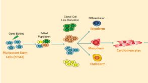
Geometric factors including the size,shape,density,and spacing of pluripotent stem cell colonies play a significant role in the maintenance of pluripotency and in cell fate determination. These factors are impossible to control using standard tissue culture methods. As such,there can be substantial batch-to-batch variability in cell line maintenance and differentiation yield. Here,we demonstrate a simple,robust technique for pluripotent stem cell expansion and cardiomyocyte differentiation by patterning cell colonies with a silicone stencil. We have observed that patterning human induced pluripotent stem cell (hiPSC) colonies improves the uniformity and repeatability of their size,density,and shape. Uniformity of colony geometry leads to improved homogeneity in the expression of pluripotency markers SSEA4 and Nanog as compared with conventional clump passaging. Patterned cell colonies are capable of undergoing directed differentiation into spontaneously beating cardiomyocyte clusters with improved yield and repeatability over unpatterned cultures seeded either as cell clumps or uniform single cell suspensions. Circular patterns result in a highly repeatable 3D ring-shaped band of cardiomyocytes which electrically couple and lead to propagating contraction waves around the ring. Because of these advantages,geometrically patterning stem cells using stencils may offer greater repeatability from batch-to-batch and person-to-person,an increase in differentiation yield,a faster experimental workflow,and a simpler protocol to communicate and follow. Furthermore,the ability to control where cardiomyocytes arise across a culture well during differentiation could greatly aid the design of electrophysiological assays for drug-screening.
View Publication
Du S-HH et al. (AUG 2015)
Journal of bioscience and bioengineering 120 2 210--217
Human iPS cell-derived fibroblast-like cells as feeder layers for iPS cell derivation and expansion
Mouse embryonic fibroblasts (MEFs) are commonly used as feeder cells for the generation of human induced pluripotent stem cells (hiPSCs). However,medical applications of cell derivatives of hiPSCs generated with a MEF feeder system run the risk of having xeno-factor contamination due to long-term cell culturing under an animal factor-containing environment. We developed a new method for the derivation of human fibroblast-like cells (FLCs) from a previously established hiPSC line in an FLC differentiation medium. The method was based on direct differentiation of hiPSCs seeded on Matrigel followed by expansion of differentiating cells on gelatin. Using inactivated FLCs as feeder layers,primary human foreskin fibroblasts were successfully reprogrammed into a state of pluripotency by Oct4,Sox2 Klf4,and c-Myc (OSKM) transcription factor genes,with a reprogramming efficiency under an optimized condition superior to that obtained on MEF feeder layers. Furthermore,the FLCs were more effective in supporting the growth of human pluripotent stem cells. The pluripotency and differentiation capability of the cells cultured on FLC feeder layers were well retained. Our results suggest that FLCs are a safe alternative to MEFs for hiPSC generation and expansion,especially in the clinical settings wherein hiPSC derivatives will be used for medical treatment.
View Publication
产品类型:
产品号#:
05850
05857
05870
05875
07923
85850
85857
85870
85875
产品名:
Dispase (1 U/mL)
mTeSR™1
mTeSR™1
Tan H-K et al. (MAY 2014)
Stem cells translational medicine 3 5 586--98
Human finger-prick induced pluripotent stem cells facilitate the development of stem cell banking.
Induced pluripotent stem cells (iPSCs) derived from somatic cells of patients can be a good model for studying human diseases and for future therapeutic regenerative medicine. Current initiatives to establish human iPSC (hiPSC) banking face challenges in recruiting large numbers of donors with diverse diseased,genetic,and phenotypic representations. In this study,we describe the efficient derivation of transgene-free hiPSCs from human finger-prick blood. Finger-prick sample collection can be performed on a do-it-yourself" basis by donors and sent to the hiPSC facility for reprogramming. We show that single-drop volumes of finger-prick samples are sufficient for performing cellular reprogramming�
View Publication


 EasySep™小鼠TIL(CD45)正选试剂盒




 产品手册Highway1™: Fast, Gentle, and Automated Cell Sorting for Every Lab
 24:25
线上讲座ISSCR Standards in Action: Advancements in Cell Line Generation and Organoid Innovation发布日期: 08/01/2024
24:25
线上讲座ISSCR Standards in Action: Advancements in Cell Line Generation and Organoid Innovation发布日期: 08/01/2024 实验方案How to Prepare and Plate Semi-Solid Methylcellulose Medium for Cell Culture
 产品手册ClonaCell™-CHO ACF Supplement for Robust Growth of CHO Cells at Low Cell Density


 沪公网安备31010102008431号