

 EasySep™小鼠TIL(CD45)正选试剂盒




搜索结果: 'gmp t cell activator'
-
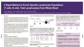 科学海报Cell Enrichment of Specific Lymphocyte Populations (T Cells, B Cells, Total Lymphocytes) from Whole Blood
产品类型:
Conference:
ASHI 2007
产品号#:
19655
21000
20155
20119
19961HLA
19961HLARF
19951HLA
19951HLARF
19954HLA
19954HLARF
19665
19655RF
产品名:
EasySep™ Direct人总淋巴细胞分选试剂盒
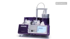
RoboSep™- S
RoboSep™分选管套装(9个塑料管)
RoboSep™ 吸头组件抛光剂
EasySep™ HLA总淋巴细胞富集: 用于处理全血的全套试剂盒
RoboSep™ HLA总淋巴细胞富集: 用于处理全血的全套试剂盒含滤芯吸头
EasySep™ HLA全血T细胞富集试剂盒
RoboSep™ HLA全血T细胞富集试剂盒含滤芯吸头
EasySep™ HLA B细胞富集: 用于处理全血的全套试剂盒
RoboSep™ HLA B细胞富集: 用于处理全血的全套试剂盒含滤芯吸头
EasySep™ Direct人NK细胞分选试剂盒
RoboSep™ Direct人总淋巴细胞分选试剂盒
-
产品类型:
产品号#:
17952
17952RF
17953
17953RF
21000
20119
20155
100-0696
100-0710
产品名:
EasySep™人CD4+ T细胞分选试剂盒
RoboSep™ 人CD4+ T细胞分选试剂盒
EasySep™人CD8+ T细胞分选试剂盒
RoboSep™ 人CD8+ T细胞分选试剂盒
RoboSep™- S
RoboSep™ 吸头组件抛光剂
RoboSep™分选管套装(9个塑料管)
EasySep™人CD4+ T细胞分离试剂盒
EasySep™人CD8+ T细胞分选试剂盒
-
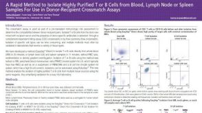 科学海报A Rapid Method to Isolate Highly Purified T or B Cells from Blood, Lymph Node or Spleen Samples For Use in Donor-Recipient Crossmatch Assays
产品类型:
Conference:
EFI 2017
产品号#:
产品名:
-
 抗人HLA-DR抗体,clone LN3 抗人、恒河猴HLA-DR的小鼠Monoclonal IgG2b抗体
 -

-
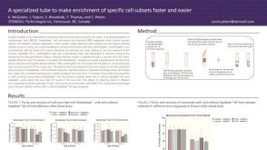 科学海报Fast and Easy Enrichment of Cell Subsets for HLA Analysis
产品类型:
Conference:
EFI 2012
产品号#:
15470
15450
15420
15460
15425
15465
15430
15415
85415
85420
85450
85460
产品名:
SepMate™-15 (IVD)
SepMate™-15 (IVD)
SepMate™-50 (IVD)
SepMate™-50 (IVD)
-
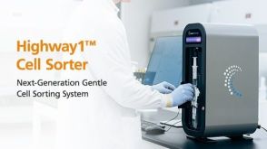
 产品手册Highway1™: Fast, Gentle, and Automated Cell Sorting for Every Lab
产品类型:
品牌:
Highway1
产品号#:
产品名:
发布日期: 04/14/2026 -
产品类型:
产品号#:
03800
03801
03802
03803
03804
03805
03806
03831
产品名:
ClonaCell™-HY杂交瘤试剂盒
ClonaCell™-HY培养基A
ClonaCell™-HY 培养基 B
ClonaCell™-HY 培养基 C
ClonaCell™-HY 培养基 D
ClonaCell™-HY 培养基 E
ClonaCell™-HY PEG
ClonaCell™-HY 液体 HAT 筛选培养基

 沪公网安备31010102008431号