Direct neural fate specification from embryonic stem cells: a primitive mammalian neural stem cell stage acquired through a default mechanism.
Little is known about how neural stem cells are formed initially during development. We investigated whether a default mechanism of neural specification could regulate acquisition of neural stem cell identity directly from embryonic stem (ES) cells. ES cells cultured in defined,low-density conditions readily acquire a neural identity. We characterize a novel primitive neural stem cell as a component of neural lineage specification that is negatively regulated by TGFbeta-related signaling. Primitive neural stem cells have distinct growth factor requirements,express neural precursor markers,generate neurons and glia in vitro,and have neural and non-neural lineage potential in vivo. These results are consistent with a default mechanism for neural fate specification and support a model whereby definitive neural stem cell formation is preceded by a primitive neural stem cell stage during neural lineage commitment.
View Publication
产品类型:
产品号#:
06902
06952
00321
00322
00323
00324
00325
产品名:
Zhu TS et al. (SEP 2011)
Cancer research 71 18 6061--72
Endothelial cells create a stem cell niche in glioblastoma by providing NOTCH ligands that nurture self-renewal of cancer stem-like cells.
One important function of endothelial cells in glioblastoma multiforme (GBM) is to create a niche that helps promote self-renewal of cancer stem-like cells (CSLC). However,the underlying molecular mechanism for this endothelial function is not known. Since activation of NOTCH signaling has been found to be required for propagation of GBM CSLCs,we hypothesized that the GBM endothelium may provide the source of NOTCH ligands. Here,we report a corroboration of this concept with a demonstration that NOTCH ligands are expressed in endothelial cells adjacent to NESTIN and NOTCH receptor-positive cancer cells in primary GBMs. Coculturing human brain microvascular endothelial cells (hBMEC) or NOTCH ligand with GBM neurospheres promoted GBM cell growth and increased CSLC self-renewal. Notably,RNAi-mediated knockdown of NOTCH ligands in hBMECs abrogated their ability to induce CSLC self-renewal and GBM tumor growth,both in vitro and in vivo. Thus,our findings establish that NOTCH activation in GBM CSLCs is driven by juxtacrine signaling between tumor cells and their surrounding endothelial cells in the tumor microenvironment,suggesting that targeting both CSLCs and their niche may provide a novel strategy to deplete CSLCs and improve GBM treatment.
View Publication
产品类型:
产品号#:
05750
05751
05752
产品名:
NeuroCult™ NS-A 基础培养基(人)
NeuroCult™ NS-A 扩增试剂盒(人)
NeuroCult™ NS-A 分化试剂盒(人)
Pineda JR et al. (APR 2013)
EMBO Molecular Medicine 5 4 548--562
Vascular-derived TGF-β increases in the stem cell niche and perturbs neurogenesis during aging and following irradiation in the adult mouse brain
Neurogenesis decreases during aging and following cranial radiotherapy,causing a progressive cognitive decline that is currently untreatable. However,functional neural stem cells remained present in the subventricular zone of high dose-irradiated and aged mouse brains. We therefore investigated whether alterations in the neurogenic niches are perhaps responsible for the neurogenesis decline. This hypothesis was supported by the absence of proliferation of neural stem cells that were engrafted into the vascular niches of irradiated host brains. Moreover,we observed a marked increase in TGF-β1 production by endothelial cells in the stem cell niche in both middle-aged and irradiated mice. In co-cultures,irradiated brain endothelial cells induced the apoptosis of neural stem/progenitor cells via TGF-β/Smad3 signalling. Strikingly,the blockade of TGF-β signalling in vivo using a neutralizing antibody or the selective inhibitor SB-505124 significantly improved neurogenesis in aged and irradiated mice,prevented apoptosis and increased the proliferation of neural stem/progenitor cells. These findings suggest that anti-TGF-β-based therapy may be used for future interventions to prevent neurogenic collapse following radiotherapy or during aging.
View Publication
Harlow DE et al. (JAN 2014)
Journal of Neuroscience 34 4 1333--1343
Expression of Proteolipid Protein Gene in Spinal Cord Stem Cells and Early Oligodendrocyte Progenitor Cells Is Dispensable for Normal Cell Migration and Myelination
Plp1 gene expression occurs very early in development,well before the onset of myelination,creating a conundrum with regard to the function of myelin proteolipid protein (PLP),one of the major proteins in compact myelin. Using PLP-EGFP mice to investigate Plp1 promoter activity,we found that,at very early time points,PLP-EGFP was expressed in Sox2+ undifferentiated precursors in the spinal cord ventricular zone (VZ),as well as in the progenitors of both neuronal and glial lineages. As development progressed,most PLP-EGFP-expressing cells gave rise to oligodendrocyte progenitor cells (OPCs). The expression of PLP-EGFP in the spinal cord was quite dynamic during development. PLP-EGFP was highly expressed as cells delaminated from the VZ. Expression was downregulated as cells moved laterally through the cord,and then robustly upregulated as OPCs differentiated into mature myelinating oligodendrocytes. The presence of PLP-EGFP expression in OPCs raises the question of its role in this migratory population. We crossed PLP-EGFP reporter mice into a Plp1-null background to investigate the role of PLP in early OPC development. In the absence of PLP,normal numbers of OPCs were generated and their distribution throughout the spinal cord was unaffected. However,the orientation and length of OPC processes during migration was abnormal in Plp1-null mice,suggesting that PLP plays a role either in the structural integrity of OPC processes or in their response to extracellular cues that orient process outgrowth.
View Publication
产品类型:
产品号#:
05707
产品名:
NeuroCult™化学解离试剂盒(小鼠)
Kang HS et al. (DEC 2015)
Journal of Korean medical science 30 12 1764--76
Advanced Properties of Urine Derived Stem Cells Compared to Adipose Tissue Derived Stem Cells in Terms of Cell Proliferation, Immune Modulation and Multi Differentiation.
Adipose tissue stem cells (ADSCs) would be an attractive autologous cell source. However,ADSCs require invasive procedures,and has potential complications. Recently,urine stem cells (USCs) have been proposed as an alternative stem cell source. In this study,we compared USCs and ADSCs collected from the same patients on stem cell characteristics and capacity to differentiate into various cell lineages to provide a useful guideline for selecting the appropriate type of cell source for use in clinical application. The urine samples were collected via urethral catheterization,and adipose tissue was obtained from subcutaneous fat tissue during elective laparoscopic kidney surgery from the same patient (n = 10). Both cells were plated for primary culture. Cell proliferation,colony formation,cell surface markers,immune modulation,chromosome stability and multi-lineage differentiation were analyzed for each USCs and ADSCs at cell passage 3,5,and 7. USCs showed high cell proliferation rate,enhanced colony forming ability,strong positive for stem cell markers expression,high efficiency for inhibition of immune cell activation compared to ADSCs at cell passage 3,5,and 7. In chromosome stability analysis,both cells showed normal karyotype through all passages. In analysis of multi-lineage capability,USCs showed higher myogenic,neurogenic,and endogenic differentiation rate,and lower osteogenic,adipogenic,and chondrogenic differentiation rate compared to ADSCs. Therefore,we expect that USC can be an alternative autologous stem cell source for muscle,neuron and endothelial tissue reconstruction instead of ADSCs.
View Publication
产品类型:
产品号#:
05752
产品名:
NeuroCult™ NS-A 分化试剂盒(人)
Dotti CG et al. (OCT 1987)
Neuroscience 23 1 121--30
The expression and distribution of the microtubule-associated proteins tau and microtubule-associated protein 2 in hippocampal neurons in the rat in situ and in cell culture.
Using a monoclonal antibody against the microtubule-associated protein tau we compared the distribution and the biochemical maturation of this protein in hippocampal pyramidal neurons in the rat in tau and in culture. In tissue sections from mature animals tau was localized heterogeneously within neurons. It was concentrated in axons; dendrites and somata showed little or no staining. In hippocampal cultures ranging from 12 h to 4 weeks in vitro tau was present in neurons but not in glial cells,as it is in situ. Within cultured neurons,however,tau was not compartmentalized but was present throughout the dendrites,axons and somata. Immunoblotting experiments showed that the biochemical maturation of tau that occurs in situ also failed to occur in culture. The young form of tau persisted,and the adult forms did not develop. In contrast the biochemical maturation and the compartmentalization of microtubule-associated protein 2 occurred normally in hippocampal cultures. These results show that the biochemical maturation and the intraneuronal compartmentalization of these two microtubule-associated proteins are independently controlled. Despite the non-restricted distribution of tau in hippocampal neurons in culture,and despite the presence of only the immature isoform which has a lessened stimulatory effect on microtubule polymerization,axons and dendrites appear to grow normally and to exhibit appropriate functional properties.
View Publication
产品类型:
产品号#:
01410
产品名:
McPherson CA et al. (JUL 2011)
Brain,behavior,and immunity 25 5 850--62
Interleukin (IL)-1 and IL-6 regulation of neural progenitor cell proliferation with hippocampal injury: differential regulatory pathways in the subgranular zone (SGZ) of the adolescent and mature mouse brain.
Current data suggests an association between elevations in interleukin 1 (IL-1)α,IL-1β,and IL-6 and the proliferation of neural progenitor cells (NPCs) following brain injury. A limited amount of work implicates changes in these pro-inflammatory responses with diminished NPC proliferation observed as a function of aging. In the current study,adolescent (21day-old) and 1year-old CD-1 male mice were injected with trimethyltin (TMT,2.3mg/kg,i.p.) to produce acute apoptosis of hippocampal dentate granule cells. In this model,fewer 5-bromo-2'-deoxyuridine (BrdU)+ NPC were observed in both naive and injured adult hippocampus as compared to the corresponding number seen in adolescent mice. At 48h post-TMT,a similar level of neuronal death was observed across ages,yet activated ameboid microglia were observed in the adolescent and hypertrophic process-bearing microglia in the adult. IL-1α mRNA levels were elevated in the adolescent hippocampus; IL-6 mRNA levels were elevated in the adult. In subgranular zone (SGZ) isolated by laser-capture microdissection,IL-1β was detected but not elevated by TMT,IL-1a was elevated at both ages,while IL-6 was elevated only in the adult. Naïve NPCs isolated from the hippocampus expressed transcripts for IL-1R1,IL-6Rα,and gp130 with significantly higher levels of IL-6Rα mRNA in the adult. In vitro,IL-1α (150pg/ml) stimulated proliferation of adolescent NPCs; IL-6 (10ng/ml) inhibited proliferation of adolescent and adult NPCs. Microarray analysis of SGZ post-TMT indicated a prominence of IL-1a/IL-1R1 signaling in the adolescent and IL-6/gp130 signaling in the adult.
View Publication


 EasySep™小鼠TIL(CD45)正选试剂盒





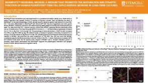 科学海报BrainPhys™ Neuronal Medium: A Medium That Promotes the Maturation and Synaptic Function of Human Pluripotent Stem Cell (hPSC)-Derived Neurons in Long-Term Cultures
 研究综述Neural Stem Cells: Identification, Function, Culture, and Isolation
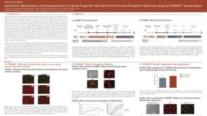 科学海报Generation, Maintenance and Cryopreservation of Neural Progenitor Cells Derived from Human Pluripotent Stem Cells Using the STEMdiff™ Neural System
 产品手册ALDEFLUOR™: For Identification and Isolation of Viable Stem & Progenitor Cells
 产品手册NeuroCult™-XF: Xeno-Free Culture Medium for the Proliferation of Human Neural Stem Cells
 沪公网安备31010102008431号