Foti SB et al. (OCT 2013)
International Journal of Developmental Neuroscience 31 6 434--447
HDAC inhibitors dysregulate neural stem cell activity in the postnatal mouse brain
The mammalian central nervous system (CNS) undergoes significant expansion postnatally,producing astrocytes,oligodendrocytes and inhibitory neurons to modulate the activity of neural circuits. This is coincident in humans with the emergence of pediatric epilepsy,a condition commonly treated with valproate/valproic acid (VPA),a potent inhibitor of histone deacetylases (HDACs). The sequential activity of specific HDACs,however,may be essential for the differentiation of distinct subpopulations of neurons and glia. Here,we show that different subsets of CNS neural stem cells (NSCs) and progenitors switch expression of HDAC1 and HDAC2 as they commit to a neurogenic lineage in the subventricular zone (SVZ) and dentate gyrus (DG). The administration of VPA for only one week from P7-P14,combined with sequential injections of thymidine analogs reveals that VPA stimulates a significant and differential decrease in the production and differentiation of progeny of NSCs in the DG,rostral migratory stream (RMS),and olfactory bulb (OB). Cross-fostering VPA-treated mice revealed,however,that a postnatal failure to thrive induced by VPA treatment had a greater effect on DG neurogenesis than VPA action directly. By one month after VPA,OB interneuron genesis was significantly and differentially reduced in both periglomerular and granule neurons. Using neurosphere assays to test if VPA directly regulates NSC activity,we found that short term treatment with VPA in vivo reduced neurosphere numbers and size,a phenotype that was also obtained in neurospheres from control mice treated with VPA and an alternative HDAC inhibitor,Trichostatin A (TSA) at 0 and 3 days in vitro (DIV). Collectively,these data show that clinically used HDAC inhibitors like VPA and TSA can perturb postnatal neurogenesis; and their use should be carefully considered,especially in individuals whose brains are actively undergoing key postnatal time windows of development.
View Publication
产品类型:
产品号#:
05700
05701
05702
产品名:
NeuroCult™ 基础培养基(小鼠和大鼠)
NeuroCult™ 扩增添加物(小鼠和大鼠)
NeuroCult™扩增试剂盒(小鼠和大鼠)
Xu X et al. (MAR 2017)
Stem Cell Reports 8 3 619--633
Reversal of Phenotypic Abnormalities by CRISPR/Cas9-Mediated Gene Correction in Huntington Disease Patient-Derived Induced Pluripotent Stem Cells
Huntington disease (HD) is a dominant neurodegenerative disorder caused by a CAG repeat expansion in HTT. Here we report correction of HD human induced pluripotent stem cells (hiPSCs) using a CRISPR-Cas9 and piggyBac transposon-based approach. We show that both HD and corrected isogenic hiPSCs can be differentiated into excitable,synaptically active forebrain neurons. We further demonstrate that phenotypic abnormalities in HD hiPSC-derived neural cells,including impaired neural rosette formation,increased susceptibility to growth factor withdrawal,and deficits in mitochondrial respiration,are rescued in isogenic controls. Importantly,using genome-wide expression analysis,we show that a number of apparent gene expression differences detected between HD and non-related healthy control lines are absent between HD and corrected lines,suggesting that these differences are likely related to genetic background rather than HD-specific effects. Our study demonstrates correction of HD hiPSCs and associated phenotypic abnormalities,and the importance of isogenic controls for disease modeling using hiPSCs.
View Publication
产品类型:
产品号#:
05790
05792
05793
05794
05795
05835
05839
08581
08582
产品名:
BrainPhys™神经元培养基
BrainPhys™神经元培养基和SM1试剂盒
BrainPhys™ 神经元培养基N2-A和SM1试剂盒
BrainPhys™原代神经元试剂盒
BrainPhys™ hPSC 神经元试剂盒
STEMdiff™ 神经诱导培养基
STEMdiff™ 神经诱导培养基
STEMdiff™SMADi神经诱导试剂盒
STEMdiff™SMADi神经诱导试剂盒,2套
Kanai R et al. (JAN 2012)
Journal of the National Cancer Institute 104 1 42--55
Oncolytic virus-mediated manipulation of DNA damage responses: synergy with chemotherapy in killing glioblastoma stem cells.
BACKGROUND: Although both the alkylating agent temozolomide (TMZ) and oncolytic viruses hold promise for treating glioblastoma,which remains uniformly lethal,the effectiveness of combining the two treatments and the mechanism of their interaction on cancer stem cells are unknown. METHODS: We investigated the efficacy of combining TMZ and the oncolytic herpes simplex virus (oHSV) G47Δ in killing glioblastoma stem cells (GSCs),using Chou-Talalay combination index analysis,immunocytochemistry and fluorescence microscopy,and neutral comet assay. The role of treatment-induced DNA double-strand breaks,activation of DNA damage responses,and virus replication in the cytotoxic interaction between G47Δ and TMZ was examined with a panel of pharmacological inhibitors and short-hairpin RNA (shRNA)-mediated knockdown of DNA repair pathways. Comparisons of cell survival and virus replication were performed using a two-sided t test (unpaired). The survival of athymic mice (n = 6-8 mice per group) bearing GSC-derived glioblastoma tumors treated with the combination of G47Δ and TMZ was analyzed by the Kaplan-Meier method and evaluated with a two-sided log-rank test. RESULTS: The combination of G47Δ and TMZ acted synergistically in killing GSCs but not neurons,with associated robust induction of DNA damage. Pharmacological and shRNA-mediated knockdown studies suggested that activated ataxia telangiectasia mutated (ATM) is a crucial mediator of synergy. Activated ATM relocalized to HSV DNA replication compartments where it likely enhanced oHSV replication and could not participate in repairing TMZ-induced DNA damage. Sensitivity to TMZ and synergy with G47Δ decreased with O(6)-methylguanine-DNA-methyltransferase (MGMT) expression and MSH6 knockdown. Combined G47Δ and TMZ treatment extended survival of mice bearing GSC-derived intracranial tumors,achieving long-term remission in four of eight mice (median survival = 228 days; G47Δ alone vs G47Δ + TMZ,hazard ratio of survival = 7.1,95% confidence interval = 1.9 to 26.1,P = .003) at TMZ doses attainable in patients. CONCLUSIONS: The combination of G47Δ and TMZ acts synergistically in killing GSCs through oHSV-mediated manipulation of DNA damage responses. This strategy is highly efficacious in representative preclinical models and warrants clinical translation.
View Publication
产品类型:
产品号#:
05707
05751
产品名:
NeuroCult™化学解离试剂盒(小鼠)
NeuroCult™ NS-A 扩增试剂盒(人)
M.-Y. Lin et al. (NOV 2017)
Scientific reports 7 1 14883
Zika Virus Infects Intermediate Progenitor Cells and Post-mitotic Committed Neurons in Human Fetal Brain Tissues.
Zika virus (ZIKV) infection is associated with microcephaly in fetuses,but the pathogenesis of ZIKV-related microcephaly is not well understood. Here we show that ZIKV infects the subventricular zone in human fetal brain tissues and that the tissue tropism broadens with the progression of gestation. Our research demonstrates also that intermediate progenitor cells (IPCs) are the main target cells for ZIKV. Post-mitotic committed neurons become susceptible to ZIKV infection as well at later stages of gestation. Furthermore,activation of microglial cells,DNA fragmentation,and apoptosis of infected or uninfected cells could be found in ZIKV-infected brain tissues. Our studies identify IPCs as the main target cells for ZIKV. They also suggest that immune activation after ZIKV infection may play an important role in the pathogenesis of ZIKV-related microcephaly.
View Publication
Yamamizu K et al. (MAY 2016)
Scientific reports 6 1 25667
Generation and gene expression profiling of 48 transcription-factor-inducible mouse embryonic stem cell lines.
Mouse embryonic stem cells (ESCs) can differentiate into a wide range - and possibly all cell types in vitro,and thus provide an ideal platform to study systematically the action of transcription factors (TFs) in cell differentiation. Previously,we have generated and analyzed 137 TF-inducible mouse ESC lines. As an extension of this NIA Mouse ESC Bank we generated and characterized 48 additional mouse ESC lines,in which single TFs in each line could be induced in a doxycycline-controllable manner. Together,with the previous ESC lines,the bank now comprises 185 TF-manipulable ESC lines (>10% of all mouse TFs). Global gene expression (transcriptome) profiling revealed that the induction of individual TFs in mouse ESCs for 48 hours shifts their transcriptomes toward specific differentiation fates (e.g.,neural lineages by Myt1 Isl1,and St18; mesodermal lineages by Pitx1,Pitx2,Barhl2,and Lmx1a; white blood cells by Myb,Etv2,and Tbx6,and ovary by Pitx1,Pitx2,and Dmrtc2). These data also provide and lists of inferred target genes of each TF and possible functions of these TFs. The results demonstrate the utility of mouse ESC lines and their transcriptome data for understanding the mechanism of cell differentiation and the function of TFs.
View Publication
产品类型:
产品号#:
05700
05703
05704
产品名:
NeuroCult™ 基础培养基(小鼠和大鼠)
NeuroCult™ 分化添加物(小鼠和大鼠)
NeuroCult™ 分化试剂盒(小鼠和大鼠)
Chua SJ et al. (FEB 2009)
Biochemical and biophysical research communications 379 2 217--21
Neural progenitors, neurons and oligodendrocytes from human umbilical cord blood cells in a serum-free, feeder-free cell culture.
We have previously demonstrated that lineage negative cells (Lin(neg)) from umbilical cord blood (UCB) develop into multipotent cells capable of differentiation into bone,muscle,endothelial and neural cells. The objective of this study was to determine the optimal conditions required for Lin(neg) UCB cells to differentiate into neuronal cells and oligodendrocytes. We demonstrate that early neural stage markers (nestin,neurofilament,A2B5 and Sox2) are expressed in Lin(neg) cells cultured in FGF4,SCF,Flt3-ligand reprogramming culture media followed by the early macroglial cell marker O4. Early stage oligodendrocyte markers CNPase,GalC,Olig2 and the late-stage marker MOSP are observed,as is the Schwann cell marker PMP22. In summary,Lin(neg) UCB cells,when appropriately cultured,are able to exhibit characteristics of neuronal and macroglial cells that can specifically differentiate into oligodendrocytes and Schwann cells and express proteins associated with myelin production after in vitro differentiation.
View Publication
产品类型:
产品号#:
09600
09650
产品名:
StemSpan™ SFEM
StemSpan™ SFEM
Begum AN et al. (JUL 2014)
Translational psychiatry 4 January e414
Women with the Alzheimer's risk marker ApoE4 lose A-specific CD4 T cells 10-20 years before men.
Adaptive immunity to self-antigens causes autoimmune disorders,such as multiple sclerosis,psoriasis and type 1 diabetes; paradoxically,T- and B-cell responses to amyloid-$\$(A$\$) reduce Alzheimer's disease (AD)-associated pathology and cognitive impairment in mouse models of the disease. The manipulation of adaptive immunity has been a promising therapeutic approach for the treatment of AD,although vaccine and anti-A$\$ approaches have proven difficult in patients,thus far. CD4(+) T cells have a central role in regulating adaptive immune responses to antigens,and A$\$-specific CD4(+) T cells have been shown to reduce AD pathology in mouse models. As these cells may facilitate endogenous mechanisms that counter AD,an evaluation of their abundance before and during AD could provide important insights. A$\$-CD4see is a new assay developed to quantify A$\$-specific CD4(+) T cells in human blood,using dendritic cells derived from human pluripotent stem cells. In tests of textgreater50 human subjects A$\$-CD4see showed an age-dependent decline of A$\$-specific CD4(+) T cells,which occurs earlier in women than men. In aggregate,men showed a 50% decline in these cells by the age of 70 years,but women reached the same level before the age of 60 years. Notably,women who carried the AD risk marker apolipoproteinE-ɛ4 (ApoE4) showed the earliest decline,with a precipitous drop between 45 and 52 years,when menopause typically begins. A$\$-CD4see requires a standard blood draw and provides a minimally invasive approach for assessing changes in A$\$ that may reveal AD-related changes in physiology by a decade. Furthermore,CD4see probes can be modified to target any peptide,providing a powerful new tool to isolate antigen-specific CD4(+) T cells from human subjects.
View Publication
Vukovic J et al. (APR 2013)
Journal of Neuroscience 33 15 6603--6613
Immature Doublecortin-Positive Hippocampal Neurons Are Important for Learning But Not for Remembering
It is now widely accepted that hippocampal neurogenesis underpins critical cognitive functions,such as learning and memory. To assess the behavioral importance of adult-born neurons,we developed a novel knock-in mouse model that allowed us to specifically and reversibly ablate hippocampal neurons at an immature stage. In these mice,the diphtheria toxin receptor (DTR) is expressed under control of the doublecortin (DCX) promoter,which allows for specific ablation of immature DCX-expressing neurons after administration of diphtheria toxin while leaving the neural precursor pool intact. Using a spatially challenging behavioral test (a modified version of the active place avoidance test),we present direct evidence that immature DCX-expressing neurons are required for successful acquisition of spatial learning,as well as reversal learning,but are not necessary for the retrieval of stored long-term memories. Importantly,the observed learning deficits were rescued as newly generated immature neurons repopulated the granule cell layer upon termination of the toxin treatment. Repeat (or cyclic) depletion of immature neurons reinstated behavioral deficits if the mice were challenged with a novel task. Together,these findings highlight the potential of stimulating neurogenesis as a means to enhance learning.
View Publication
产品类型:
产品号#:
05700
05701
05702
产品名:
NeuroCult™ 基础培养基(小鼠和大鼠)
NeuroCult™ 扩增添加物(小鼠和大鼠)
NeuroCult™扩增试剂盒(小鼠和大鼠)
de Boer AS et al. (AUG 2014)
Science Translational Medicine 6 248 248ra104--248ra104
Genetic validation of a therapeutic target in a mouse model of ALS
AbstractBack to TopbackslashnNeurons produced from stem cells have emerged as a tool to identify new therapeutic targets for neurological diseases such as amyotrophic lateral sclerosis (ALS). However,it remains unclear to what extent these new mechanistic insights will translate to animal models,an important step in the validation of new targets. Previously,we found that glia from mice carrying the SOD1G93A mutation,a model of ALS,were toxic to stem cell–derived human motor neurons. We use pharmacological and genetic approaches to demonstrate that the prostanoid receptor DP1 mediates this glial toxicity. Furthermore,we validate the importance of this mechanism for neural degeneration in vivo. Genetic ablation of DP1 in SOD1G93A mice extended life span,decreased microglial activation,and reduced motor neuron loss. Our findings suggest that blocking DP1 may be a therapeutic strategy in ALS and demonstrate that discoveries from stem cell models of disease can be corroborated in vivo.
View Publication


 EasySep™小鼠TIL(CD45)正选试剂盒





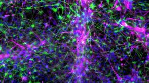 实验方案How to Co-Culture Astrocytes and NGN2 mRNA-Driven Induced Forebrain Neurons Derived from Human Pluripotent Stem Cells
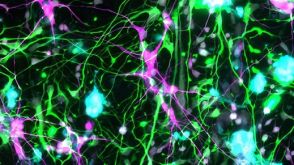 实验方案How to Tri-Culture Astrocytes, Microglia, and NGN2 mRNA-LNP-Induced Forebrain Neurons Derived from Human Pluripotent Stem Cells
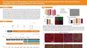 科学海报The Downstream Differentiation Potential of Human Induced Pluripotent Stem Cell-Derived Neural Progenitor Cells to Forebrain Neurons and Astrocytes
 沪公网安备31010102008431号