Liu Y et al. (MAY 2011)
Nature protocols 6 5 640--55
OLIG gene targeting in human pluripotent stem cells for motor neuron and oligodendrocyte differentiation.
Pluripotent stem cells can be genetically labeled to facilitate differentiation studies. In this paper,we describe a gene-targeting protocol to knock in a GFP cassette into key gene loci in human pluripotent stem cells (hPSCs),and then use the genetically tagged hPSCs to guide in vitro differentiation,immunocytochemical and electrophysiological profiling and in vivo characterization after cell transplantation. The Olig transcription factors have key roles in the transcription regulatory pathways for the genesis of motor neurons (MNs) and oligodendrocytes (OLs). We have generated OLIG2-GFP hPSC reporter lines that reliably mark MNs and OLs for monitoring their sequential differentiation from hPSCs. The expression of the GFP reporter recapitulates the endogenous expression of OLIG genes. The in vitro characterization of fluorescence-activated cell sorting-purified cells is consistent with cells of the MN or OL lineages,depending on the stages at which they are collected. This protocol is efficient and reliable and usually takes 5-7 months to complete. The genetic tagging-differentiation methodology used herein provides a general framework for similar work for differentiation of hPSCs into other lineages.
View Publication
产品类型:
产品号#:
05850
05857
05870
05875
85850
85857
85870
85875
产品名:
mTeSR™1
mTeSR™1
Swartz EW et al. (NOV 2016)
STEM CELLS Translational Medicine 5 11 1461--1472
A Novel Protocol for Directed Differentiation of C9orf72-Associated Human Induced Pluripotent Stem Cells Into Contractile Skeletal Myotubes
Induced pluripotent stem cells (iPSCs) offer an unlimited resource of cells to be used for the study of underlying molecular biology of disease,therapeutic drug screening,and transplant-based regenerative medicine. However,methods for the directed differentiation of skeletal muscle for these purposes remain scarce and incomplete. Here,we present a novel,small molecule-based protocol for the generation of multinucleated skeletal myotubes using eight independent iPSC lines. Through combinatorial inhibition of phosphoinositide 3-kinase (PI3K) and glycogen synthase kinase 3β (GSK3β) with addition of bone morphogenic protein 4 (BMP4) and fibroblast growth factor 2 (FGF2),we report up to 64% conversion of iPSCs into the myogenic program by day 36 as indicated by MYOG+ cell populations. These cells began to exhibit spontaneous contractions as early as 34 days in vitro in the presence of a serum-free medium formulation. We used this protocol to obtain iPSC-derived muscle cells from frontotemporal dementia (FTD) patients harboring C9orf72 hexanucleotide repeat expansions (rGGGGCC),sporadic FTD,and unaffected controls. iPSCs derived from rGGGGCC carriers contained RNA foci but did not vary in differentiation efficiency when compared to unaffected controls nor display mislocalized TDP-43 after as many as 120 days in vitro. This study presents a rapid,efficient,and transgene-free method for generating multinucleated skeletal myotubes from iPSCs and a resource for further modeling the role of skeletal muscle in amyotrophic lateral sclerosis and other motor neuron diseases. SIGNIFICANCE Protocols to produce skeletal myotubes for disease modeling or therapy are scarce and incomplete. The present study efficiently generates functional skeletal myotubes from human induced pluripotent stem cells using a small molecule-based approach. Using this strategy,terminal myogenic induction of up to 64% in 36 days and spontaneously contractile myotubes within 34 days were achieved. Myotubes derived from patients carrying the C9orf72 repeat expansion show no change in differentiation efficiency and normal TDP-43 localization after as many as 120 days in vitro when compared to unaffected controls. This study provides an efficient,novel protocol for the generation of skeletal myotubes from human induced pluripotent stem cells that may serve as a valuable tool in drug discovery and modeling of musculoskeletal and neuromuscular diseases.
View Publication
产品类型:
产品号#:
05832
72302
72304
72307
72308
78006
78006.1
78006.2
78005
78005.1
78005.2
78005.3
34811
34815
34850
34821
34825
34860
05835
05839
100-1044
产品名:
STEMdiff™ 神经花环选择试剂
Y-27632(二盐酸盐)
Y-27632(二盐酸盐)
Y-27632(二盐酸盐)
Y-27632(二盐酸盐)
重组人EGF
重组人EGF
重组人EGF
重组人BDNF
重组人BDNF
重组人BDNF
重组人BDNF
AggreWell™ 800 24孔板,1个
AggreWell™ 800 24孔板,5个
AggreWell™ 800 24孔板启动套装
AggreWell™ 800 6孔板,1个
AggreWell™ 800 6孔板,5个
AggreWell™ 800 6孔板启动套装
STEMdiff™ 神经诱导培养基
STEMdiff™ 神经诱导培养基
Y-27632(二盐酸盐)
Robinson M et al. (AUG 2016)
Stem Cell Reviews and Reports 12 4 476--483
Functionalizing Ascl1 with Novel Intracellular Protein Delivery Technology for Promoting Neuronal Differentiation of Human Induced Pluripotent Stem Cells
Pluripotent stem cells can become any cell type found in the body. Accordingly,one of the major challenges when working with pluripotent stem cells is producing a highly homogenous population of differentiated cells,which can then be used for downstream applications such as cell therapies or drug screening. The transcription factor Ascl1 plays a key role in neural development and previous work has shown that Ascl1 overexpression using viral vectors can reprogram fibroblasts directly into neurons. Here we report on how a recombinant version of the Ascl1 protein functionalized with intracellular protein delivery technology (Ascl1-IPTD) can be used to rapidly differentiate human induced pluripotent stem cells (hiPSCs) into neurons. We first evaluated a range of Ascl1-IPTD concentrations to determine the most effective amount for generating neurons from hiPSCs cultured in serum free media. Next,we looked at the frequency of Ascl1-IPTD supplementation in the media on differentiation and found that one time supplementation is sufficient enough to trigger the neural differentiation process. Ascl1-IPTD was efficiently taken up by the hiPSCs and enabled rapid differentiation into TUJ1-positive and NeuN-positive populations with neuronal morphology after 8 days. After 12 days of culture,hiPSC-derived neurons produced by Ascl1-IPTD treatment exhibited greater neurite length and higher numbers of branch points compared to neurons derived using a standard neural progenitor differentiation protocol. This work validates Ascl1-IPTD as a powerful tool for engineering neural tissue from pluripotent stem cells.
View Publication
产品类型:
产品号#:
05832
05833
05838
05872
05873
05940
07174
07180
07183
07190
27147
07191
36254
07930
07931
07940
07955
07956
07959
07954
27845
27945
27840
27865
27940
27965
05835
05839
08581
08582
100-0483
100-0484
100-1061
07952
100-0763
100-0485
100-1077
产品名:
STEMdiff™ 神经花环选择试剂
STEMdiff™神经前体细胞培养基
STEMdiff™神经祖细胞冻存液
Vitronectin XF™
CellAdhere™ 稀释缓冲液
DMEM/F-12 with 15 mM HEPES
CryoStor® CS10
CryoStor® CS10
CryoStor® CS10
CryoStor® CS10
CryoStor® CS10
STEMdiff™ 神经诱导培养基
STEMdiff™ 神经诱导培养基
STEMdiff™SMADi神经诱导试剂盒
STEMdiff™SMADi神经诱导试剂盒,2套
Hausser Scientificᵀᴹ 明线血球计数板
ReLeSR™
CryoStor® CS10
CryoStor® CS10
Vitronectin XF™
温和细胞解离试剂
ReLeSR™
Kim J et al. (NOV 2013)
Stem Cell Research 11 3 978--989
Alginate microcapsule as a 3D platform for the efficient differentiation of human embryonic stem cells to dopamine neurons
Human embryonic stem cells (hESCs) are emerging as an attractive alternative source for cell replacement therapy since the cells can be expanded in culture indefinitely and differentiated into any cell types in the body. In order to optimize cell-to-cell interaction,cell proliferation and differentiation into specific lineages as well as tissue organization,it is important to provide a microenvironment for the hESCs which mimics the stem cell niche. One approach is to provide a three-dimensional (3D) environment such as encapsulation. We present an approach to culture and differentiate hESCs into midbrain dopamine (mdDA) neurons in a 3D microenvironment using alginate microcapsules for the first time. A detailed gene and protein expression analysis during neuronal differentiation showed an increased gene and protein expression of various specific DA neuronal markers,particularly tyrosine hydroxylase (TH) by textgreater100 folds after 2weeks and at least 50% higher expression after 4weeks respectively,compared to cells differentiated under conventional two-dimensional (2D) platform. The encapsulated TH+ cells co-expressed mdDA neuronal markers,forkhead box protein A-2 (FOXA2) and pituitary homeobox-3 (PITX3) after 4weeks and secreted approximately 60pg/ml/106 cells higher DA level when induced. We propose that the 3D platform facilitated an early onset of DA neuronal generation compared to that with conventional 2D system which also secretes more DA under potassium-induction. It is a very useful model to study the proliferation and directed differentiation of hESCs to various lineages,particularly to mdDA neurons. This 3D system also allows the separation of feeder cells from hESCs during the process of differentiation and also has potential for immune-isolation during transplantation studies. ?? 2013 Elsevier B.V.
View Publication
Azari H et al. (JAN 2011)
Journal of visualized experiments : JoVE 56 e3633
Isolation and expansion of human glioblastoma multiforme tumor cells using the neurosphere assay.
Stem-like cells have been isolated in tumors such as breast,lung,colon,prostate and brain. A critical issue in all these tumors,especially in glioblastoma mutliforme (GBM),is to identify and isolate tumor initiating cell population(s) to investigate their role in tumor formation,progression,and recurrence. Understanding tumor initiating cell populations will provide clues to finding effective therapeutic approaches for these tumors. The neurosphere assay (NSA) due to its simplicity and reproducibility has been used as the method of choice for isolation and propagation of many of this tumor cells. This protocol demonstrates the neurosphere culture method to isolate and expand stem-like cells in surgically resected human GBM tumor tissue. The procedures include an initial chemical digestion and mechanical dissociation of tumor tissue,and subsequently plating the resulting single cell suspension in NSA culture. After 7-10 days,primary neurospheres of 150-200 μm in diameter can be observed and are ready for further passaging and expansion.
View Publication
产品类型:
产品号#:
05751
05752
产品名:
NeuroCult™ NS-A 扩增试剂盒(人)
NeuroCult™ NS-A 分化试剂盒(人)
Chailangkarn T et al. (AUG 2016)
Nature 536 7616 338--343
A human neurodevelopmental model for Williams syndrome.
Williams syndrome is a genetic neurodevelopmental disorder characterized by an uncommon hypersociability and a mosaic of retained and compromised linguistic and cognitive abilities. Nearly all clinically diagnosed individuals with Williams syndrome lack precisely the same set of genes,with breakpoints in chromosome band 7q11.23 (refs 1-5). The contribution of specific genes to the neuroanatomical and functional alterations,leading to behavioural pathologies in humans,remains largely unexplored. Here we investigate neural progenitor cells and cortical neurons derived from Williams syndrome and typically developing induced pluripotent stem cells. Neural progenitor cells in Williams syndrome have an increased doubling time and apoptosis compared with typically developing neural progenitor cells. Using an individual with atypical Williams syndrome,we narrowed this cellular phenotype to a single gene candidate,frizzled 9 (FZD9). At the neuronal stage,layer V/VI cortical neurons derived from Williams syndrome were characterized by longer total dendrites,increased numbers of spines and synapses,aberrant calcium oscillation and altered network connectivity. Morphometric alterations observed in neurons from Williams syndrome were validated after Golgi staining of post-mortem layer V/VI cortical neurons. This model of human induced pluripotent stem cells fills the current knowledge gap in the cellular biology of Williams syndrome and could lead to further insights into the molecular mechanism underlying the disorder and the human social brain.
View Publication
产品类型:
产品号#:
05850
05857
05870
05875
85850
85857
85870
85875
产品名:
mTeSR™1
mTeSR™1
Su H et al. (JUL 2013)
Stem Cell Research 11 1 529--539
Transplanted motoneurons derived from human induced pluripotent stem cells form functional connections with target muscle
Induced pluripotent stem cells (iPSCs) hold promise for the treatment of motoneuron diseases because of their distinct features including pluripotency,self-derivation and potential ability to differentiate into motoneurons. However,it is still unknown whether human iPSC-derived motoneurons can functionally innervate target muscles in vivo,which is the definitive sign of successful cell therapy for motoneuron diseases. In the present study,we demonstrated that human iPSCs derived from mesenchymal cells of the umbilical cord possessed a high yield in neural differentiation. Using a chemically-defined in vitro system,human iPSCs efficiently differentiated into motoneurons which displayed typical morphology,expressed specific molecules,and generated repetitive trains of action potentials. When transplanted into the injured musculocutaneous nerve of rats,they survived robustly,extended axons along the nerve,and formed functional connections with the target muscle (biceps brachii),thereby protecting the muscle from atrophy. Our study provides evidence for the first time that human iPSC-derived motoneurons are truly functional not only in vitro but also in vivo,and they have potential for stem cell-based therapies for motoneuron diseases. textcopyright 2013 Elsevier B.V.
View Publication
产品类型:
产品号#:
05850
05857
05870
05875
85850
85857
85870
85875
产品名:
mTeSR™1
mTeSR™1
M.-Y. Lin et al. (NOV 2017)
Scientific reports 7 1 14883
Zika Virus Infects Intermediate Progenitor Cells and Post-mitotic Committed Neurons in Human Fetal Brain Tissues.
Zika virus (ZIKV) infection is associated with microcephaly in fetuses,but the pathogenesis of ZIKV-related microcephaly is not well understood. Here we show that ZIKV infects the subventricular zone in human fetal brain tissues and that the tissue tropism broadens with the progression of gestation. Our research demonstrates also that intermediate progenitor cells (IPCs) are the main target cells for ZIKV. Post-mitotic committed neurons become susceptible to ZIKV infection as well at later stages of gestation. Furthermore,activation of microglial cells,DNA fragmentation,and apoptosis of infected or uninfected cells could be found in ZIKV-infected brain tissues. Our studies identify IPCs as the main target cells for ZIKV. They also suggest that immune activation after ZIKV infection may play an important role in the pathogenesis of ZIKV-related microcephaly.
View Publication

 EasySep™小鼠TIL(CD45)正选试剂盒


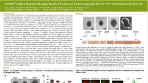 科学海报STEMdiff Cerebral Organoid Kit: A New Tool for the Culture of 3D Brain Organoids Derived from Human Pluripotent Stem Cells

 沪公网安备31010102008431号