Organic cation transporter-mediated ergothioneine uptake in mouse neural progenitor cells suppresses proliferation and promotes differentiation into neurons.
The aim of the present study is to clarify the functional expression and physiological role in neural progenitor cells (NPCs) of carnitine/organic cation transporter OCTN1/SLC22A4,which accepts the naturally occurring food-derived antioxidant ergothioneine (ERGO) as a substrate in vivo. Real-time PCR analysis revealed that mRNA expression of OCTN1 was much higher than that of other organic cation transporters in mouse cultured cortical NPCs. Immunocytochemical analysis showed colocalization of OCTN1 with the NPC marker nestin in cultured NPCs and mouse embryonic carcinoma P19 cells differentiated into neural progenitor-like cells (P19-NPCs). These cells exhibited time-dependent [(3)H]ERGO uptake. These results demonstrate that OCTN1 is functionally expressed in murine NPCs. Cultured NPCs and P19-NPCs formed neurospheres from clusters of proliferating cells in a culture time-dependent manner. Exposure of cultured NPCs to ERGO or other antioxidants (edaravone and ascorbic acid) led to a significant decrease in the area of neurospheres with concomitant elimination of intracellular reactive oxygen species. Transfection of P19-NPCs with small interfering RNA for OCTN1 markedly promoted formation of neurospheres with a concomitant decrease of [(3)H]ERGO uptake. On the other hand,exposure of cultured NPCs to ERGO markedly increased the number of cells immunoreactive for the neuronal marker βIII-tubulin,but decreased the number immunoreactive for the astroglial marker glial fibrillary acidic protein (GFAP),with concomitant up-regulation of neuronal differentiation activator gene Math1. Interestingly,edaravone and ascorbic acid did not affect such differentiation of NPCs,in contrast to the case of proliferation. Knockdown of OCTN1 increased the number of cells immunoreactive for GFAP,but decreased the number immunoreactive for βIII-tubulin,with concomitant down-regulation of Math1 in P19-NPCs. Thus,OCTN1-mediated uptake of ERGO in NPCs inhibits cellular proliferation via regulation of oxidative stress,and also promotes cellular differentiation by modulating the expression of basic helix-loop-helix transcription factors via an unidentified mechanism different from antioxidant action.
View Publication
产品类型:
产品号#:
05707
产品名:
NeuroCult™化学解离试剂盒(小鼠)
Kayama T et al. (JAN 2018)
Biochemical and Biophysical Research Communications 495 1 1028--1033
Temporally coordinated spiking activity of human induced pluripotent stem cell-derived neurons co-cultured with astrocytes
In culture conditions,human induced-pluripotent stem cells (hiPSC)-derived neurons form synaptic connections with other cells and establish neuronal networks,which are expected to be an in vitro model system for drug discovery screening and toxicity testing. While early studies demonstrated effects of co-culture of hiPSC-derived neurons with astroglial cells on survival and maturation of hiPSC-derived neurons,the population spiking patterns of such hiPSC-derived neurons have not been fully characterized. In this study,we analyzed temporal spiking patterns of hiPSC-derived neurons recorded by a multi-electrode array system. We discovered that specific sets of hiPSC-derived neurons co-cultured with astrocytes showed more frequent and highly coherent non-random synchronized spike trains and more dynamic changes in overall spike patterns over time. These temporally coordinated spiking patterns are physiological signs of organized circuits of hiPSC-derived neurons and suggest benefits of co-culture of hiPSC-derived neurons with astrocytes.
View Publication
On-demand optogenetic activation of human stem-cell-derived neurons
The widespread application of human stem-cell-derived neurons for functional studies is impeded by complicated differentiation protocols,immaturity,and deficient optogene expression as stem cells frequently lose transgene expression over time. Here we report a simple but precise Cre-loxP-based strategy for generating conditional,and thereby stable,optogenetic human stem-cell lines. These cells can be easily and efficiently differentiated into functional neurons,and optogene expression can be triggered by administering Cre protein to the cultures. This conditional expression system may be applied to stem-cell-derived neurons whenever timed transgene expression could help to overcome silencing at the stem-cell level.
View Publication
产品类型:
产品号#:
05711
05790
05792
05793
05794
05795
100-1281
产品名:
NeuroCult™ SM1 神经添加物
BrainPhys™神经元培养基
BrainPhys™神经元培养基和SM1试剂盒
BrainPhys™ 神经元培养基N2-A和SM1试剂盒
BrainPhys™原代神经元试剂盒
BrainPhys™ hPSC 神经元试剂盒
NeuroCult™ SM1 神经添加物
Lawn S et al. (FEB 2015)
The Journal of biological chemistry 290 6 3814--24
Neurotrophin signaling via TrkB and TrkC receptors promotes the growth of brain tumor-initiating cells.
Neurotrophins and their receptors are frequently expressed in malignant gliomas,yet their functions are largely unknown. Previously,we have shown that p75 neurotrophin receptor is required for glioma invasion and proliferation. However,the role of Trk receptors has not been examined. In this study,we investigated the importance of TrkB and TrkC in survival of brain tumor-initiating cells (BTICs). Here,we show that human malignant glioma tissues and also tumor-initiating cells isolated from fresh human malignant gliomas express the neurotrophin receptors TrkB and TrkC,not TrkA,and they also express neurotrophins NGF,BDNF,and neurotrophin 3 (NT3). Specific activation of TrkB and TrkC receptors by ligands BDNF and NT3 enhances tumor-initiating cell viability through activation of ERK and Akt pathways. Conversely,TrkB and TrkC knockdown or pharmacologic inhibition of Trk signaling decreases neurotrophin-dependent ERK activation and BTIC growth. Further,pharmacological inhibition of both ERK and Akt pathways blocked BDNF,and NT3 stimulated BTIC survival. Importantly,attenuation of BTIC growth by EGFR inhibitors could be overcome by activation of neurotrophin signaling,and neurotrophin signaling is sufficient for long term BTIC growth as spheres in the absence of EGF and FGF. Our results highlight a novel role for neurotrophin signaling in brain tumor and suggest that Trks could be a target for combinatorial treatment of malignant glioma.
View Publication
产品类型:
产品号#:
05750
05751
产品名:
NeuroCult™ NS-A 基础培养基(人)
NeuroCult™ NS-A 扩增试剂盒(人)
Lin H et al. (JAN 2017)
Neuro-oncology 19 1 43--54
Fatty acid oxidation is required for the respiration and proliferation of malignant glioma cells.
BACKGROUND Glioma is the most common form of primary malignant brain tumor in adults,with approximately 4 cases per 100 000 people each year. Gliomas,like many tumors,are thought to primarily metabolize glucose for energy production; however,the reliance upon glycolysis has recently been called into question. In this study,we aimed to identify the metabolic fuel requirements of human glioma cells. METHODS We used database searches and tissue culture resources to evaluate genotype and protein expression,tracked oxygen consumption rates to study metabolic responses to various substrates,performed histochemical techniques and fluorescence-activated cell sorting-based mitotic profiling to study cellular proliferation rates,and employed an animal model of malignant glioma to evaluate a new therapeutic intervention. RESULTS We observed the presence of enzymes required for fatty acid oxidation within human glioma tissues. In addition,we demonstrated that this metabolic pathway is a major contributor to aerobic respiration in primary-cultured cells isolated from human glioma and grown under serum-free conditions. Moreover,inhibiting fatty acid oxidation reduces proliferative activity in these primary-cultured cells and prolongs survival in a syngeneic mouse model of malignant glioma. CONCLUSIONS Fatty acid oxidation enzymes are present and active within glioma tissues. Targeting this metabolic pathway reduces energy production and cellular proliferation in glioma cells. The drug etomoxir may provide therapeutic benefit to patients with malignant glioma. In addition,the expression of fatty acid oxidation enzymes may provide prognostic indicators for clinical practice.
View Publication
Delivery of Functional Anti-miR-9 by Mesenchymal Stem Cellderived Exosomes to Glioblastoma Multiforme Cells Conferred Chemosensitivity
Glioblastoma multiforme (GBM),the most common and lethal tumor of the adult brain,generally shows chemo- and radioresistance. MicroRNAs (miRs) regulate physiological processes,such as resistance of GBM cells to temozolomide (TMZ). Although miRs are attractive targets for cancer therapeutics,the effectiveness of this approach requires targeted delivery. Mesenchymal stem cells (MSCs) can migrate to the sites of cancers,including GBM. We report on an increase in miR-9 in TMZ-resistant GBM cells. miR-9 was involved in the expression of the drug efflux transporter,P-glycoprotein. To block miR-9,methods were developed with Cy5-tagged anti-miR-9. Dye-transfer studies indicated intracellular communication between GBM cells and MSCs. This occurred by gap junctional intercellular communication and the release of microvesicles. In both cases,anti-miR-9 was transferred from MSCs to GBM cells. However,the major form of transfer occurred with the microvesicles. The delivery of anti-miR-9 to the resistant GBM cells reversed the expression of the multidrug transporter and sensitized the GBM cells to TMZ,as shown by increased cell death and caspase activity. The data showed a potential role for MSCs in the functional delivery of synthetic anti-miR-9 to reverse the chemoresistance of GBM cells.Molecular Therapy-Nucleic Acids (2013) 2,e126; doi:10.1038/mtna.2013.60; published online 1 October 2013.
View Publication
产品类型:
产品号#:
05750
05751
产品名:
NeuroCult™ NS-A 基础培养基(人)
NeuroCult™ NS-A 扩增试剂盒(人)
Rosa AI et al. (DEC 2016)
Frontiers in cellular neuroscience 10 284
Heterocellular Contacts with Mouse Brain Endothelial Cells Via Laminin and α6β1 Integrin Sustain Subventricular Zone (SVZ) Stem/Progenitor Cells Properties.
Neurogenesis in the subventricular zone (SVZ) is regulated by diffusible factors and cell-cell contacts. In vivo,SVZ stem cells are associated with the abluminal surface of blood vessels and such interactions are thought to regulate their neurogenic capacity. SVZ neural stem cells (NSCs) have been described to contact endothelial-derived laminin via α6β1 integrin. To elucidate whether heterocellular contacts with brain endothelial cells (BEC) regulate SVZ cells neurogenic capacities,cocultures of SVZ neurospheres and primary BEC,both obtained from C57BL/6 mice,were performed. The involvement of laminin-integrin interactions in SVZ homeostasis was tested in three ways. Firstly,SVZ cells were analyzed following incubation of BEC with the protein synthesis inhibitor cycloheximide (CHX) prior to coculture,a treatment expected to decrease membrane proteins. Secondly,SVZ cells were cocultured with BEC in the presence of an anti-α6 integrin neutralizing antibody. Thirdly,BEC were cultured with β1-/- SVZ cells. We showed that contact with BEC supports,at least in part,proliferation and stemness of SVZ cells,as evaluated by the number of BrdU positive (+) and Sox2+ cells in contact with BEC. These effects are dependent on BEC-derived laminin binding to α6β1 integrin and are decreased in cocultures incubated with anti-α6 integrin neutralizing antibody and in cocultures with SVZ β1-/- cells. Moreover,BEC-derived laminin sustains stemness in SVZ cell cultures via activation of the Notch and mTOR signaling pathways. Our results show that BEC/SVZ interactions involving α6β1 integrin binding to laminin,contribute to SVZ cell proliferation and stemness.
View Publication
产品类型:
产品号#:
05707
产品名:
NeuroCult™化学解离试剂盒(小鼠)
Swartz EW et al. (NOV 2016)
STEM CELLS Translational Medicine 5 11 1461--1472
A Novel Protocol for Directed Differentiation of C9orf72-Associated Human Induced Pluripotent Stem Cells Into Contractile Skeletal Myotubes
Induced pluripotent stem cells (iPSCs) offer an unlimited resource of cells to be used for the study of underlying molecular biology of disease,therapeutic drug screening,and transplant-based regenerative medicine. However,methods for the directed differentiation of skeletal muscle for these purposes remain scarce and incomplete. Here,we present a novel,small molecule-based protocol for the generation of multinucleated skeletal myotubes using eight independent iPSC lines. Through combinatorial inhibition of phosphoinositide 3-kinase (PI3K) and glycogen synthase kinase 3β (GSK3β) with addition of bone morphogenic protein 4 (BMP4) and fibroblast growth factor 2 (FGF2),we report up to 64% conversion of iPSCs into the myogenic program by day 36 as indicated by MYOG+ cell populations. These cells began to exhibit spontaneous contractions as early as 34 days in vitro in the presence of a serum-free medium formulation. We used this protocol to obtain iPSC-derived muscle cells from frontotemporal dementia (FTD) patients harboring C9orf72 hexanucleotide repeat expansions (rGGGGCC),sporadic FTD,and unaffected controls. iPSCs derived from rGGGGCC carriers contained RNA foci but did not vary in differentiation efficiency when compared to unaffected controls nor display mislocalized TDP-43 after as many as 120 days in vitro. This study presents a rapid,efficient,and transgene-free method for generating multinucleated skeletal myotubes from iPSCs and a resource for further modeling the role of skeletal muscle in amyotrophic lateral sclerosis and other motor neuron diseases. SIGNIFICANCE Protocols to produce skeletal myotubes for disease modeling or therapy are scarce and incomplete. The present study efficiently generates functional skeletal myotubes from human induced pluripotent stem cells using a small molecule-based approach. Using this strategy,terminal myogenic induction of up to 64% in 36 days and spontaneously contractile myotubes within 34 days were achieved. Myotubes derived from patients carrying the C9orf72 repeat expansion show no change in differentiation efficiency and normal TDP-43 localization after as many as 120 days in vitro when compared to unaffected controls. This study provides an efficient,novel protocol for the generation of skeletal myotubes from human induced pluripotent stem cells that may serve as a valuable tool in drug discovery and modeling of musculoskeletal and neuromuscular diseases.
View Publication
Rapid and Efficient Direct Conversion of Human Adult Somatic Cells into Neural Stem Cells by HMGA2/let-7b.
A recent study has suggested that fibroblasts can be converted into mouse-induced neural stem cells (miNSCs) through the expression of defined factors. However,successful generation of human iNSCs (hiNSCs) has proven challenging to achieve. Here,using microRNA (miRNA) expression profile analyses,we showed that let-7 microRNA has critical roles for the formation of PAX6/NESTIN-positive colonies from human adult fibroblasts and the proliferation and self-renewal of hiNSCs. HMGA2,a let-7-targeting gene,enables induction of hiNSCs that displayed morphological/molecular features and in vitro/in vivo differentiation potential similar to H9-derived NSCs. Interestingly,HMGA2 facilitated the efficient conversion of senescent somatic cells or blood CD34+ cells into hiNSCs through an interaction with SOX2,whereas other combinations or SOX2 alone showed a limited conversion ability. Taken together,these findings suggest that HMGA2/let-7 facilitates direct reprogramming toward hiNSCs in minimal conditions and maintains hiNSC self-renewal,providing a strategy for the clinical treatment of neurological diseases.
View Publication


 EasySep™小鼠TIL(CD45)正选试剂盒




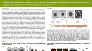 科学海报STEMdiff Cerebral Organoid Kit: A New Tool for the Culture of 3D Brain Organoids Derived from Human Pluripotent Stem Cells

 沪公网安备31010102008431号