Gallia GL et al. (FEB 2009)
Molecular cancer therapeutics 8 2 386--93
Inhibition of Akt inhibits growth of glioblastoma and glioblastoma stem-like cells.
A commonly activated signaling cascade in many human malignancies,including glioblastoma multiforme,is the Akt pathway. This pathway can be activated via numerous upstream alterations including genomic amplification of epidermal growth factor receptor,PTEN deletion,or PIK3CA mutations. In this study,we screened phosphatidylinositol 3-kinase/Akt small-molecule inhibitors in an isogenic cell culture system with an activated Akt pathway secondary to a PIK3CA mutation. One small molecule,A-443654,showed the greatest selective inhibition of cells with the mutant phenotype. Based on these findings,this inhibitor was screened in vitro against a panel of glioblastoma multiforme cell lines. All cell lines tested were sensitive to A-443654 with a mean IC(50) of approximately 150 nmol/L. An analogue of A-443654,methylated at a region that blocks Akt binding,was on average 36-fold less active. Caspase assays and dual flow cytometric analysis showed an apoptotic mechanism of cell death. A-443654 was further tested in a rat intracranial model of glioblastoma multiforme. Animals treated intracranially with polymers containing A-443654 had significantly extended survival compared with control animals; animals survived 79% and 43% longer than controls when A-443654-containing polymers were implanted simultaneously or in a delayed fashion,respectively. This small molecule also inhibited glioblastoma multiforme stem-like cells with similar efficacy compared with traditionally cultured glioblastoma multiforme cell lines. These results suggest that local delivery of an Akt small-molecule inhibitor is effective against experimental intracranial glioma,with no observed resistance to glioblastoma multiforme cells grown in stem cell conditions.
View Publication
产品类型:
产品号#:
05751
产品名:
NeuroCult™ NS-A 扩增试剂盒(人)
Wagner JP et al. (AUG 2014)
Journal of pediatric surgery 49 8 1319--24; discussion 1324--5
INTRODUCTION Hirschsprung's disease is characterized by a developmental arrest of neural crest cell migration,causing distal aganglionosis. Transplanted cells derived from the neural crest may regenerate enteric ganglia in this condition. We investigated the potential of skin-derived precursor cells (SKPs) to engraft and to differentiate into enteric ganglia in aganglionic rat intestine in vivo. METHODS Adult Lewis rat jejunal segments were separated from intestinal continuity and treated with benzalkonium chloride to induce aganglionosis. Ganglia were identified via immunohistochemical stains for S100 and β-III tubulin (TUJ1). SKPs were procured from neonatal Lewis rats expressing enhanced green fluorescent protein (GFP) and cultured in neuroglial-selective media. SKP cell line expansion was quantified,and immunophenotypes were assessed by immunocytochemistry. Aganglionic segments underwent SKP transplantation 21-79days after benzalkonium chloride treatment. The presence of GFP+cells,mature neurons,and mature glia was evaluated at posttransplant days 1,6,and 9. RESULTS Benzalkonium chloride-induced aganglionosis persisted for at least 85days. Prior to differentiation,SKPs expressed S100,denoting neural crest lineage,and nestin,a marker of neuronal precursors. Differentiated SKPs in vitro expressed GFAP,a marker of glial differentiation,as well as TUJ1 and several enteric neurotransmitters. After transplantation,GFP+structures resembling ganglia were identified between longitudinal and circular smooth muscle layers. CONCLUSION SKPs are capable of engraftment,migration,and differentiation within aganglionic rodent intestine in vivo. Differentiated SKPs generate structures that resemble enteric ganglia. Our observations suggest that SKPs represent a potential gangliogenic therapeutic agent for Hirschsprung's disease.
View Publication
K. B. Langer et al. (APR 2018)
Stem cell reports 10 4 1282--1293
Retinal Ganglion Cell Diversity and Subtype Specification from Human Pluripotent Stem Cells.
Retinal ganglion cells (RGCs) are the projection neurons of the retina and transmit visual information to postsynaptic targets in the brain. While this function is shared among nearly all RGCs,this class of cell is remarkably diverse,comprised of multiple subtypes. Previous efforts have identified numerous RGC subtypes in animal models,but less attention has been paid to human RGCs. Thus,efforts of this study examined the diversity of RGCs differentiated from human pluripotent stem cells (hPSCs) and characterized defined subtypes through the expression of subtype-specific markers. Further investigation of these subtypes was achieved using single-cell transcriptomics,confirming the combinatorial expression of molecular markers associated with these subtypes,and also provided insight into more subtype-specific markers. Thus,the results of this study describe the derivation of RGC subtypes from hPSCs and will support the future exploration of phenotypic and functional diversity within human RGCs.
View Publication
产品类型:
产品号#:
05790
05792
05793
05794
05795
85850
85857
产品名:
BrainPhys™神经元培养基
BrainPhys™神经元培养基和SM1试剂盒
BrainPhys™ 神经元培养基N2-A和SM1试剂盒
BrainPhys™原代神经元试剂盒
BrainPhys™ hPSC 神经元试剂盒
mTeSR™1
mTeSR™1
I. Canals et al. (SEP 2018)
Nature methods 15 9 693--696
Rapid and efficient induction of functional astrocytes from human pluripotent stem cells.
The derivation of astrocytes from human pluripotent stem cells is currently slow and inefficient. We demonstrate that overexpression of the transcription factors SOX9 and NFIB in human pluripotent stem cells rapidly and efficiently yields homogeneous populations of induced astrocytes. In our study these cells exhibited molecular and functional properties resembling those of adult human astrocytes and were deemed suitable for disease modeling. Our method provides new possibilities for the study of human astrocytes in health and disease.
View Publication
Oikawa T et al. (OCT 2015)
Nature communications 6 8070
Model of fibrolamellar hepatocellular carcinomas reveals striking enrichment in cancer stem cells.
The aetiology of human fibrolamellar hepatocellular carcinomas (hFL-HCCs),cancers occurring increasingly in children to young adults,is poorly understood. We present a transplantable tumour line,maintained in immune-compromised mice,and validate it as a bona fide model of hFL-HCCs by multiple methods. RNA-seq analysis confirms the presence of a fusion transcript (DNAJB1-PRKACA) characteristic of hFL-HCC tumours. The hFL-HCC tumour line is highly enriched for cancer stem cells as indicated by limited dilution tumourigenicity assays,spheroid formation and flow cytometry. Immunohistochemistry on the hFL-HCC model,with parallel studies on 27 primary hFL-HCC tumours,provides robust evidence for expression of endodermal stem cell traits. Transcriptomic analyses of the tumour line and of multiple,normal hepatic lineage stages reveal a gene signature for hFL-HCCs closely resembling that of biliary tree stem cells--newly discovered precursors for liver and pancreas. This model offers unprecedented opportunities to investigate mechanisms underlying hFL-HCCs pathogenesis and potential therapies.
View Publication
产品类型:
产品号#:
05707
产品名:
NeuroCult™化学解离试剂盒(小鼠)
Ma I and Allan AL (JUN 2011)
Stem cell reviews 7 2 292--306
The role of human aldehyde dehydrogenase in normal and cancer stem cells.
Normal stem cells and cancer stem cells (CSCs) share similar properties,in that both have the capacity to self-renew and differentiate into multiple cell types. In both the normal stem cell and cancer stem cell fields,there has been a great need for a universal marker that can effectively identify and isolate these rare populations of cells in order to characterize them and use this information for research and therapeutic purposes. Currently,it would appear that certain isoenzymes of the aldehyde dehydrogenase (ALDH) superfamily may be able to fulfill this role as a marker for both normal and cancer stem cells. ALDH has been identified as an important enzyme in the protection of normal hematopoietic stem cells,and is now also widely used as a marker to identify and isolate various types of normal stem cells and CSCs. In addition,emerging evidence suggests that ALDH1 is not only a marker for stem cells,but may also play important functional roles related to self-protection,differentiation,and expansion. This comprehensive review discusses the role that ALDH plays in normal stem cells and CSCs,with focus on ALDH1 and ALDH3A1. Discrepancies in the functional themes between cell types and future perspectives for therapeutic applications will also be discussed.
View Publication
产品类型:
产品号#:
01700
01705
01702
产品名:
ALDEFLUOR™ 试剂盒
ALDEFLUOR™ DEAB试剂, 1.5 mM, 1 mL
ALDEFLUOR™检测缓冲液
Li Y et al. (MAR 2015)
PLoS ONE 10 3 e0118266
A comprehensive library of familial human amyotrophic lateral sclerosis induced pluripotent stem cells
Amyotrophic lateral sclerosis is a progressive disease characterized by the loss of upper and lower motor neurons,leading to paralysis of voluntary muscles. About 10% of all ALS cases are familial (fALS),among which 15-20% are linked to Cu/Zn superoxide dismutase (SOD1) mutations,usually inherited in an autosomal dominant manner. To date only one FDA approved drug is available which increases survival moderately. Our understanding of ALS disease mechanisms is largely derived from rodent model studies,however due to the differences between rodents and humans,it is necessary to have humanized models for studies of disease pathogenesis as well as drug development. Therefore,we generated a comprehensive library of a total 22 of fALS patient-specific induced pluripotent stem cell (iPSC) lines. These cells were thoroughly characterized before being deposited into the library. The library of cells includes a variety of C9orf72 mutations,sod1 mutations,FUS,ANG and FIG4 mutations. Certain mutations are represented with more than one line,which allows for studies of variable genetic backgrounds. In addition,these iPSCs can be successfully differentiated to astroglia,a cell type known to play a critical role in ALS disease progression. This library represents a comprehensive resource that can be used for ALS disease modeling and the development of novel therapeutics.
View Publication
产品类型:
产品号#:
05850
05857
05870
05875
07923
85850
85857
85870
85875
产品名:
Dispase (1 U/mL)
mTeSR™1
mTeSR™1
Duan S et al. (DEC 2015)
Nature communications 6 10068
PTEN deficiency reprogrammes human neural stem cells towards a glioblastoma stem cell-like phenotype.
PTEN is a tumour suppressor frequently mutated in many types of cancers. Here we show that targeted disruption of PTEN leads to neoplastic transformation of human neural stem cells (NSCs),but not mesenchymal stem cells. PTEN-deficient NSCs display neoplasm-associated metabolic and gene expression profiles and generate intracranial tumours in immunodeficient mice. PTEN is localized to the nucleus in NSCs,binds to the PAX7 promoter through association with cAMP responsive element binding protein 1 (CREB)/CREB binding protein (CBP) and inhibits PAX7 transcription. PTEN deficiency leads to the upregulation of PAX7,which in turn promotes oncogenic transformation of NSCs and instates 'aggressiveness' in human glioblastoma stem cells. In a large clinical database,we find increased PAX7 levels in PTEN-deficient glioblastoma. Furthermore,we identify that mitomycin C selectively triggers apoptosis in NSCs with PTEN deficiency. Together,we uncover a potential mechanism of how PTEN safeguards NSCs,and establish a cellular platform to identify factors involved in NSC transformation,potentially permitting personalized treatment of glioblastoma.
View Publication
产品类型:
产品号#:
05700
05701
05702
05750
05850
05857
05870
05875
85850
85857
85870
85875
产品名:
NeuroCult™ 基础培养基(小鼠和大鼠)
NeuroCult™ 扩增添加物(小鼠和大鼠)
NeuroCult™扩增试剂盒(小鼠和大鼠)
NeuroCult™ NS-A 基础培养基(人)
mTeSR™1
mTeSR™1
Piccirillo SGM et al. (DEC 2006)
Nature 444 7120 761--5
Bone morphogenetic proteins inhibit the tumorigenic potential of human brain tumour-initiating cells.
Transformed,oncogenic precursors,possessing both defining neural-stem-cell properties and the ability to initiate intracerebral tumours,have been identified in human brain cancers. Here we report that bone morphogenetic proteins (BMPs),amongst which BMP4 elicits the strongest effect,trigger a significant reduction in the stem-like,tumour-initiating precursors of human glioblastomas (GBMs). Transient in vitro exposure to BMP4 abolishes the capacity of transplanted GBM cells to establish intracerebral GBMs. Most importantly,in vivo delivery of BMP4 effectively blocks the tumour growth and associated mortality that occur in 100% of mice after intracerebral grafting of human GBM cells. We demonstrate that BMPs activate their cognate receptors (BMPRs) and trigger the Smad signalling cascade in cells isolated from human glioblastomas (GBMs). This is followed by a reduction in proliferation,and increased expression of markers of neural differentiation,with no effect on cell viability. The concomitant reduction in clonogenic ability,in the size of the CD133+ population and in the growth kinetics of GBM cells indicates that BMP4 reduces the tumour-initiating cell pool of GBMs. These findings show that the BMP-BMPR signalling system--which controls the activity of normal brain stem cells--may also act as a key inhibitory regulator of tumour-initiating,stem-like cells from GBMs and the results also identify BMP4 as a novel,non-cytotoxic therapeutic effector,which may be used to prevent growth and recurrence of GBMs in humans.
View Publication
产品类型:
产品号#:
05751
产品名:
NeuroCult™ NS-A 扩增试剂盒(人)
Thirant C et al. (JAN 2011)
PloS one 6 1 e16375
Clinical relevance of tumor cells with stem-like properties in pediatric brain tumors.
BACKGROUND: Primitive brain tumors are the leading cause of cancer-related death in children. Tumor cells with stem-like properties (TSCs),thought to account for tumorigenesis and therapeutic resistance,have been isolated from high-grade gliomas in adults. Whether TSCs are a common component of pediatric brain tumors and are of clinical relevance remains to be determined. METHODOLOGY/PRINCIPAL FINDINGS: Tumor cells with self-renewal properties were isolated with cell biology techniques from a majority of 55 pediatric brain tumors samples,regardless of their histopathologies and grades of malignancy (57% of embryonal tumors,57% of low-grade gliomas and neuro-glial tumors,70% of ependymomas,91% of high-grade gliomas). Most high-grade glioma-derived oncospheres (10/12) sustained long-term self-renewal akin to neural stem cells (textgreater7 self-renewals),whereas cells with limited renewing abilities akin to neural progenitors dominated in all other tumors. Regardless of tumor entities,the young age group was associated with self-renewal properties akin to neural stem cells (P = 0.05,chi-square test). Survival analysis of the cohort showed an association between isolation of cells with long-term self-renewal abilities and a higher patient mortality rate (P = 0.013,log-rank test). Sampling of low- and high-grade glioma cultures showed that self-renewing cells forming oncospheres shared a molecular profile comprising embryonic and neural stem cell markers. Further characterization performed on subsets of high-grade gliomas and one low-grade glioma culture showed combination of this profile with mesenchymal markers,the radio-chemoresistance of the cells and the formation of aggressive tumors after intracerebral grafting. CONCLUSIONS/SIGNIFICANCE: In brain tumors affecting adult patients,TSCs have been isolated only from high-grade gliomas. In contrast,our data show that tumor cells with stem cell-like or progenitor-like properties can be isolated from a wide range of histological sub-types and grades of pediatric brain tumors. They suggest that cellular mechanisms fueling tumor development differ between adult and pediatric brain tumors.
View Publication

 EasySep™小鼠TIL(CD45)正选试剂盒


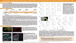 科学海报Generation of Functional 3D Spinal Cord Organoids from Human Pluripotent Stem Cells
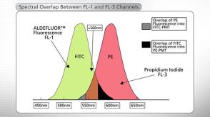

 沪公网安备31010102008431号