Kim MY et al. (MAR 2017)
Oncology letters 13 3 1767--1774
Accumulation of low-dose BIX01294 promotes metastatic potential of U251 glioblastoma cells.
BIX01294 (Bix) is known to be a euchromatic histone-lysine N-methyltransferase 2 inhibitor and treatment with Bix suppresses cancer cell survival and proliferation. In the present study,it was observed that sequential treatment with low-dose Bix notably increases glioblastoma cell migration and metastasis. It was demonstrated that U251 cells sequentially treated with low-dose Bix exhibited induced characteristic changes in critical epithelial-mesenchymal transition (EMT) markers,including E-cadherin,N-cadherin,β-catenin and zinc finger protein SNAI2. Notably,sequential treatment with Bix also increased the expression of cancer stem cell-associated markers,including sex determining region Y-box 2,octamer-binding transcription factor 4 and cluster of differentiation 133. Neurosphere formation was significantly enhanced in cells sequentially treated with Bix,compared with control cells (control: P=0.011; single treatment of Bix,P=0.045). The results of the present study suggest that accumulation of low-dose Bix enhanced the migration and metastatic potential of glioblastoma cells by regulating EMT-associated gene expression,which may be the cause of the altered properties of glioblastoma stem cells.
View Publication
产品类型:
产品号#:
05750
产品名:
NeuroCult™ NS-A 基础培养基(人)
Stipcevic T et al. (DEC 2013)
Acta Neurologica Belgica 113 4 501--506
Stimulation of adult neural stem cells with a novel glycolipid biosurfactant
Glycolipids are amphipathic molecules which are highly expressed on cell membranes in skin and brain where they mediate several key cellular processes. Neural stem cells are defined as undifferentiated,proliferative,multipotential cells with extensive self-renewal and are responsive to brain injury. Di-rhamnolipid: α-L-rhamnopyranosyl-(1-2)α-L-rhamnopyranosyl-3-hydroxydecanoyl-3-hydroxydecanoic acid,also referred to as di-rhamnolipid BAC-3,is a glycolipid isolated from the bacteria Pseudomonas aeruginosa. In the previous studies,di-rhamnolipid enhanced dermal tissue healing and regeneration. The present study provides the first assessment of di-rhamnolipid,and glycolipid biosurfactants in general,on the nervous system. Treatment of neural stem cells isolated from the lateral ventricle of adult mice and cultured in defined media containing growth factors at 0.5 and 1 μg/ml of di-rhamnolipid increased the number of neurospheres (2.7- and 2.8-fold,respectively) compared to controls and this effect remained even after passaging in the absence of di-rhamnolipid. In addition,neural stem cells treated with di-rhamnolipid at 50 and 100 μg/ml in defined media supplemented with fetal calf serum and without growth factors exhibited increased cell viability,indicating an interaction between di-rhamnolipid and serum components in the regulation of neural stem cells and neuroprogenitors. Intracerebroventricular administration of di-rhamnolipid at 300 and 120 ng/day increased the number of neurospheres (1.3- and 1.63-fold,respectively) that could be derived from the anterior lateral ventricles of adult mice. These results indicate that di-rhamnolipid stimulates proliferation of neural stem cells and increases their endogenous pools which may have therapeutic potential in managing neurodegenerative or neuropsychiatric disorders and promoting nervous tissue regeneration following injury.
View Publication
产品类型:
产品号#:
05700
产品名:
NeuroCult™ 基础培养基(小鼠和大鼠)
Xu G et al. (MAY 2013)
Neuroscience 238 195--208
Functional analysis of platelet-derived growth factor receptor-β in neural stem/progenitor cells
Activation of neural stem/progenitor cells (NSPCs) is a potential therapeutic strategy of neurological disorders. In this study,NSPCs of subventricular zone were isolated and cultured from platelet-derived growth factor-β-receptor-knockout (PDGFR-β(-/-)) mice of postnatal day 1 (P1) and P28,and the roles of PDGFR-β were examined in these cells. In PDGFR-β-preserving control NSPCs,stem cell activities,such as numbers and diameters of secondary neurospheres,cell proliferation and survival rates,were significantly higher in P1 NSPCs than those in P28 NSPCs. In PDGFR-β(-/-) NSPCs,most of these parameters were decreased as compared with age-matched controls. Among them,the decrease of secondary neurosphere formation was most striking in P1 and P28 PDGFR-β(-/-) NSPCs and in P28 control NSPCs as compared with P1 control NSPCs. PCR-array and following quantitative real-time PCR (qRT-PCR) analyses demonstrated that expressions of fibroblast growth factor-2 (FGF2) and exons IV-IX of brain-derived neurotrophic factor (BDNF) were decreased,and noggin was increased in P1 PDGFR-β(-/-) as compared with P1 controls. Addition of BDNF rescued the number and diameter of secondary neurospheres in P1 PDGFR-β(-/-) NSPCs to similar levels as controls. The expressions of PDGFs and PDGFRs in control NSPCs were increased along with the differentiation-induction,where phosphorylated PDGFR-β was co-localized with neuronal and astrocyte differentiation markers. In controls,the neuronal differentiation was decreased,and the glial differentiation was increased from P1 to P28 NSPCs. Compared with P1 controls,neuronal differentiation was reduced in P1 PDGFR-β(-/-) NSPCs,whereas glial differentiation was comparable between the two genotypes. These results suggest that PDGFR-β signaling is important for the self-renewal and multipotency of NSPCs,particularly in neonatal NSPCs. BDNF,FGF2,and noggin may be involved in the effects of PDGFR-β signaling in these cells. Accordingly,the activation of PDGFR-β in NSPCs may be a novel therapeutic strategy of neurological diseases.
View Publication
Although human induced pluripotent stem cells (hiPSCs) hold great potential for the study of human diseases affecting disparate cell types,they have been underutilized in seeking mechanistic insights into the pathogenesis of congenital craniofacial disorders. Craniofrontonasal syndrome (CFNS) is a rare X-linked disorder caused by mutations in EFNB1 and characterized by craniofacial,skeletal,and neurological anomalies. Heterozygous females are more severely affected than hemizygous males,a phenomenon termed cellular interference that involves mosaicism for EPHRIN-B1 function. Although the mechanistic basis for cellular interference in CFNS has been hypothesized to involve Eph/ephrin-mediated cell segregation,no direct evidence for this has been demonstrated. Here,by generating hiPSCs from CFNS patients,we demonstrate that mosaicism for EPHRIN-B1 expression induced by random X inactivation in heterozygous females results in robust cell segregation in human neuroepithelial cells,thus supplying experimental evidence that Eph/ephrin-mediated cell segregation is relevant to pathogenesis in human CFNS patients.
View Publication
产品类型:
产品号#:
05835
05839
08581
08582
产品名:
STEMdiff™ 神经诱导培养基
STEMdiff™ 神经诱导培养基
STEMdiff™SMADi神经诱导试剂盒
STEMdiff™SMADi神经诱导试剂盒,2套
Rasper M et al. (OCT 2010)
Neuro-oncology 12 10 1024--33
Glioblastoma (GBM) is the most aggressive primary brain tumor and is resistant to all therapeutic regimens. Relapse occurs regularly and might be caused by a poorly characterized tumor stem cell (TSC) subpopulation escaping therapy. We suggest aldehyde dehydrogenase 1 (ALDH1) as a novel stem cell marker in human GBM. Using the neurosphere formation assay as a functional method to identify brain TSCs,we show that high protein levels of ALDH1 facilitate neurosphere formation in established GBM cell lines. Even single ALDH1 positive cells give rise to colonies and neurospheres. Consequently,the inhibition of ALDH1 in vitro decreases both the number of neurospheres and their size. Cell lines without expression of ALDH1 do not form tumor spheroids under the same culturing conditions. High levels of ALDH1 seem to keep tumor cells in an undifferentiated,stem cell-like state indicated by the low expression of beta-III-tubulin. In contrast,ALDH1 inhibition induces premature cellular differentiation and reduces clonogenic capacity. Primary cell cultures obtained from fresh tumor samples approve the established GBM cell line results.
View Publication
产品类型:
产品号#:
01700
01705
01701
01702
产品名:
ALDEFLUOR™ 试剂盒
ALDEFLUOR™ DEAB试剂, 1.5 mM, 1 mL
ALDEFLUOR™检测缓冲液
Mathieu C et al. (AUG 2008)
Molecular and cellular neurosciences 38 4 569--77
Endothelial cell-derived bone morphogenetic proteins control proliferation of neural stem/progenitor cells.
Neurogenesis persists in the adult brain subventricular zone where neural stem/progenitor cells (NSPCs) lie close to brain endothelial cells (BECs). We show in mouse that BECs produce bone morphogenetic proteins (BMPs). Coculture of embryonic and adult NSPCs with BECs activated the canonical BMP/Smad pathway and reduced their proliferation. We demonstrate that coculture with BECs in the presence of EGF and FGF2 induced a reversible cell cycle exit of NSPCs (LeX+) and an increase in the amount of GFAP/LeX-expressing progenitors thought to be stem cells. Levels of the phosphatidylinositol phosphatase PTEN were upregulated in NSPCs after coculture with BECs,or treatment with recombinant BMP4,with a concomitant reduction in Akt phosphorylation. Silencing Smad5 with siRNA or treatment with Noggin,a BMP antagonist,demonstrated that upregulation of PTEN in NSPCs required BMP/Smad signaling and that this pathway regulated cell cycle exit of NSPCs. Therefore,BECs may provide a feedback mechanism to control the proliferation of NSPCs.
View Publication
产品类型:
产品号#:
05700
05701
05702
产品名:
NeuroCult™ 基础培养基(小鼠和大鼠)
NeuroCult™ 扩增添加物(小鼠和大鼠)
NeuroCult™扩增试剂盒(小鼠和大鼠)
Yoshikawa K et al. (FEB 2013)
Biochemical and biophysical research communications 431 1 104--10
Multipotent stem cells are effectively collected from adult human cheek skin.
Skin-derived precursor (SKP) cells are a valuable resource for tissue engineering and regenerative medicine,because they represent multipotent stem cells that differentiate into neural and mesodermal progenies. Previous studies suggest that the stem cell pool decreases with age. Here,we show that human multipotent SKP cells can be efficiently collected from adult cheek/chin skin,even in aged individuals of 70-78years. SKP cells were isolated from 38 skin samples by serum-free sphere culture and examined for the ability to differentiate into neural and mesodermal lineages. The number of spheres obtained from adult facial skin was significantly higher than that of trunk or extremity skin. SKP cells derived from cheek/chin skin exhibited a high ability to differentiate into neural and mesodermal cells relative to those derived from eyelid,trunk,or extremity skin. Furthermore,cheek/chin skin SKP cells were shown to express markers for undifferentiated stem cells,including a high expression level of the Sox9 gene. These results indicate that cheek/chin skin is useful for the recovery of multipotent stem cells for tissue engineering and regenerative therapy.
View Publication
产品类型:
产品号#:
05752
产品名:
NeuroCult™ NS-A 分化试剂盒(人)
Kaur G et al. (JUL 2013)
Journal of Clinical Neuroscience 20 7 1014--1018
Glioblastoma multiforme (GBM) is a grade IV malignant brain tumor with high mortality and has been well known to involve many molecular pathways,including G-protein coupled receptor (GPCR)-mediated signaling (such as epithelial growth factor receptor [EGFR] and platelet derived growth factor receptor [PDGFR]). G protein-coupled receptor kinases (GRK) directly regulate GPCR activity by phosphorylating activated agonist-bound receptors to desensitize signaling and internalize receptors through beta-arrestins. Recent studies in various cancers,including prostate and breast cancer,have highlighted the role of change in GRK expression to oncogenesis and tumor proliferation. In this study,we evaluated the expression of GRK5 in grade II to grade IV glioma specimens using immunohistochemistry and found that GRK5 expression levels are highly correlated with aggressiveness of glioma. We used culture conditions to selectively promote the growth of either glioblastoma cells with stem cell markers (GSC) or differentiated glioblastoma cells (DGC) from fresh GBM specimens. GSC are known to be highly invasive and mobile,and have the capacity to self-renew and are more resistant to chemotherapy and radiation compared to differentiated populations of GBM. We examined the expression of GRK5 in these two sets of culturing conditions for GBM cells and found that GRK5 expression is upregulated in GSC compared to differentiated GBM cells. To better understand the role of GRK5 in GBM-derived stem cells,we created stable GRK5 knockdown and evaluated the proliferation rate. Using an ATP chemiluminescence assay,we show,for the first time,that knocking down the expression of GRK5 decreased the proliferation rate of GSC in contrast to control.
View Publication
产品类型:
产品号#:
05750
05751
产品名:
NeuroCult™ NS-A 基础培养基(人)
NeuroCult™ NS-A 扩增试剂盒(人)
Drago D et al. (SEP 2016)
Journal of neuroinflammation 13 1 232
Metabolic determinants of the immune modulatory function of neural stem cells.
BACKGROUND Neural stem cells (NSCs) display tissue trophic and immune modulatory therapeutic activities after transplantation in central nervous system disorders. The intercellular interplay between stem cells and target immune cells is increased in NSCs exposed to inflammatory cues. Here,we hypothesize that inflammatory cytokine signalling leads to metabolic reprogramming of NSCs regulating some of their immune modulatory effects. METHODS NSC lines were prepared from the subventricular zone (SVZ) of 7-12-week-old mice. Whole secretome-based screening and analysis of intracellular small metabolites was performed in NSCs exposed to cocktails of either Th1-like (IFN-γ,500 U/ml; TNF-α,200 U/ml; IL-1β,100 U/ml) or Th2-like (IL-4,IL-5 and IL-13; 10 ng/ml) inflammatory cytokines for 16 h in vitro. Isotopologues distribution of arginine and downstream metabolites was assessed by liquid chromatography/mass spectrometry in NSCs incubated with U-(13)C6 L-arginine in the presence or absence of Th1 or Th2 cocktails (Th1 NSCs or Th2 NSCs). The expression of arginase I and II was investigated in vitro in Th1 NSCs and Th2 NSCs and in vivo in the SVZ of mice with experimental autoimmune encephalomyelitis,as prototypical model of Th1 cell-driven brain inflammatory disease. The effects of the inflammatory cytokine signalling were studied in NSC-lymph node cells (LNC) co-cultures by flow cytometry-based analysis of cell proliferation following pan-arginase inhibition with N(ω)-hydroxy-nor-arginine (nor-NOHA). RESULTS Cytokine-primed NSCs showed significantly higher anti-proliferative effect in co-cultures vs. control NSCs. Metabolomic analysis of intracellular metabolites revealed alteration of arginine metabolism and increased extracellular arginase I activity in cytokine-primed NSCs. Arginase inhibition by nor-NOHA partly rescued the anti-proliferative effects of cytokine-primed NSCs. CONCLUSIONS Our work underlines the use of metabolic profiling as hypothesis-generating tools that helps unravelling how stem cell-mediated mechanisms of tissue restoration become affected by local inflammatory responses. Among different therapeutic candidates,we identify arginase signalling as novel metabolic determinant of the NSC-to-immune system communication.
View Publication
Badizadegan K et al. (NOV 2014)
AJP: Gastrointestinal and Liver Physiology 307 10 G1002--G1012
Presence of intramucosal neuroglial cells in normal and aganglionic human colon
The enteric nervous system (ENS) is composed of neural crest-derived neurons (also known as ganglion cells) the cell bodies of which are located in the submucosal and myenteric plexuses of the intestinal wall. Intramucosal ganglion cells are known to exist but are rare and often considered ectopic. Also derived from the neural crest are enteric glial cells that populate the ganglia and the associated nerves,as well as the lamina propria of the intestinal mucosa. In Hirschsprung disease (HSCR),ganglion cells are absent from the distal gut because of a failure of neural crest-derived progenitor cells to complete their rostrocaudal migration during embryogenesis. The fate of intramucosal glial cells in human HSCR is essentially unknown. We demonstrate a network of intramucosal cells that exhibit dendritic morphology typical of neurons and glial cells. These dendritic cells are present throughout the human gut and express Tuj1,S100,glial fibrillary acidic protein,CD56,synaptophysin,and calretinin,consistent with mixed or overlapping neuroglial differentiation. The cells are present in aganglionic colon from patients with HSCR,but with an altered immunophenotype. Coexpression of Tuj1 and HNK1 in this cell population supports a neural crest origin. These findings extend and challenge the current understanding of ENS microanatomy and suggest the existence of an intramucosal population of neural crest-derived cells,present in HSCR,with overlapping immunophenotype of neurons and glia. Intramucosal neuroglial cells have not been previously recognized,and their presence in HSCR poses new questions about ENS development and the pathobiology of HSCR that merit further investigation.
View Publication


 EasySep™小鼠TIL(CD45)正选试剂盒





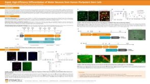 科学海报Rapid, High-Efficiency Differentiation of Motor Neurons from Human Pluripotent Stem Cells
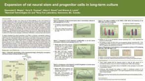 科学海报Expansion of Rat Neural Stem and Progenitor Cells in Long-Term Culture
 沪公网安备31010102008431号