Sensitivity of human embryonic stem cells to different conditions during cryopreservation
Low cell recovery rate of human embryonic stem cells (hESCs) resulting from cryopreservation damages leads to the difficulty in their successful commercialization of clinical applications. Hence in this study,sensitivity of human embryonic stem cells (hESCs) to different cooling rates,ice seeding and cryoprotective agent (CPA) types was compared and cell viability and recovery after cryopreservation under different cooling conditions were assessed. Both extracellular and intracellular ice formation were observed. Reactive oxidative species (ROS) accumulation of hESCs was determined. Cryopreservation of hESCs at 1 °C/min with the ice seeding and at the theoretically predicted optimal cooling rate (TPOCR) led to lower level of intracellular ROS,and prevented irregular and big ice clump formation compared with cryopreservation at 1 °C/min. This strategy further resulted in a significant increase in the hESC recovery when glycerol and 1,2-propanediol were used as the CPAs,but no increase for Me2SO. hESCs after cryopreservation under all the tested conditions still maintained their pluripotency. Our results provide guidance for improving the hESC cryopreservation recovery through the combination of CPA type,cooling rate and ice seeding.
View Publication
产品类型:
产品号#:
05835
05839
产品名:
STEMdiff™ 神经诱导培养基
STEMdiff™ 神经诱导培养基
Kim YY et al. (SEP 2016)
PLOS ONE 11 9 e0163812
Alcohol-Induced Molecular Dysregulation in Human Embryonic Stem Cell-Derived Neural Precursor Cells
Adverse effect of alcohol on neural function has been well documented. Especially,the teratogenic effect of alcohol on neurodevelopment during embryogenesis has been demonstrated in various models,which could be a pathologic basis for fetal alcohol spectrum disorders (FASDs). While the developmental defects from alcohol abuse during gestation have been described,the specific mechanisms by which alcohol mediates these injuries have yet to be determined. Recent studies have shown that alcohol has significant effect on molecular and cellular regulatory mechanisms in embryonic stem cell (ESC) differentiation including genes involved in neural development. To test our hypothesis that alcohol induces molecular alterations during neural differentiation we have derived neural precursor cells from pluripotent human ESCs in the presence or absence of ethanol treatment. Genome-wide transcriptomic profiling identified molecular alterations induced by ethanol exposure during neural differentiation of hESCs into neural rosettes and neural precursor cell populations. The Database for Annotation,Visualization and Integrated Discovery (DAVID) functional analysis on significantly altered genes showed potential ethanol's effect on JAK-STAT signaling pathway,neuroactive ligand-receptor interaction,Toll-like receptor (TLR) signaling pathway,cytokine-cytokine receptor interaction and regulation of autophagy. We have further quantitatively verified ethanol-induced alterations of selected candidate genes. Among verified genes we further examined the expression of P2RX3,which is associated with nociception,a peripheral pain response. We found ethanol significantly reduced the level of P2RX3 in undifferentiated hESCs,but induced the level of P2RX3 mRNA and protein in hESC-derived NPCs. Our result suggests ethanol-induced dysregulation of P2RX3 along with alterations in molecules involved in neural activity such as neuroactive ligand-receptor interaction may be a molecular event associated with alcohol-related peripheral neuropathy of an enhanced nociceptive response.
View Publication
产品类型:
产品号#:
05850
05857
05870
05875
85850
85857
85870
85875
05835
05839
产品名:
mTeSR™1
mTeSR™1
STEMdiff™ 神经诱导培养基
STEMdiff™ 神经诱导培养基
Bershteyn M et al. (MAR 2014)
Nature 507 7490 99--103
Cell-autonomous correction of ring chromosomes in human induced pluripotent stem cells.
Ring chromosomes are structural aberrations commonly associated with birth defects,mental disabilities and growth retardation. Rings form after fusion of the long and short arms of a chromosome,and are sometimes associated with large terminal deletions. Owing to the severity of these large aberrations that can affect multiple contiguous genes,no possible therapeutic strategies for ring chromosome disorders have been proposed. During cell division,ring chromosomes can exhibit unstable behaviour leading to continuous production of aneuploid progeny with low viability and high cellular death rate. The overall consequences of this chromosomal instability have been largely unexplored in experimental model systems. Here we generated human induced pluripotent stem cells (iPSCs) from patient fibroblasts containing ring chromosomes with large deletions and found that reprogrammed cells lost the abnormal chromosome and duplicated the wild-type homologue through the compensatory uniparental disomy (UPD) mechanism. The karyotypically normal iPSCs with isodisomy for the corrected chromosome outgrew co-existing aneuploid populations,enabling rapid and efficient isolation of patient-derived iPSCs devoid of the original chromosomal aberration. Our results suggest a fundamentally different function for cellular reprogramming as a means of /`chromosome therapy/' to reverse combined loss-of-function across many genes in cells with large-scale aberrations involving ring structures. In addition,our work provides an experimentally tractable human cellular system for studying mechanisms of chromosomal number control,which is of critical relevance to human development and disease.
View Publication
产品类型:
产品号#:
05850
05857
05870
05875
85850
85857
85870
85875
产品名:
mTeSR™1
mTeSR™1
Huat T et al. (APR 2015)
International Journal of Molecular Sciences 16 5 9693--9718
MicroRNA Expression Profile of Neural Progenitor-Like Cells Derived from Rat Bone Marrow Mesenchymal Stem Cells under the Influence of IGF-1, bFGF and EGF
Insulin-like growth factor 1 (IGF-1) enhances cellular proliferation and reduces apoptosis during the early differentiation of bone marrow derived mesenchymal stem cells (BMSCs) into neural progenitor-like cells (NPCs) in the presence of epidermal growth factor (EGF) and basic fibroblast growth factor (bFGF). BMSCs were differentiated in three groups of growth factors: (A) EGF + bFGF,(B) EGF + bFGF + IGF-1,and (C) without growth factor. To unravel the molecular mechanisms of the NPCs derivation,microarray analysis using GeneChip miRNA arrays was performed. The profiles were compared among the groups. Annotated microRNA fingerprints (GSE60060) delineated 46 microRNAs temporally up-regulated or down-regulated compared to group C. The expressions of selected microRNAs were validated by real-time PCR. Among the 46 microRNAs,30 were consistently expressed for minimum of two consecutive time intervals. In Group B,only miR-496 was up-regulated and 12 microRNAs,including the let-7 family,miR-1224,miR-125a-3p,miR-214,miR-22,miR-320,miR-708,and miR-93,were down-regulated. Bioinformatics analysis reveals that some of these microRNAs (miR-22,miR-214,miR-125a-3p,miR-320 and let-7 family) are associated with reduction of apoptosis. Here,we summarize the roles of key microRNAs associated with IGF-1 in the differentiation of BMSCs into NPCs. These findings may provide clues to further our understanding of the mechanisms and roles of microRNAs as key regulators of BMSC-derived NPC maintenance.
View Publication
Cells enriched in markers of neural tissue-committed stem cells reside in the bone marrow and are mobilized into the peripheral blood following stroke.
The concept that bone marrow (BM)-derived cells participate in neural regeneration remains highly controversial and the identity of the specific cell type(s) involved remains unknown. We recently reported that the BM contains a highly mobile population of CXCR4+ cells that express mRNA for various markers of early tissue-committed stem cells (TCSCs),including neural TCSCs. Here,we report that these cells not only express neural lineage markers (beta-III-tubulin,Nestin,NeuN,and GFAP),but more importantly form neurospheres in vitro. These neural TCSCs are present in significant amounts in BM harvested from young mice but their abundance and responsiveness to gradients of motomorphogens,such as SDF-1,HGF,and LIF,decreases with age. FACS analysis,combined with analysis of neural markers at the mRNA and protein levels,revealed that these cells reside in the nonhematopoietic CXCR4+/Sca-1+/lin-/CD45 BM mononuclear cell fraction. Neural TCSCs are mobilized into the peripheral-blood following stroke and chemoattracted to the damaged neural tissue in an SDF-1-CXCR4-,HGF-c-Met-,and LIF-LIF-R-dependent manner. Based on these data,we hypothesize that the postnatal BM harbors a nonhematopoietic population of cells that express markers of neural TCSCs that may account for the beneficial effects of BM-derived cells in neural regeneration.
View Publication
产品类型:
产品号#:
05700
05701
05702
05703
05704
产品名:
NeuroCult™ 基础培养基(小鼠和大鼠)
NeuroCult™ 扩增添加物(小鼠和大鼠)
NeuroCult™扩增试剂盒(小鼠和大鼠)
NeuroCult™ 分化添加物(小鼠和大鼠)
NeuroCult™ 分化试剂盒(小鼠和大鼠)
Sareen D et al. (AUG 2014)
Journal of Comparative Neurology 522 12 2707--2728
Human induced pluripotent stem cells are a novel source of neural progenitor cells (iNPCs) that migrate and integrate in the rodent spinal cord
Transplantation of human neural progenitor cells (NPCs) into the brain or spinal cord to replace lost cells,modulate the injury environment,or create a permissive milieu to protect and regenerate host neurons is a promising therapeutic strategy for neurological diseases. Deriving NPCs from human fetal tissue is feasible,although problematic issues include limited sources and ethical concerns. Here we describe a new and abundant source of NPCs derived from human induced pluripotent stem cells (iPSCs). A novel chopping technique was used to transform adherent iPSCs into free-floating spheres that were easy to maintain and were expandable (EZ spheres) (Ebert et al. [2013] Stem Cell Res 10:417–427). These EZ spheres could be differentiated towards NPC spheres with a spinal cord phenotype using a combination of all-trans retinoic acid (RA) and epidermal growth factor (EGF) and fibroblast growth factor-2 (FGF-2) mitogens. Suspension cultures of NPCs derived from human iPSCs or fetal tissue have similar characteristics,although they were not similar when grown as adherent cells. In addition,iPSC-derived NPCs (iNPCs) survived grafting into the spinal cord of athymic nude rats with no signs of overgrowth and with a very similar profile to human fetal-derived NPCs (fNPCs). These results suggest that human iNPCs behave like fNPCs and could thus be a valuable alternative for cellular regenerative therapies of neurological diseases. J. Comp. Neurol. 522:2707–2728,2014. textcopyright 2014 Wiley Periodicals,Inc.
View Publication
产品类型:
产品号#:
05850
05857
05870
05875
85850
85857
85870
85875
产品名:
mTeSR™1
mTeSR™1
Zhou P et al. (MAY 2016)
Biomaterials 87 1--17
Simple and versatile synthetic polydopamine-based surface supports reprogramming of human somatic cells and long-term self-renewal of human pluripotent stem cells under defined conditions
Human pluripotent stem cells (hPSCs) possess great value in the aspect of cellular therapies due to its self-renewal and potential to differentiate into all somatic cell types. A few defined synthetic surfaces such as polymers and adhesive biological materials conjugated substrata were established for the self-renewal of hPSCs. However,none of them was effective in the generation of human induced pluripotent stem cells (hiPSCs) and long-term maintenance of multiple hPSCs,and most of them required complicated manufacturing processes. Polydopamine has good biocompatibility,is able to form a stable film on nearly all solid substrates surface,and can immobilize adhesive biomolecules. In this manuscript,a polydopamine-mediated surface was developed,which not only supported the reprogramming of human somatic cells into hiPSCs under defined conditions,but also sustained the growth of hiPSCs on diverse substrates. Moreover,the proliferation and pluripotency of hPSCs cultured on the surface were comparable to Matrigel for more than 20 passages. Besides,hPSCs were able to differentiate to cardiomyocytes and neural cells on the surface. This polydopamine-based synthetic surface represents a chemically-defined surface extensively applicable both for fundamental research and cell therapies of hPSCs.
View Publication
产品类型:
产品号#:
05833
05835
05839
产品名:
STEMdiff™神经前体细胞培养基
STEMdiff™ 神经诱导培养基
STEMdiff™ 神经诱导培养基
Zhou et al. ( 2013)
Neural Regeneration Research 8 16 1455
Novel nanometer scaffolds regulate the biological behaviors of neural stem cells
Abstract
Ideal tissue-engineered scaffold materials regulate proliferation,apoptosis and differentiation of cells seeded on them by regulating gene expression. In this study,aligned and randomly oriented collagen nanofiber scaffolds were prepared using electronic spinning technology. Their diameters and appearance reached the standards of tissue-engineered nanometer scaffolds. The nanofiber scaffolds were characterized by a high swelling ratio,high porosity and good mechanical properties. The proliferation of spinal cord-derived neural stem cells on novel nanofiber scaffolds was obviously enhanced. The proportions of cells in the S and G2/M phases noticeably increased. Moreover,the proliferation rate of neural stem cells on the aligned collagen nanofiber scaffolds was high. The expression levels of cyclin D1 and cyclin-dependent kinase 2 were increased. Bcl-2 expression was significantly increased,but Bax and caspase-3 gene expressions were obviously decreased. There was no significant difference in the differentiation of neural stem cells into neurons on aligned and randomly oriented collagen nanofiber scaffolds. These results indicate that novel nanofiber scaffolds could promote the proliferation of spinal cord-derived neural stem cells and inhibit apoptosis without inducing differentiation. Nanofiber scaffolds regulate apoptosis and proliferation in neural stem cells by altering gene expression.
Research Highlights
(1) Electronic spinning technology was used to obtain randomly oriented nanofiber membranes and aligned nanofiber membranes. The aligned and randomly oriented collagen nanometer scaffolds were shown to alter the biological behaviors of neural stem cells and induce changes in gene expression.
(2) The effects of the aligned nanofiber membranes on promoting neural stem cell proliferation and on inhibiting apoptosis of neural stem cells were better than those of the randomly oriented nanofiber membranes. Aligned and randomly oriented collagen nanometer scaffolds did not significantly induce apoptosis or differentiation in stem cells.
(3) Aligned and randomly oriented collagen nanometer scaffolds regulated the expression of apoptosis and cell cycle genes in neural stem cells.
Cheng LS et al. (OCT 2015)
Neurogastroenterology and motility : the official journal of the European Gastrointestinal Motility Society 27 10 1509--14
Endoscopic delivery of enteric neural stem cells to treat Hirschsprung disease.
BACKGROUND Transplantation of enteric neural stem cells (ENSC) holds promise as a potential therapy for enteric neuropathies,including Hirschsprung disease. Delivery of transplantable cells via laparotomy has been described,but we propose a novel,minimally invasive endoscopic method of cell delivery. METHODS Enteric neural stem cells for transplantation were cultured from dissociated gut of postnatal donor mice. Twelve recipient mice,including Ednrb(-/-) mice with distal colonic aganglionosis,underwent colonoscopic injection of ENSC under direct vision using a 30-gauge Hamilton needle passed through a rigid cystoureteroscope. Cell engraftment,survival,and neuroglial differentiation were studied 1-4 weeks after the procedure. KEY RESULTS All recipient mice tolerated the procedure without complications and survived to sacrifice. Transplanted cells were found within the colonic wall in 9 of 12 recipient mice with differentiation into enteric neurons and glia. CONCLUSIONS & INFERENCES Endoscopic injection of ENSC is a safe and reliable method for cell delivery,and can be used to deliver a large number of cells to a specific area of disease. This minimally invasive endoscopic approach may prove beneficial to future human applications of cell therapy for neurointestinal disease.
View Publication
产品类型:
产品号#:
05700
05701
05702
产品名:
NeuroCult™ 基础培养基(小鼠和大鼠)
NeuroCult™ 扩增添加物(小鼠和大鼠)
NeuroCult™扩增试剂盒(小鼠和大鼠)
Zhu X et al. (JUL 2010)
Molecular cancer therapeutics 9 7 2131--41
Identification of internalizing human single-chain antibodies targeting brain tumor sphere cells.
Glioblastoma multiforme (GBM) is the most common and aggressive form of primary brain tumor for which there is no curative treatment to date. Resistance to conventional therapies and tumor recurrence pose major challenges to treatment and management of this disease,and therefore new therapeutic strategies need to be developed. Previous studies by other investigators have shown that a subpopulation of GBM cells can grow as neurosphere-like cells when cultured in restrictive medium and exhibits enhanced tumor-initiating ability and resistance to therapy. We report here the identification of internalizing human single-chain antibodies (scFv) targeting GBM tumor sphere cells. We selected a large naive phage antibody display library on the glycosylation-dependent CD133 epitope-positive subpopulation of GBM cells grown as tumor spheres and identified internalizing scFvs that target tumor sphere cells broadly,as well as scFvs that target the CD133-positive subpopulation. These scFvs were found to be efficiently internalized by GBM tumor sphere cells. One scFv GC4 inhibited self-renewal of GBM tumor sphere cells in vitro. We have further developed a full-length human IgG1 based on this scFv,and found that it potently inhibits proliferation of GBM tumor sphere cells and GBM cells grown in regular nonselective medium. Taken together,these results show that internalizing human scFvs targeting brain tumor sphere cells can be readily identified from a phage antibody display library,which could be useful for further development of novel therapies that target subpopulations of GBM cells to combat recurrence and resistance to treatment.
View Publication


 EasySep™小鼠TIL(CD45)正选试剂盒





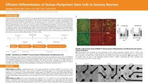 科学海报Efficient Differentiation of Human Pluripotent Stem Cells to Sensory Neurons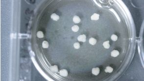

 沪公网安备31010102008431号