Gallo M et al. (JAN 2013)
Cancer Research 73 1 417--427
A Tumorigenic MLL-Homeobox Network in Human Glioblastoma Stem Cells
Glioblastoma growth is driven by cancer cells that have stem cell properties,but molecular determinants of their tumorigenic behavior are poorly defined. In cancer,altered activity of the epigenetic modifiers Polycomb and Trithorax complexes may contribute to the neoplastic phenotype. Here,we provide the first mechanistic insights into the role of the Trithorax protein mixed lineage leukemia (MLL) in maintaining cancer stem cell characteristics in human glioblastoma. We found that MLL directly activates the Homeobox gene HOXA10. In turn,HOXA10 activates a downstream Homeobox network and other genes previously characterized for their role in tumorigenesis. The MLL-Homeobox axis we identified significantly contributes to the tumorigenic potential of glioblastoma stem cells. Our studies suggest a role for MLL in contributing to the epigenetic heterogeneity between tumor-initiating and non-tumor-initiating cells in glioblastoma.
View Publication
产品类型:
产品号#:
05750
产品名:
NeuroCult™ NS-A 基础培养基(人)
Flanagan LA et al. (MAR 2008)
Stem cells (Dayton,Ohio) 26 3 656--65
Unique dielectric properties distinguish stem cells and their differentiated progeny.
The relatively new field of stem cell biology is hampered by a lack of sufficient means to accurately determine the phenotype of cells. Cell-type-specific markers,such as cell surface proteins used for flow cytometry or fluorescence-activated cell sorting,are limited and often recognize multiple members of a stem cell lineage. We sought to develop a complementary approach that would be less dependent on the identification of particular markers for the subpopulations of cells and would instead measure their overall character. We tested whether a microfluidic system using dielectrophoresis (DEP),which induces a frequency-dependent dipole in cells,would be useful for characterizing stem cells and their differentiated progeny. We found that populations of mouse neural stem/precursor cells (NSPCs),differentiated neurons,and differentiated astrocytes had different dielectric properties revealed by DEP. By isolating NSPCs from developmental ages at which they are more likely to generate neurons,or astrocytes,we were able to show that a shift in dielectric property reflecting their fate bias precedes detectable marker expression in these cells and identifies specific progenitor populations. In addition,experimental data and mathematical modeling suggest that DEP curve parameters can indicate cell heterogeneity in mixed cultures. These findings provide evidence for a whole cell property that reflects stem cell fate bias and establish DEP as a tool with unique capabilities for interrogating,characterizing,and sorting stem cells.
View Publication
产品类型:
产品号#:
05707
产品名:
NeuroCult™化学解离试剂盒(小鼠)
Lavasani M et al. (APR 2014)
The Journal of clinical investigation 124 4 1745--56
Human muscle-derived stem/progenitor cells promote functional murine peripheral nerve regeneration.
Peripheral nerve injuries and neuropathies lead to profound functional deficits. Here,we have demonstrated that muscle-derived stem/progenitor cells (MDSPCs) isolated from adult human skeletal muscle (hMDSPCs) can adopt neuronal and glial phenotypes in vitro and ameliorate a critical-sized sciatic nerve injury and its associated defects in a murine model. Transplanted hMDSPCs surrounded the axonal growth cone,while hMDSPCs infiltrating the regenerating nerve differentiated into myelinating Schwann cells. Engraftment of hMDSPCs into the area of the damaged nerve promoted axonal regeneration,which led to functional recovery as measured by sustained gait improvement. Furthermore,no adverse effects were observed in these animals up to 18 months after transplantation. Following hMDSPC therapy,gastrocnemius muscles from mice exhibited substantially less muscle atrophy,an increase in muscle mass after denervation,and reorganization of motor endplates at the postsynaptic sites compared with those from PBS-treated mice. Evaluation of nerve defects in animals transplanted with vehicle-only or myoblast-like cells did not reveal histological or functional recovery. These data demonstrate the efficacy of hMDSPC-based therapy for peripheral nerve injury and suggest that hMDSPC transplantation has potential to be translated for use in human neuropathies.
View Publication
产品类型:
产品号#:
05750
05751
产品名:
NeuroCult™ NS-A 基础培养基(人)
NeuroCult™ NS-A 扩增试剂盒(人)
Xia G and Ashizawa T (JUN 2015)
Histochemistry and cell biology 143 6 557--64
Dynamic changes of nuclear RNA foci in proliferating DM1 cells.
Nuclear RNA foci are molecular hallmarks of myotonic dystrophy type 1 (DM1). However,no designated study has investigated their formation and changes in proliferating cells. Proliferating cells,as stem cells,consist of an important cellular pool in the human body. The revelation of foci changes in these cells might shed light on the effects of the mutation on these specific cells and tissues. In this study,we used human DM1 iPS-cell-derived neural stem cells (NSCs) as cellular models to investigate the formation and dynamic changes of RNA foci in proliferating cells. Human DM1 NSCs derived from human DM1 iPS cells were cultured under proliferation conditions and nonproliferation conditions following mitomycin C treatment. The dynamic changes of foci during the cell cycle were investigated by fluorescence in situ hybridization. We found RNA foci formed and dissociated during the cell cycle. Nuclear RNA foci were most prominent in number and size just prior to entering mitosis (early prophase). During mitosis,most foci disappeared. After entering interphase,RNA foci accumulated again in the nuclei. After stopping cell dividing by treatment of mitomycin C,the number of nuclear RNA foci increased significantly. In summary,DM1 NSC nuclear RNA foci undergo dynamic changes during cell cycle,and mitosis is a mechanism to decrease foci load in the nuclei,which may explain why dividing cells are less affected by the mutation. The dynamic changes need to be considered when using foci as a marker to monitor the effects of therapeutic drugs.
View Publication
产品类型:
产品号#:
05750
05751
产品名:
NeuroCult™ NS-A 基础培养基(人)
NeuroCult™ NS-A 扩增试剂盒(人)
Relañ et al. (AUG 2013)
PLoS Pathogens 9 8 e1003485
Prion Replication Occurs in Endogenous Adult Neural Stem Cells and Alters Their Neuronal Fate: Involvement of Endogenous Neural Stem Cells in Prion Diseases
Prion diseases are irreversible progressive neurodegenerative diseases,leading to severe incapacity and death. They are characterized in the brain by prion amyloid deposits,vacuolisation,astrocytosis,neuronal degeneration,and by cognitive,behavioural and physical impairments. There is no treatment for these disorders and stem cell therapy therefore represents an interesting new approach. Gains could not only result from the cell transplantation,but also from the stimulation of endogenous neural stem cells (NSC) or by the combination of both approaches. However,the development of such strategies requires a detailed knowledge of the pathology,particularly concerning the status of the adult neurogenesis and endogenous NSC during the development of the disease. During the past decade,several studies have consistently shown that NSC reside in the adult mammalian central nervous system (CNS) and that adult neurogenesis occurs throughout the adulthood in the subventricular zone of the lateral ventricle or the Dentate Gyrus of the hippocampus. Adult NSC are believed to constitute a reservoir for neuronal replacement during normal cell turnover or after brain injury. However,the activation of this system does not fully compensate the neuronal loss that occurs during neurodegenerative diseases and could even contribute to the disease progression. We investigated here the status of these cells during the development of prion disorders. We were able to show that NSC accumulate and replicate prions. Importantly,this resulted in the alteration of their neuronal fate which then represents a new pathologic event that might underlie the rapid progression of the disease.
View Publication
产品类型:
产品号#:
05700
05701
05702
05715
产品名:
NeuroCult™ 基础培养基(小鼠和大鼠)
NeuroCult™ 扩增添加物(小鼠和大鼠)
NeuroCult™扩增试剂盒(小鼠和大鼠)
NeuroCult™成年中枢神经系统(CNS)组织酶解试剂盒(小鼠和大鼠)
Mizutani E et al. (DEC 2006)
Reproduction (Cambridge,England) 132 6 849--57
Developmental ability of cloned embryos from neural stem cells.
The success rate is generally higher when cloning mice from embryonic stem (ES) cell nuclei than from somatic cell nuclei,suggesting that the embryonic nature or the undifferentiated state of the donor cell increases cloning efficiency. We assessed the developmental ability of cloned embryos derived from cultured neural stem cell (NSC) nuclei and compared the success rate with that of embryos cloned from other donor cells such as differentiated NSCs,cumulus cells,Sertoli cells and ES cells in the mouse. The transfer of two-cell cloned embryos derived from cultured NSC nuclei into surrogate mothers produced five live cloned mice. However,the success rate (0.5%) was higher in embryos cloned from cultured NSC nuclei than from differentiated NSCs (0%),but lower than that obtained by cloning mice from other cell nuclei (2.2-3.5%). Although the in vitro developmental potential to the two-cell stage of the cloned embryos derived from NSC nuclei (73%) was similar to that of the cloned embryos derived from other somatic cell nuclei (e.g.,85% in Sertoli cells and 75% in cumulus cells),the developmental rate to the morula-blastocyst stage was only 7%. This rate is remarkably lower than that produced from other somatic cells (e.g.,50% in Sertoli cells and 54% in cumulus cells). These results indicate that the undifferentiated state of neural cells does not enhance the cloning efficiency in mice and that the arrest point for in vitro development of cloned embryos depends on the donor cell type.
View Publication
产品类型:
产品号#:
05700
05701
05702
05703
05704
产品名:
NeuroCult™ 基础培养基(小鼠和大鼠)
NeuroCult™ 扩增添加物(小鼠和大鼠)
NeuroCult™扩增试剂盒(小鼠和大鼠)
NeuroCult™ 分化添加物(小鼠和大鼠)
NeuroCult™ 分化试剂盒(小鼠和大鼠)
Ito N et al. (APR 2016)
Disease models & mechanisms 9 4 451--462
Decreased N-TAF1 expression in X-linked dystonia-parkinsonism patient-specific neural stem cells.
X-linked dystonia-parkinsonism (XDP) is a hereditary neurodegenerative disorder involving a progressive loss of striatal medium spiny neurons. The mechanisms underlying neurodegeneration are not known,in part because there have been few cellular models available for studying the disease. The XDP haplotype consists of multiple sequence variations in a region of the X chromosome containingTAF1,a large gene with at least 38 exons,and a multiple transcript system (MTS) composed of five unconventional exons. A previous study identified an XDP-specific insertion of a SINE-VNTR-Alu (SVA)-type retrotransposon in intron 32 ofTAF1,as well as a neural-specific TAF1 isoform,N-TAF1,which showed decreased expression in post-mortem XDP brain compared with control tissue. Here,we generated XDP patient and control fibroblasts and induced pluripotent stem cells (iPSCs) in order to further probe cellular defects associated with this disease. As initial validation of the model,we compared expression ofTAF1and MTS transcripts in XDP versus control fibroblasts and iPSC-derived neural stem cells (NSCs). Compared with control cells,XDP fibroblasts exhibited decreased expression ofTAF1transcript fragments derived from exons 32-36,a region spanning the SVA insertion site. N-TAF1,which incorporates an alternative exon (exon 34'),was not expressed in fibroblasts,but was detectable in iPSC-differentiated NSCs at levels that were ∼threefold lower in XDP cells than in controls. These results support the previous findings that N-TAF1 expression is impaired in XDP,but additionally indicate that this aberrant transcription might occur in neural cells at relatively early stages of development that precede neurodegeneration.
View Publication
产品类型:
产品号#:
产品名:
Paulsen BdS et al. (APR 2014)
Schizophrenia Research 154 1-3 30--35
Valproate reverts zinc and potassium imbalance in schizophrenia-derived reprogrammed cells
Schizophrenia has been considered a devastating clinical syndrome rather than a single disease. Nevertheless,the mechanisms behind the onset of schizophrenia have been only partially elucidated. Several studies propose that levels of trace elements are abnormal in schizophrenia; however,conflicting data generated from different biological sources prevent conclusions being drawn. In this work,we used synchrotron radiation X-ray microfluorescence spectroscopy to compare trace element levels in neural progenitor cells (NPCs) derived from two clones of induced pluripotent stem cell lines of a clozapine-resistant schizophrenic patient and two controls. Our data reveal the presence of elevated levels of potassium and zinc in schizophrenic NPCs. Neural cells treated with valproate,an adjunctive medication for schizophrenia,brought potassium and zinc content back to control levels. These results expand the understanding of atomic element imbalance related to schizophrenia and may provide novel insights for the screening of drugs to treat mental disorders. ?? 2014 Elsevier B.V.
View Publication
产品类型:
产品号#:
05850
05857
05870
05875
85850
85857
85870
85875
产品名:
mTeSR™1
mTeSR™1
Kang HS et al. (DEC 2015)
Journal of Korean medical science 30 12 1764--76
Advanced Properties of Urine Derived Stem Cells Compared to Adipose Tissue Derived Stem Cells in Terms of Cell Proliferation, Immune Modulation and Multi Differentiation.
Adipose tissue stem cells (ADSCs) would be an attractive autologous cell source. However,ADSCs require invasive procedures,and has potential complications. Recently,urine stem cells (USCs) have been proposed as an alternative stem cell source. In this study,we compared USCs and ADSCs collected from the same patients on stem cell characteristics and capacity to differentiate into various cell lineages to provide a useful guideline for selecting the appropriate type of cell source for use in clinical application. The urine samples were collected via urethral catheterization,and adipose tissue was obtained from subcutaneous fat tissue during elective laparoscopic kidney surgery from the same patient (n = 10). Both cells were plated for primary culture. Cell proliferation,colony formation,cell surface markers,immune modulation,chromosome stability and multi-lineage differentiation were analyzed for each USCs and ADSCs at cell passage 3,5,and 7. USCs showed high cell proliferation rate,enhanced colony forming ability,strong positive for stem cell markers expression,high efficiency for inhibition of immune cell activation compared to ADSCs at cell passage 3,5,and 7. In chromosome stability analysis,both cells showed normal karyotype through all passages. In analysis of multi-lineage capability,USCs showed higher myogenic,neurogenic,and endogenic differentiation rate,and lower osteogenic,adipogenic,and chondrogenic differentiation rate compared to ADSCs. Therefore,we expect that USC can be an alternative autologous stem cell source for muscle,neuron and endothelial tissue reconstruction instead of ADSCs.
View Publication
产品类型:
产品号#:
05752
产品名:
NeuroCult™ NS-A 分化试剂盒(人)
Nakamura H et al. (OCT 2013)
Herpesviridae 4 1 2
Human cytomegalovirus induces apoptosis in neural stem/progenitor cells derived from induced pluripotent stem cells by generating mitochondrial dysfunction and endoplasmic reticulum stress
BACKGROUND Congenital human cytomegalovirus (HCMV) infection,a leading cause of birth defects,is most often manifested as neurological disorders. The pathogenesis of HCMV-induced neurological disorders is,however,largely unresolved,primarily because of limited availability of model systems to analyze the effects of HCMV infection on neural cells. METHODS An induced pluripotent stem cell (iPSC) line was established from the human fibroblast line MRC5 by introducing the Yamanaka's four factors and then induced to differentiate into neural stem/progenitor cells (NSPCs) by dual inhibition of the SMAD signaling pathway using Noggin and SB-431542. RESULTS iPSC-derived NSPCs (NSPC/iPSCs) were susceptible to HCMV infection and allowed the expression of both early and late viral gene products. HCMV-infected NSPC/iPSCs underwent apoptosis with the activation of caspase-3 and -9 as well as positive staining by the terminal deoxynucleotidyl transferase-mediated dUTP nick-end labeling (TUNEL). Cytochrome c release from mitochondria to cytosol was observed in these cells,indicating the involvement of mitochondrial dysfunction in their apoptosis. In addition,phosphorylation of proteins involved in the unfolded protein response (UPR),such as PKR-like eukaryotic initiation factor 2a kinase (PERK),c-Jun NH2-terminal kinase (JNK),inositol-requiring enzyme 1 (IRE1),and the alpha subunit of eukaryotic initiation factor 2 (eIF2$$) was observed in HCMV-infected NSPC/iPSCs. These results,coupled with the finding of increased expression of mRNA encoding the C/EBP-homologous protein (CHOP) and the detection of a spliced form of X-box binding protein 1 (XBP1) mRNA,suggest that endoplasmic reticulum (ER) stress is also involved in HCMV-induced apoptosis of these cells. CONCLUSIONS iPSC-derived NSPCs are thought to be a useful model to study HCMV neuropathogenesis and to analyze the mechanisms of HCMV-induced apoptosis in neural cells.
View Publication
Scalable Production of Glioblastoma Tumor-initiating Cells in 3 Dimension Thermoreversible Hydrogels.
There is growing interest in developing drugs that specifically target glioblastoma tumor-initiating cells (TICs). Current cell culture methods,however,cannot cost-effectively produce the large numbers of glioblastoma TICs required for drug discovery and development. In this paper we report a new method that encapsulates patient-derived primary glioblastoma TICs and grows them in 3 dimension thermoreversible hydrogels. Our method allows long-term culture (˜50 days,10 passages tested,accumulative ˜>10(10)-fold expansion) with both high growth rate (˜20-fold expansion/7 days) and high volumetric yield (˜2.0%A-%10(7)%cells/ml) without the loss of stemness. The scalable method can be used to produce sufficient,affordable glioblastoma TICs for drug discovery.
View Publication

 EasySep™小鼠TIL(CD45)正选试剂盒


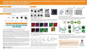 科学海报A Human Pluripotent Stem Cell-Derived Organoid Model for Recapitulation of Central Nervous System (CNS) Barrier and Fluid Secretion Functions of the Choroid Plexus
 沪公网安备31010102008431号