Booth L et al. (AUG 2015)
Journal of cellular physiology 230 8 1982--98
OSU-03012 and Viagra Treatment Inhibits the Activity of Multiple Chaperone Proteins and Disrupts the Blood-Brain Barrier: Implications for Anti-Cancer Therapies.
We examined the interaction between OSU-03012 (also called AR-12) with phosphodiesterase 5 (PDE5) inhibitors to determine the role of the chaperone glucose-regulated protein (GRP78)/BiP/HSPA5 in the cellular response. Sildenafil (Viagra) interacted in a greater than additive fashion with OSU-03012 to kill stem-like GBM cells. Treatment of cells with OSU-03012/sildenafil: abolished the expression of multiple oncogenic growth factor receptors and plasma membrane drug efflux pumps and caused a rapid degradation of GRP78 and other HSP70 and HSP90 family chaperone proteins. Decreased expression of plasma membrane receptors and drug efflux pumps was dependent upon enhanced PERK-eIF2α-ATF4-CHOP signaling and was blocked by GRP78 over-expression. In vivo OSU-03012/sildenafil was more efficacious than treatment with celecoxib and sildenafil at killing tumor cells without damaging normal tissues and in parallel reduced expression of ABCB1 and ABCG2 in the normal brain. The combination of OSU-03012/sildenafil synergized with low concentrations of sorafenib to kill tumor cells,and with lapatinib to kill ERBB1 over-expressing tumor cells. In multiplex assays on plasma and human tumor tissue from an OSU-03012/sildenafil treated mouse,we noted a profound reduction in uPA signaling and identified FGF and JAK1/2 as response biomarkers for potentially suppressing the killing response. Inhibition of FGFR signaling and to a lesser extent JAK1/2 signaling profoundly enhanced OSU-03012/sildenafil lethality.
View Publication
Zhu TS et al. (SEP 2011)
Cancer research 71 18 6061--72
Endothelial cells create a stem cell niche in glioblastoma by providing NOTCH ligands that nurture self-renewal of cancer stem-like cells.
One important function of endothelial cells in glioblastoma multiforme (GBM) is to create a niche that helps promote self-renewal of cancer stem-like cells (CSLC). However,the underlying molecular mechanism for this endothelial function is not known. Since activation of NOTCH signaling has been found to be required for propagation of GBM CSLCs,we hypothesized that the GBM endothelium may provide the source of NOTCH ligands. Here,we report a corroboration of this concept with a demonstration that NOTCH ligands are expressed in endothelial cells adjacent to NESTIN and NOTCH receptor-positive cancer cells in primary GBMs. Coculturing human brain microvascular endothelial cells (hBMEC) or NOTCH ligand with GBM neurospheres promoted GBM cell growth and increased CSLC self-renewal. Notably,RNAi-mediated knockdown of NOTCH ligands in hBMECs abrogated their ability to induce CSLC self-renewal and GBM tumor growth,both in vitro and in vivo. Thus,our findings establish that NOTCH activation in GBM CSLCs is driven by juxtacrine signaling between tumor cells and their surrounding endothelial cells in the tumor microenvironment,suggesting that targeting both CSLCs and their niche may provide a novel strategy to deplete CSLCs and improve GBM treatment.
View Publication
Comparative expression study of the endo-G protein coupled receptor (GPCR) repertoire in human glioblastoma cancer stem-like cells, U87-MG cells and non malignant cells of neural origin unveils new potential therapeutic targets.
Glioblastomas (GBMs) are highly aggressive,invasive brain tumors with bad prognosis and unmet medical need. These tumors are heterogeneous being constituted by a variety of cells in different states of differentiation. Among these,cells endowed with stem properties,tumor initiating/propagating properties and particularly resistant to chemo- and radiotherapies are designed as the real culprits for tumor maintenance and relapse after treatment. These cells,termed cancer stem-like cells,have been designed as prominent targets for new and more efficient cancer therapies. G-protein coupled receptors (GPCRs),a family of membrane receptors,play a prominent role in cell signaling,cell communication and crosstalk with the microenvironment. Their role in cancer has been highlighted but remains largely unexplored. Here,we report a descriptive study of the differential expression of the endo-GPCR repertoire in human glioblastoma cancer stem-like cells (GSCs),U-87 MG cells,human astrocytes and fetal neural stem cells (f-NSCs). The endo-GPCR transcriptome has been studied using Taqman Low Density Arrays. Of the 356 GPCRs investigated,138 were retained for comparative studies between the different cell types. At the transcriptomic level,eight GPCRs were specifically expressed/overexpressed in GSCs. Seventeen GPCRs appeared specifically expressed in cells with stem properties (GSCs and f-NSCs). Results of GPCR expression at the protein level using mass spectrometry and proteomic analysis are also presented. The comparative GPCR expression study presented here gives clues for new pathways specifically used by GSCs and unveils novel potential therapeutic targets.
View Publication
Ostrakhovitch EA et al. (DEC 2012)
Archives of biochemistry and biophysics 528 1 21--31
Directed differentiation of embryonic P19 cells and neural stem cells into neural lineage on conducting PEDOT-PEG and ITO glass substrates.
Differentiation of pluripotent and lineage restricted stem cells such as neural stem cells (NSCs) was studied on conducting substrates of various nature without perturbation of the genome with exogenous genetic material or chemical stimuli. Primary mouse adult neural stem cells (NSCs) and P19 pluripotent embryonal (P19 EC) carcinoma cells were used. Expression levels of neuronal markers β-III-tubulin and neurofilament were evaluated by immunochemistry and flow cytometry. It was shown that the ability of the substrate to induce differentiation directly correlated with its conductivity. Conducting substrates (conducting oxides or doped pi-conjugated organic polymers) with different morphology,structure,and conductivity mechanisms all promoted differentiation of NSC and P19 cells into neuronal lineage to a similar degree without use of additional factors such as poly-L-ornithine coating or retinoic acid,as verified by their morphology and upregulation of the neuronal markers but not astrocyte marker GFAP. However,substrates with low conductance below ca. 10(-4) S cm(-2) did not show this ability. Morphology of differentiating cells was visualized by atomic force microscopy. NSCs cells increased β-III-tubulin expression by 95% and P19 cells by over 30%. Our results suggest that the substrate conductivity is a key factor governing the cell fate. Differentiation of P19 cells into neuronal lineage on conducting substrates was attributed to downregualtion of Akt signaling pathway and increase in expression of dual oxidase 1 (DUOX 1).
View Publication
Bain G et al. (APR 1995)
Developmental biology 168 2 342--57
Embryonic stem cells express neuronal properties in vitro.
Mouse embryonic stem (ES) cells cultured as aggregates and exposed to retinoic acid are induced to express multiple phenotypes normally associated with neurons. A large percentage of treated aggregates produce a rich neuritic outgrowth. Dissociating the induced aggregates with trypsin and plating the cells as a monolayer results in cultures in which a sizable percentage of the cells have a neuronal appearance. These neuron-like cells express class III beta-tubulin and the neurofilament M subunit. Induced cultures express transcripts for neural-associated genes including the neurofilament L subunit,glutamate receptor subunits,the transcription factor Brn-3,and GFAP. Levels of neurofilament L and GAD67 and GAD65 transcripts rise dramatically upon induction. Physiological studies show that the neuron-like cells generate action potentials and express TTX-sensitive sodium channels,as well as voltage-gated potassium channels and calcium channels. We conclude that a complex system of neuronal gene expression can be activated in cultured ES cells. This system should be favorable for investigating some of the mechanisms that regulate neuronal differentiation.
View Publication
产品类型:
产品号#:
06902
06952
00321
00322
00323
00324
00325
产品名:
Badr CE et al. (MAY 2013)
JNCI: Journal of the National Cancer Institute 105 9 643--653
Targeting Cancer Cells With the Natural Compound Obtusaquinone
BACKGROUND Tumor cells present high levels of oxidative stress. Cancer therapeutics exploiting such biochemical changes by increasing reactive oxygen species (ROS) production or decreasing intracellular ROS scavengers could provide a powerful treatment strategy. METHODS To test the effect of our compound,obtusaquinone (OBT),we used several cell viability assays on seven different glioblastoma (GBM) cell lines and primary cells and on 12 different cell lines representing various cancer types in culture as well as on subcutaneous (n = 7 mice per group) and two intracranial GBM (n = 6-8 mice per group) and breast cancer (n = 6 mice per group) tumor models in vivo. Immunoblotting,immunostaining,flow cytometry,and biochemical assays were used to investigate the OBT mechanism of action. Histopathological analysis (n = 2 mice per group) and blood chemistry (n = 2 mice per group) were used to test for any compound-related toxicity. Statistical tests were two-sided. RESULTS OBT induced rapid increase in intracellular ROS levels,downregulation of cellular glutathione levels and increase in its oxidized form,and activation of cellular stress pathways and DNA damage,subsequently leading to apoptosis. Oxidative stress is believed to be the main mechanism through which this compounds targets cancer cells. OBT was well tolerated in mice,slowed tumor growth,and statistically prolonged survival in GBM tumor models. The ratio of median survival in U251 intracranial model in OBT vs control was 1.367 (95% confidence interval [CI] of ratio = 1.031 to 1.367,P = .008). Tumor growth inhibition was also observed in a mouse breast cancer model (average tumor volume per mouse,OBT vs control: 36.3 vs 200.4mm(3),difference = 164.1mm(3),95% CI =72.6 to 255.6mm(3),P = .005). CONCLUSIONS Given its properties and efficacy in cancer killing,our results suggest that OBT is a promising cancer therapeutic.
View Publication


 EasySep™小鼠TIL(CD45)正选试剂盒





 技术公告Identification of ALDH-Expressing Cancer Stem Cells Using ALDEFLUOR™
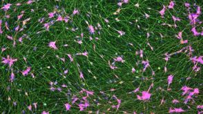 实验方案How to Culture Human Pluripotent Stem Cell (hPSC)-Derived Induced Forebrain Neurons for MEA Analysis Using the Maestro MEA™ System
 研究综述Neural Stem Cells: Identification, Function, Culture, and Isolation
 挂图Derivation and Applications of Human Pluripotent Stem Cells Overview of the derivation of human embryonic stem cells (hESCs) and induced pluripotent stem cells (iPSCs)发布日期: 11/26/2020
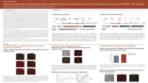 科学海报Generation, Maintenance and Cryopreservation of Neural Progenitor Cells Derived from Human Pluripotent Stem Cells Using the STEMdiff™ Neural System

 沪公网安备31010102008431号