Rahman M et al. (SEP 2013)
Future Oncology 9 9 1389--1396
Controlling tumor invasion: bevacizumab and BMP4 for glioblastoma
AIM Bevacizumab has been reported to result in increased tumor invasion when used to treat malignant glioma. We hypothesized that BMP4 would prevent diffuse tumor infiltration induced by bevacizumab for malignant glioma in a xenograft model. METHODS Human glioblastoma (GBM) tumor cells were implanted in the striatum of immunocompromised mice. The animals were treated with bevacizumab and BMP4. Tumor growth and invasion were measured. RESULTS The bevacizumab-treated mice had increased survival compared with control animals (p = 0.02). BMP4 alone did not result in improved survival (p = 1.0). The bevacizumab (p = 0.006) and bevacizumab plus BMP4 (p = 0.006) groups demonstrated significantly decreased total tumor size compared with control. Tumor invasion was significantly decreased in the bevacizumab (p = 0.005),BMP4 (p = 0.04) alone and bevacizumab plus BMP4 (p = 0.002) groups compared with control. No synergistic effect between bevacizumab and BMP4 was observed. CONCLUSION Bevacizumab treatment did not result in diffuse infiltration of human GBM in a mouse xenograft model. BMP4 did have an independent favorable effect on GBM that was not synergistic with bevacizumab treatment.
View Publication
Er JC et al. (FEB 2015)
Angewandte Chemie - International Edition 54 8 2442--2446
Neuo: A fluorescent chemical probe for live neuron labeling
To address existing limitations in live neuron imaging,we have developed NeuO,a novel cell-permeable fluorescent probe with an unprecedented ability to label and image live neurons selectively over other cells in the brain. NeuO enables robust live neuron imaging and isolation in vivo and in vitro across species; its versatility and ease of use sets the basis for its development in a myriad of neuronal targeting applications.
View Publication
Chakrabarti L et al. (JAN 2012)
Frontiers in oncology 2 82
Reversible adaptive plasticity: a mechanism for neuroblastoma cell heterogeneity and chemo-resistance.
We describe a novel form of tumor cell plasticity characterized by reversible adaptive plasticity in murine and human neuroblastoma. Two cellular phenotypes were defined by their ability to exhibit adhered,anchorage dependent (AD) or sphere forming,anchorage independent (AI) growth. The tumor cells could transition back and forth between the two phenotypes and the transition was dependent on the culture conditions. Both cell phenotypes exhibited stem-like features such as expression of nestin,self-renewal capacity,and mesenchymal differentiation potential. The AI tumorspheres were found to be more resistant to chemotherapy and proliferated slower in vitro compared to the AD cells. Identification of specific molecular markers like MAP2,β-catenin,and PDGFRβ enabled us to characterize and observe both phenotypes in established mouse tumors. Irrespective of the phenotype originally implanted in mice,tumors grown in vivo show phenotypic heterogeneity in molecular marker signatures and are indistinguishable in growth or histologic appearance. Similar molecular marker heterogeneity was demonstrated in primary human tumor specimens. Chemotherapy or growth factor receptor inhibition slowed tumor growth in mice and promoted initial loss of AD or AI heterogeneity,respectively. Simultaneous targeting of both phenotypes led to further tumor growth delay with emergence of new unique phenotypes. Our results demonstrate that neuroblastoma cells are plastic,dynamic,and may optimize their ability to survive by changing their phenotype. Phenotypic switching appears to be an adaptive mechanism to unfavorable selection pressure and could explain the phenotypic and functional heterogeneity of neuroblastoma.
View Publication
产品类型:
产品号#:
05700
05701
05702
产品名:
NeuroCult™ 基础培养基(小鼠和大鼠)
NeuroCult™ 扩增添加物(小鼠和大鼠)
NeuroCult™扩增试剂盒(小鼠和大鼠)
Di Cristofori A et al. (JUL 2015)
Oncotarget 6 19 17514--31
The vacuolar H+ ATPase is a novel therapeutic target for glioblastoma.
The vacuolar H+ ATPase (V-ATPase) is a proton pump responsible for acidification of cellular microenvironments,an activity exploited by tumors to survive,proliferate and resist to therapy. Despite few observations,the role of V-ATPase in human tumorigenesis remains unclear.We investigated the expression of ATP6V0C,ATP6V0A2,encoding two subunits belonging to the V-ATPase V0 sector and ATP6V1C,ATP6V1G1,ATPT6V1G2,ATP6V1G3,which are part of the V1 sector,in series of adult gliomas and in cancer stem cell-enriched neurospheres isolated from glioblastoma (GBM) patients. ATP6V1G1 expression resulted significantly upregulated in tissues of patients with GBM and correlated with shorter patients' overall survival independent of clinical variables.ATP6V1G1 knockdown in GBM neurospheres hampered sphere-forming ability,induced cell death,and decreased matrix invasion,a phenotype not observed in GBM monolayer cultures. Treating GBM organotypic cultures or neurospheres with the selective V-ATPase inhibitor bafilomycin A1 reproduced the effects of ATP6V1G1 siRNA and strongly suppressed expression of the stem cell markers Nestin,CD133 and transcription factors SALL2 and POU3F2 in neurospheres.These data point to ATP6V1G1 as a novel marker of poor prognosis in GBM patients and identify V-ATPase inhibition as an innovative therapeutic strategy for GBM.
View Publication
产品类型:
产品号#:
05750
05751
产品名:
NeuroCult™ NS-A 基础培养基(人)
NeuroCult™ NS-A 扩增试剂盒(人)
Villa GR et al. (NOV 2016)
Cancer cell 30 5 683--693
An LXR-Cholesterol Axis Creates a Metabolic Co-Dependency for Brain Cancers.
Small-molecule inhibitors targeting growth factor receptors have failed to show efficacy for brain cancers,potentially due to their inability to achieve sufficient drug levels in the CNS. Targeting non-oncogene tumor co-dependencies provides an alternative approach,particularly if drugs with high brain penetration can be identified. Here we demonstrate that the highly lethal brain cancer glioblastoma (GBM) is remarkably dependent on cholesterol for survival,rendering these tumors sensitive to Liver X receptor (LXR) agonist-dependent cell death. We show that LXR-623,a clinically viable,highly brain-penetrant LXRα-partial/LXRβ-full agonist selectively kills GBM cells in an LXRβ- and cholesterol-dependent fashion,causing tumor regression and prolonged survival in mouse models. Thus,a metabolic co-dependency provides a pharmacological means to kill growth factor-activated cancers in the CNS.
View Publication
Polyglutamine Disease Modeling: Epitope Based Screen for Homologous Recombination using CRISPR/Cas9 System.
We have previously reported the genetic correction of Huntington's disease (HD) patient-derived induced pluripotent stem cells using traditional homologous recombination (HR) approaches. To extend this work,we have adopted a CRISPR-based genome editing approach to improve the efficiency of recombination in order to generate allelic isogenic HD models in human cells. Incorporation of a rapid antibody-based screening approach to measure recombination provides a powerful method to determine relative efficiency of genome editing for modeling polyglutamine diseases or understanding factors that modulate CRISPR/Cas9 HR.
View Publication

 EasySep™小鼠TIL(CD45)正选试剂盒


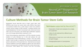 产品手册NeuroCult™: Reagents for Brain Tumor Stem Cell Research
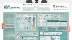 挂图Building Three-Dimensional Human Brain Organoids Overview of brain organogenesis and the applications of brain organoids in studying the development and maturation of the nervous system发布日期: 05/11/2018


 沪公网安备31010102008431号