J. Yun et al. (Jan 2023)
Nature communications 14 156
Senescent cells perturb intestinal stem cell differentiation through Ptk7 induced noncanonical Wnt and YAP signaling.
Cellular senescence and the senescence-associated secretory phenotype (SASP) are implicated in aging and age-related disease,and SASP-related inflammation is thought to contribute to tissue dysfunction in aging and diseased animals. However,whether and how SASP factors influence the regenerative capacity of tissues remains unclear. Here,using intestinal organoids as a model of tissue regeneration,we show that SASP factors released by senescent fibroblasts deregulate stem cell activity and differentiation and ultimately impair crypt formation. We identify the secreted N-terminal domain of Ptk7 as a key component of the SASP that activates non-canonical Wnt / Ca2+ signaling through FZD7 in intestinal stem cells (ISCs). Changes in cytosolic [Ca2+] elicited by Ptk7 promote nuclear translocation of YAP and induce expression of YAP/TEAD target genes,impairing symmetry breaking and stem cell differentiation. Our study discovers secreted Ptk7 as a factor released by senescent cells and provides insight into the mechanism by which cellular senescence contributes to tissue dysfunction in aging and disease.
View Publication
产品类型:
产品号#:
06005
产品名:
IntestiCult™ 类器官生长培养基 (小鼠)
E. A. Davis et al. (JUN 2018)
Physiological reports 6 12 e13745
Evidence for a direct effect of the autonomic nervous system on intestinal epithelial stem cell proliferation.
The sympathetic (SNS) and parasympathetic (PNS) branches of the autonomic nervous system have been implicated in the modulation of the renewal of many tissues,including the intestinal epithelium. However,it is not known whether these mechanisms are direct,requiring an interaction between autonomic neurotransmitters and receptors on proliferating epithelial cells. To evaluate the existence of a molecular framework for a direct effect of the SNS or PNS on intestinal epithelial renewal,we measured gene expression for the main autonomic neurotransmitter receptors in this tissue. We separately evaluated intestinal epithelial regions comprised of the stem,progenitor,and mature cells,which allowed us to investigate the distinct contributions of each cell population to this proposed autonomic effect. Notably,we found that the stem cells expressed the receptors for the SNS-associated alpha2A adrenoreceptor and the PNS-associated muscarinic acetylcholine receptors (M1 and M3). In a separate experiment,we found that the application of norepinephrine or acetylcholine decreases the expression of cyclin D1,a gene necessary for cell cycle progression,in intestinal epithelial organoids compared with controls (P {\textless} 0.05). Together,these results provide evidence of a direct mechanism for the autonomic nervous system influence on intestinal epithelial stem cell proliferation.
View Publication
产品类型:
产品号#:
06005
产品名:
IntestiCult™ 类器官生长培养基 (小鼠)
Y. Sei et al. (MAY 2018)
American journal of physiology. Gastrointestinal and liver physiology
Mature Enteroendocrine Cells Contributes to Basal and Pathological Stem Cell Dynamics in the Small Intestine.
Lgr5-expressing intestinal stem cells (ISCs) maintain continuous and rapid generation of the intestinal epithelium. Here we present evidence that dedifferentiation of committed enteroendocrine cells (EECs) contributes to maintenance of the epithelium under both basal conditions and in response to injury. Lineage tracing studies identified a subset of EECs that reside at +4 position for more than 2 weeks,most of which were BrdU-label-retaining cells. Under basal conditions,cells derived from these EECs grow from the bottom of the crypt to generate intestinal epithelium according to neutral drift kinetics that is consistent with dedifferentiation of mature EECs to ISCs. The lineage tracing of EECs demonstrated reserve stem cell properties in response to radiation-induced injury with the generation of reparative EEC-derived epithelial patches. Finally,the enterochromaffin (EC) cell was the predominant EEC type participating in these stem cell dynamics. These results provide novel insights into the +4 reserve ISC hypothesis,stem cell dynamics of the intestinal epithelium and novel insight in the development of EC-derived small intestinal tumors.
View Publication
Okkelman IA et al. ( 2016)
PloS one 11 12 e0167385
Use of Fluorescence Lifetime Imaging Microscopy (FLIM) as a Timer of Cell Cycle S Phase.
Incorporation of thymidine analogues in replicating DNA,coupled with antibody and fluorophore staining,allows analysis of cell proliferation,but is currently limited to monolayer cultures,fixed cells and end-point assays. We describe a simple microscopy imaging method for live real-time analysis of cell proliferation,S phase progression over several division cycles,effects of anti-proliferative drugs and other applications. It is based on the prominent (˜ 1.7-fold) quenching of fluorescence lifetime of a common cell-permeable nuclear stain,Hoechst 33342 upon the incorporation of 5-bromo-2'-deoxyuridine (BrdU) in genomic DNA and detection by fluorescence lifetime imaging microscopy (FLIM). We show that quantitative and accurate FLIM technique allows high-content,multi-parametric dynamic analyses,far superior to the intensity-based imaging. We demonstrate its uses with monolayer cell cultures,complex 3D tissue models of tumor cell spheroids and intestinal organoids,and in physiological study with metformin treatment.
View Publication
产品类型:
产品号#:
06005
产品名:
IntestiCult™ 类器官生长培养基 (小鼠)
Lindvall C et al. (NOV 2006)
The Journal of biological chemistry 281 46 35081--7
The Wnt signaling receptor Lrp5 is required for mammary ductal stem cell activity and Wnt1-induced tumorigenesis.
Canonical Wnt signaling has emerged as a critical regulatory pathway for stem cells. The association between ectopic activation of Wnt signaling and many different types of human cancer suggests that Wnt ligands can initiate tumor formation through altered regulation of stem cell populations. Here we have shown that mice deficient for the Wnt co-receptor Lrp5 are resistant to Wnt1-induced mammary tumors,which have been shown to be derived from the mammary stem/progenitor cell population. These mice exhibit a profound delay in tumorigenesis that is associated with reduced Wnt1-induced accumulation of mammary progenitor cells. In addition to the tumor resistance phenotype,loss of Lrp5 delays normal mammary development. The ductal trees of 5-week-old Lrp5-/- females have fewer terminal end buds,which are structures critical for juvenile ductal extension presumed to be rich in stem/progenitor cells. Consequently,the mature ductal tree is hypomorphic and does not completely fill the fat pad. Furthermore,Lrp5-/- ductal cells from mature females exhibit little to no stem cell activity in limiting dilution transplants. Finally,we have shown that Lrp5-/- embryos exhibit substantially impaired canonical Wnt signaling in the primitive stem cell compartment of the mammary placodes. These findings suggest that Lrp5-mediated canonical signaling is required for mammary ductal stem cell activity and for tumor development in response to oncogenic Wnt effectors.
View Publication
产品类型:
产品号#:
05601
产品名:
EpiCult™-B 人培养基
Bhushal S et al. ( 2017)
Frontiers in immunology 8 JUN 671
Cell Polarization and Epigenetic Status Shape the Heterogeneous Response to Type III Interferons in Intestinal Epithelial Cells.
Type I and type III interferons (IFNs) are crucial components of the first-line antiviral host response. While specific receptors for both IFN types exist,intracellular signaling shares the same Jak-STAT pathway. Due to its receptor expression,IFN-λ responsiveness is restricted mainly to epithelial cells. Here,we display IFN-stimulated gene induction at the single cell level to comparatively analyze the activities of both IFN types in intestinal epithelial cells and mini-gut organoids. Initially,we noticed that the response to both types of IFNs at low concentrations is based on a single cell decision-making determining the total cell intrinsic antiviral activity. We identified histone deacetylase (HDAC) activity as a crucial restriction factor controlling the cell frequency of IFN-stimulated gene (ISG) induction upon IFN-λ but not IFN-β stimulation. Consistently,HDAC blockade confers antiviral activity to an elsewise non-responding subpopulation. Second,in contrast to the type I IFN system,polarization of intestinal epithelial cells strongly enhances their ability to respond to IFN-λ signaling and raises the kinetics of gene induction. Finally,we show that ISG induction in mini-gut organoids by low amounts of IFN is characterized by a scattered heterogeneous responsiveness of the epithelial cells and HDAC activity fine-tunes exclusively IFN-λ activity. This study provides a comprehensive description of the differential response to type I and type III IFNs and demonstrates that cell polarization in gut epithelial cells specifically increases IFN-λ activity.
View Publication
产品类型:
产品号#:
06005
产品名:
IntestiCult™ 类器官生长培养基 (小鼠)
Yu C et al. ( )
In vivo (Athens,Greece) 25 1 69--76
ALDH activity indicates increased tumorigenic cells, but not cancer stem cells, in prostate cancer cell lines.
BACKGROUND: Cancer stem cells (CSCs) have been shown to be a small stem cell-like cell population which appears to drive tumorigenesis,tumor recurrence and metastasis. Thus,identification and characterization of CSCs may be critical to defining effective anticancer therapies. In prostate cancer (PCa),the CD44(+) cell population appears to have stem cell-like properties including being tumorigenic. The enzyme aldehyde dehydrogenase (ALDH) has been found to identify hematopoietic stem cells and our aim was to determine the utility of ALDH activity and CD44 in identifying PCa stem cell-like cells in PCa cell lines. MATERIALS AND METHODS: LNCaP cells and PC-3 cells were sorted based on their expression of CD44 and ALDH activity. The cell populations were investigated using colony-forming assays,invasion assays,sphere formation experiments in a non-adherent environment and 3-D Matrigel matrix culture to observe the in vitro stem-cell like properties. Different sorted cell populations were injected subcutaneously into NOD/SCID mice to determine the corresponding tumorigenic capacities. RESULTS: ALDH(hi) CD44(+) cells exhibit a higher proliferative,clonogenic and metastatic capacity in vitro and demonstrate higher tumorigenicity capacity in vivo than did ALDH(lo) CD44(-) cells. The tumors recapitulated the population of the original cell line. However,ALDHlo CD44(-) cells were able to develop tumors,albeit with longer latency periods. CONCLUSION: ALDH activity and CD44 do not appear to identify PCa stem cells; however,they do indicate increased tumorigenic and metastatic potential,indicating their potential importance for further exploration.
View Publication
产品类型:
产品号#:
01700
01705
01702
产品名:
ALDEFLUOR™ 试剂盒
ALDEFLUOR™ DEAB试剂, 1.5 mM, 1 mL
ALDEFLUOR™检测缓冲液
Aladegbami B et al. (JUL 2017)
Scientific reports 7 1 5580
Epithelial cell specific Raptor is required for initiation of type 2 mucosal immunity in small intestine.
Intestinal tuft cells are one of 4 secretory cell linages in the small intestine and the source of IL-25,a critical initiator of the type 2 immune response to parasite infection. When Raptor,a critical scaffold protein for mammalian target of rapamycin complex 1 (mTORC1),was acutely deleted in intestinal epithelium via Tamoxifen injection in Tritrichomonas muris (Tm) infected mice,tuft cells,IL-25 in epithelium and IL-13 in the mesenchyme were significantly reduced,but Tm burden was not affected. When Tm infected mice were treated with rapamycin,DCLK1 and IL-25 expression in enterocytes and IL-13 expression in mesenchyme were diminished. After massive small bowel resection,tuft cells and Tm were diminished due to the diet used postoperatively. The elimination of Tm and subsequent re-infection of mice with Tm led to type 2 immune response only in WT,but Tm colonization in both WT and Raptor deficient mice. When intestinal organoids were stimulated with IL-4,tuft cells and IL-25 were induced in both WT and Raptor deficient organoids. In summary,our study reveals that enterocyte specific Raptor is required for initiating a type 2 immune response which appears to function through the regulation of mTORC1 activity.
View Publication
产品类型:
产品号#:
06005
产品名:
IntestiCult™ 类器官生长培养基 (小鼠)
Pond AC et al. ( 2013)
Stem cells (Dayton,Ohio) 31 1 10.1002/stem.1266
Fibroblast Growth Factor Receptor Signaling Is Essential for Normal Mammary Gland Development and Stem Cell Function
Fibroblast growth factor (FGF) signaling plays an important role in embryonic stem cells and adult tissue homeostasis,but the function of FGFs in mammary gland stem cells is less well defined. Both FGFR1 and FGFR2 are expressed in basal and luminal mammary epithelial cells (MECs),suggesting that together they might play a role in mammary gland development and stem cell dynamics. Previous studies have demonstrated that the deletion of FGFR2 resulted only in transient developmental defects in branching morphogenesis. Using a conditional deletion strategy,we investigated the consequences of FGFR1 deletion alone and then the simultaneous deletion of both FGFR1 and FGFR2 in the mammary epithelium. FGFR1 deletion using a keratin 14 promoter-driven Cre-recombinase resulted in an early,yet transient delay in development. However,no reduction in functional outgrowth potential was observed following limiting dilution transplantation analysis. In contrast,a significant reduction in outgrowth potential was observed upon the deletion of both FGFR1 and FGFR2 in MECs using adenovirus-Cre. Additionally,using a fluorescent reporter mouse model to monitor Cre-mediated recombination,we observed a competitive disadvantage following transplantation of both FGFR1/R2-null MECs,most prominently in the basal epithelial cells. This correlated with the complete loss of the mammary stem cell repopulating population in the FGFR1/R2-attenuated epithelium. FGFR1/R2-null MECs were partially rescued in chimeric outgrowths containing wild-type MECs,suggesting the potential importance of paracrine mechanisms involved in the maintenance of the basal epithelial stem cells. These studies document the requirement for functional FGFR signaling in mammary stem cells during development.
View Publication
产品类型:
产品号#:
19758
60099
60099.1
60099AD
60099AD.1
60099AZ
60099AZ.1
60099BT
60099BT.1
60099FI
60099FI.1
60099PE
60099PE.1
60099PS
60099PS.1
60037
60037AD
60037AD.1
60037AZ
60037AZ.1
60037BT
60037BT.1
60037FI
60037FI.1
60037PE
60037PE.1
60037PB
60037PB.1
产品名:
抗小鼠CD24抗体,clone M1/69
抗小鼠CD24抗体,clone M1/69
抗小鼠CD24抗体,clone M1/69,Alexa Fluor® 488
抗小鼠CD24抗体,clone M1/69,Alexa Fluor® 488
抗小鼠CD24抗体,clone M1/69,APC
抗小鼠CD24抗体,clone M1/69,Biotin
抗小鼠CD24抗体,clone M1/69,Biotin
抗小鼠CD24抗体,clone M1/69,PE
抗小鼠CD24抗体,clone M1/69,PE
抗小鼠CD24抗体,clone M1/69,PerCP-Cy5.5
抗小鼠CD24抗体,clone M1/69,PerCP-Cy5.5
抗小鼠CD49f抗体,clone GoH3
抗小鼠CD49f抗体,clone GoH3,Alexa Fluor® 488
抗小鼠CD49f抗体,clone GoH3,Alexa Fluor® 488
抗小鼠CD49f抗体,clone GoH3,APC
抗小鼠CD49f抗体,clone GoH3,Biotin
抗小鼠CD49f抗体,clone GoH3,FITC
抗小鼠CD49f抗体,clone GoH3,PE
抗小鼠CD49f抗体,clone GoH3,PE
抗小鼠CD49f抗体,clone GoH3,Pacific Blue™
抗小鼠CD49f抗体,clone GoH3,Pacific Blue™
Deng S et al. (JAN 2010)
PloS one 5 4 e10277
Distinct expression levels and patterns of stem cell marker, aldehyde dehydrogenase isoform 1 (ALDH1), in human epithelial cancers.
Aldehyde dehydrogenase isoform 1 (ALDH1) has been proved useful for the identification of cancer stem cells. However,our knowledge of the expression and activity of ALDH1 in common epithelial cancers and their corresponding normal tissues is still largely absent. Therefore,we characterized ALDH1 expression in 24 types of normal tissues and a large collection of epithelial tumor specimens (six cancer types,n = 792) by immunohistochemical staining. Using the ALDEFUOR assay,ALDH1 activity was also examined in 16 primary tumor specimens and 43 established epithelial cancer cell lines. In addition,an ovarian cancer transgenic mouse model and 7 murine ovarian cancer cell lines were analyzed. We found that the expression levels and patterns of ALDH1 in epithelial cancers are remarkably distinct,and they correlate with their corresponding normal tissues. ALDH1 protein expression levels are positively correlated with ALDH1 enzymatic activity measured by ALDEFLUOR assay. Long-term in vitro culture doesn't significantly affect ALDH1 activity in epithelial tumor cells. Consistent with research on other cancers,we found that high ALDH1 expression is significantly associated with poor clinical outcomes in serous ovarian cancer patients (n = 439,p = 0.0036). Finally,ALDH(br) tumor cells exhibit cancer stem cell properties and are resistant to chemotherapy. As a novel cancer stem cell marker,ALDH1 can be used for tumors whose corresponding normal tissues express ALDH1 in relatively restricted or limited levels such as breast,lung,ovarian or colon cancer.
View Publication


 EasySep™小鼠TIL(CD45)正选试剂盒




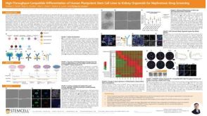 科学海报High-Throughput-Compatible Differentiation of Human Pluripotent Stem Cell Lines to Kidney Organoids for Nephrotoxic Drug Screening

 沪公网安备31010102008431号