Wittman VP et al. (SEP 2006)
The Journal of Immunology 177 6 4187--95
Antibody targeting to acClass I MHC-peptide epitope promotes tumor cell death
Therapeutic mAbs that target tumor-associated Ags on the surface of malignant cells have proven to be an effective and specific option for the treatment of certain cancers. However,many of these protein markers of carcinogenesis are not expressed on the cells' surface. Instead these tumor-associated Ags are processed into peptides that are presented at the cell surface,in the context of MHC class I molecules,where they become targets for T cells. To tap this vast source of tumor Ags,we generated a murine IgG2a mAb,3.2G1,endowed with TCR-like binding specificity for peptide-HLA-A*0201 (HLA-A2) complex and designated this class of Ab as TCR mimics (TCRm). The 3.2G1 TCRm recognizes the GVL peptide (GVLPALPQV) from human chorionic gonadotropin beta presented by the peptide-HLA-A*0201 complex. When used in immunofluorescent staining reactions using GVL peptide-loaded T2 cells,the 3.2G1 TCRm specifically stained the cells in a peptide and Ab concentration-dependent manner. Staining intensity correlated with the extent of cell lysis by complement-dependent cytotoxicity (CDC),and a peptide concentration-dependent threshold level existed for the CDC reaction. Staining of human tumor lines demonstrated that 3.2G1 TCRm was able to recognize endogenously processed peptide and that the breast cancer cell line MDA-MB-231 highly expressed the target epitope. The 3.2G1 TCRm-mediated CDC and Ab-dependent cellular cytotoxicity of a human breast carcinoma line in vitro and inhibited in vivo tumor implantation and growth in nude mice. These results provide validation for the development of novel TCRm therapeutic reagents that specifically target and kill tumors via recognition and binding to MHC-peptide epitopes.
View Publication
Inhibition of osteoclast function reduces hematopoietic stem cell numbers in vivo.
Osteoblasts play a crucial role in the hematopoietic stem cell (HSC) niche; however,an overall increase in their number does not necessarily promote hematopoiesis. Because the activity of osteoblasts and osteoclasts is coordinately regulated,we hypothesized that active bone-resorbing osteoclasts would participate in HSC niche maintenance. Mice treated with bisphosphonates exhibited a decrease in proportion and absolute number of Lin(-)cKit(+)Sca1(+) Flk2(-) (LKS Flk2(-)) and long-term culture-initiating cells in bone marrow (BM). In competitive transplantation assays,the engraftment of treated BM cells was inferior to that of controls,confirming a decrease in HSC numbers. Accordingly,bisphosphonates abolished the HSC increment produced by parathyroid hormone. In contrast,the number of colony-forming-unit cells in BM was increased. Because a larger fraction of LKS in the BM of treated mice was found in the S/M phase of the cell cycle,osteoclast impairment makes a proportion of HSCs enter the cell cycle and differentiate. To prove that HSC impairment was a consequence of niche manipulation,a group of mice was treated with bisphosphonates and then subjected to BM transplantation from untreated donors. Treated recipient mice experienced a delayed hematopoietic recovery compared with untreated controls. Our findings demonstrate that osteoclast function is fundamental in the HSC niche.
View Publication
Raya A et al. (JAN 2008)
Cold Spring Harbor Symposia on Quantitative Biology 73 127--135
Generation of cardiomyocytes from new human embryonic stem cell lines derived from poor-quality blastocysts
Human embryonic stem (hES) cells represent a potential source for cell replacement therapy of many degenerative diseases. Most frequently,hES cell lines are derived from surplus embryos from assisted reproduction cycles,independent of their quality or morphology. Here,we show that hES cell lines can be obtained from poor-quality blastocysts with the same efficiency as that obtained from good- or intermediate-quality blastocysts. Furthermore,we show that the self-renewal,pluripotency,and differentiation ability of hES cell lines derived from either source are comparable. Finally,we present a simple and reproducible embryoid body-based protocol for the differentiation of hES cells into functional cardiomyocytes. The five new hES cell lines derived here should widen the spectrum of available resources for investigating the biology of hES cells and advancing toward efficient strategies of regenerative medicine.
View Publication


 EasySep™小鼠TIL(CD45)正选试剂盒




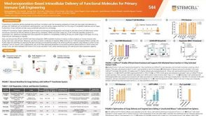 科学海报Mechanoporation-Based Intracellular Delivery of Functional Molecules for Primary Immune Cell Engineering
 24:25
线上讲座ISSCR Standards in Action: Advancements in Cell Line Generation and Organoid Innovation发布日期: 08/01/2024
24:25
线上讲座ISSCR Standards in Action: Advancements in Cell Line Generation and Organoid Innovation发布日期: 08/01/2024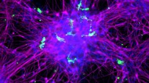 实验方案How to Co-Culture Human Pluripotent Stem Cell (hPSC)-Derived Forebrain Neurons and Microglia
 实验方案How to Prepare and Plate Semi-Solid Methylcellulose Medium for Cell Culture
 实验方案How to Process Leukocyte Reduction System (LRS) Cones/Chambers for Downstream Cell Isolation
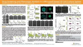 科学海报Drug Screening and Phenotypic Analysis in a Microwell-based 3D Cell Culture System


 沪公网安备31010102008431号