Schiedlmeier B et al. (MAR 2003)
Blood 101 5 1759--68
High-level ectopic HOXB4 expression confers a profound in vivo competitive growth advantage on human cord blood CD34+ cells, but impairs lymphomyeloid differentiation.
Ectopic retroviral expression of homeobox B4 (HOXB4) causes an accelerated and enhanced regeneration of murine hematopoietic stem cells (HSCs) and is not known to compromise any program of lineage differentiation. However,HOXB4 expression levels for expansion of human stem cells have still to be established. To test the proposed hypothesis that HOXB4 could become a prime tool for in vivo expansion of genetically modified human HSCs,we retrovirally overexpressed HOXB4 in purified cord blood (CB) CD34+ cells together with green fluorescent protein (GFP) as a reporter protein,and evaluated the impact of ectopic HOXB4 expression on proliferation and differentiation in vitro and in vivo. When injected separately into nonobese diabetic-severe combined immunodeficient (NOD/SCID) mice or in competition with control vector-transduced cells,HOXB4-overexpressing cord blood CD34+ cells had a selective growth advantage in vivo,which resulted in a marked enhancement of the primitive CD34+ subpopulation (P =.01). However,high HOXB4 expression substantially impaired the myeloerythroid differentiation program,and this was reflected in a severe reduction of erythroid and myeloid progenitors in vitro (P textless.03) and in vivo (P =.01). Furthermore,HOXB4 overexpression also significantly reduced B-cell output (P textless.01). These results show for the first time unwanted side effects of ectopic HOXB4 expression and therefore underscore the need to carefully determine the therapeutic window of HOXB4 expression levels before initializing clinical trials.
View Publication
产品类型:
产品号#:
04434
04444
09600
09650
产品名:
MethoCult™ H4434 Classic
MethoCult™ H4434 Classic
StemSpan™ SFEM
StemSpan™ SFEM
Hase H et al. (MAR 2004)
Blood 103 6 2257--65
BAFF/BLyS can potentiate B-cell selection with the B-cell coreceptor complex.
The tumor necrosis factor (TNF)-like ligand BAFF/BLyS (B-cell activating factor of the TNF family/B-lymphocyte stimulator) is a potent B-cell survival factor,yet its functional relationship with other B-cell surface molecules such as CD19 and CD40 is poorly understood. We found that follicular dendritic cells (FDCs) in human lymph nodes expressed BAFF abundantly. BAFF up-regulated a B cell-specific transcription factor Pax5/BSAP (Pax5/B cell-specific activator protein) activity and its target CD19,a major component of the B-cell coreceptor complex,and synergistically enhanced CD19 phosphorylation by B-cell antigen receptor (BCR). BAFF further enhanced B-cell proliferation,immunoglobulin G (IgG) production,and reactivity to CD154 by BCR/CD19 coligation and interleukin-15 (IL-15). Our results suggest that BAFF may play an important role in FDC-B-cell interactions through the B-cell coreceptor complex and a possibly sequential link between the T cell-independent and -dependent B-cell responses in the germinal centers.
View Publication
产品类型:
产品号#:
03800
03801
03802
03803
03804
03805
03806
产品名:
ClonaCell™-HY杂交瘤试剂盒
ClonaCell™-HY培养基A
ClonaCell™-HY 培养基 B
ClonaCell™-HY 培养基 C
ClonaCell™-HY 培养基 D
ClonaCell™-HY 培养基 E
ClonaCell™-HY PEG
Charrier S et al. (AUG 2004)
Blood 104 4 978--85
Inhibition of angiotensin I-converting enzyme induces radioprotection by preserving murine hematopoietic short-term reconstituting cells.
Angiotensin I-converting enzyme (ACE) inhibitors can affect hematopoiesis by several mechanisms including inhibition of angiotensin II formation and increasing plasma concentrations of AcSDKP (acetyl-N-Ser-Asp-Lys-Pro),an ACE substrate and a negative regulator of hematopoiesis. We tested whether ACE inhibition could decrease the hematopoietic toxicity of lethal or sublethal irradiation protocols. In all cases,short treatment with the ACE inhibitor perindopril protected against irradiation-induced death. ACE inhibition accelerated hematopoietic recovery and led to a significant increase in platelet and red cell counts. Pretreatment with perindopril increased bone marrow cellularity and the number of hematopoietic progenitors (granulocyte macrophage colony-forming unit [CFU-GM],erythroid burst-forming unit [BFU-E],and megakaryocyte colony-forming unit [CFU-MK]) from day 7 to 28 after irradiation. Perindopril also increased the number of hematopoietic stem cells with at least a short-term reconstitutive activity in animals that recovered from irradiation. To determine the mechanism of action involved,we evaluated the effects of increasing AcSDKP plasma concentrations and of an angiotensin II type 1 (AT1) receptor antagonist (telmisartan) on radioprotection. We found that the AT1-receptor antagonism mediated similar radioprotection as the ACE inhibitor. These results suggest that ACE inhibitors and AT1-receptor antagonists could be used to decrease the hematopoietic toxicity of irradiation.
View Publication
产品类型:
产品号#:
03134
产品名:
MethoCult™ M3134
Yang J et al. (SEP 2007)
Blood 110 6 2034--40
AZD1152, a novel and selective aurora B kinase inhibitor, induces growth arrest, apoptosis, and sensitization for tubulin depolymerizing agent or topoisomerase II inhibitor in human acute leukemia cells in vitro and in vivo.
Aurora kinases play an important role in chromosome alignment,segregation,and cytokinesis during mitosis. We have recently shown that hematopoietic malignant cells including those from acute myeloid leukemia (AML) and acute lymphoblastic leukemia (ALL) aberrantly expressed Aurora A and B kinases,and ZM447439,a potent inhibitor of Aurora kinases,effectively induced growth arrest and apoptosis of a variety of leukemia cells. The present study explored the effect of AZD1152,a highly selective inhibitor of Aurora B kinase,on various types of human leukemia cells. AZD1152 inhibited the proliferation of AML lines (HL-60,NB4,MOLM13),ALL line (PALL-2),biphenotypic leukemia (MV4-11),acute eosinophilic leukemia (EOL-1),and the blast crisis of chronic myeloid leukemia K562 cells with an IC50 ranging from 3 nM to 40 nM,as measured by thymidine uptake on day 2 of culture. These cells had 4N/8N DNA content followed by apoptosis,as measured by cell-cycle analysis and annexin V staining,respectively. Of note,AZD1152 synergistically enhanced the antiproliferative activity of vincristine,a tubulin depolymerizing agent,and daunorubicin,a topoisomerase II inhibitor,against the MOLM13 and PALL-2 cells in vitro. Furthermore,AZD1152 potentiated the action of vincristine and daunorubicin in a MOLM13 murine xenograft model. Taken together,AZD1152 is a promising new agent for treatment of individuals with leukemia. The combined administration of AZD1152 and conventional chemotherapeutic agent to patients with leukemia warrants further investigation.
View Publication
产品类型:
产品号#:
04564
04534
04544
产品名:
MethoCult™ H4534 Classic 无 EPO 入门试剂盒
MethoCult™ H4534 Classic(不含 EPO)
MethoCult™ H4534 Classic(不含 EPO)
Ortiz-Sá et al. (JAN 2009)
Leukemia 23 1 59--70
Enhanced cytotoxicity of an anti-transferrin receptor IgG3-avidin fusion protein in combination with gambogic acid against human malignant hematopoietic cells: functional relevance of iron, the receptor, and reactive oxygen species.
The human transferrin receptor (hTfR) is a target for cancer immunotherapy due to its overexpression on the surface of cancer cells. We previously developed an antibody-avidin fusion protein that targets hTfR (anti-hTfR IgG3-Av) and exhibits intrinsic cytotoxicity against certain malignant cells. Gambogic acid (GA),a drug that also binds hTfR,induces cytotoxicity in several malignant cell lines. We now report that anti-hTfR IgG3-Av and GA induce cytotoxicity in a new broader panel of hematopoietic malignant cell lines. Our results show that the effect of anti-hTfR IgG3-Av is iron-dependent whereas that of GA is iron-independent in all cells tested. In addition,we observed that GA exerts a TfR-independent cytotoxicity. We also found that GA increases the generation of reactive oxygen species that may play a role in the cytotoxicity induced by this drug. Additive cytotoxicity was observed by simultaneous combination treatment with these drugs and synergy by using anti-hTfR IgG3-Av as a chemosensitizing agent. In addition,we found a concentration of GA that is toxic to malignant hematopoietic cells but not to human hematopoietic progenitor cells. Our results suggest that these two compounds may be effective,alone or in combination,for the treatment of human hematopoietic malignancies.
View Publication
产品类型:
产品号#:
04434
04444
产品名:
MethoCult™ H4434 Classic
MethoCult™ H4434 Classic
Sun Y et al. (MAR 2010)
Blood 115 9 1709--17
Slug deficiency enhances self-renewal of hematopoietic stem cells during hematopoietic regeneration.
Both extrinsic and intrinsic mechanisms tightly govern hematopoietic stem cell (HSC) decisions of self-renewal and differentiation. However,transcription factors that can selectively regulate HSC self-renewal division after stress remain to be identified. Slug is an evolutionarily conserved zinc-finger transcription factor that is highly expressed in primitive hematopoietic cells and is critical for the radioprotection of these key cells. We studied the effect of Slug in the regulation of HSCs in Slug-deficient mice under normal and stress conditions using serial functional assays. Here,we show that Slug deficiency does not disturb hematopoiesis or alter HSC homeostasis and differentiation in bone marrow but increases the numbers of primitive hematopoietic cells in the extramedullary spleen site. Deletion of Slug enhances HSC repopulating potential but not its homing and differentiation ability. Furthermore,Slug deficiency increases HSC proliferation and repopulating potential in vivo after myelosuppression and accelerates HSC expansion during in vitro culture. Therefore,we propose that Slug is essential for controlling the transition of HSCs from relative quiescence under steady-state condition to rapid proliferation under stress conditions. Our data suggest that inhibition of Slug in HSCs may present a novel strategy for accelerating hematopoietic recovery,thus providing therapeutic benefits for patients after clinical myelosuppressive treatment.
View Publication
产品类型:
产品号#:
09600
09650
28600
产品名:
StemSpan™ SFEM
StemSpan™ SFEM
L-Calc™有限稀释软件
Wang X et al. (DEC 2010)
Blood 116 26 5972--82
Sequential treatment of CD34+ cells from patients with primary myelofibrosis with chromatin-modifying agents eliminate JAK2V617F-positive NOD/SCID marrow repopulating cells.
Because primary myelofibrosis (PMF) originates at the level of the pluripotent hematopoietic stem cell (HSC),we examined the effects of various therapeutic agents on the in vitro and in vivo behavior of PMF CD34(+) cells. Treatment of PMF CD34(+) cells with chromatin-modifying agents (CMAs) but not hydroxyurea,Janus kinase 2 (JAK2) inhibitors,or low doses of interferon-α led to the generation of greater numbers of CD34(+) chemokine (C-X-C motif) receptor (CXCR)4(+) cells,which were capable of migrating in response to chemokine (C-X-C motif) ligand (CXCL)12 and resulted in a reduction in the proportion of hematopoietic progenitor cells (HPCs) that were JAK2V617F(+). Furthermore,sequential treatment of PMF CD34(+) cells but not normal CD34(+) cells with decitabine (5-aza-2'-deoxycytidine [5azaD]),followed by suberoylanilide hydroxamic acid (SAHA; 5azaD/SAHA),or trichostatin A (5azaD/TSA) resulted in a higher degree of apoptosis. Two to 6 months after the transplantation of CMAs treated JAK2V617F(+) PMF CD34(+) cells into nonobese diabetic/severe combined immunodeficient (SCID)/IL-2Rγ(null) mice,the percentage of JAK2V617F/JAK2(total) in human CD45(+) marrow cells was dramatically reduced. These findings suggest that both PMF HPCs,short-term and long-term SCID repopulating cells (SRCs),are JAK2V617F(+) and that JAK2V617F(+) HPCs and SRCs can be eliminated by sequential treatment with CMAs. Sequential treatment with CMAs,therefore,represents a possible effective means of treating PMF at the level of the malignant SRC.
View Publication
产品类型:
产品号#:
04230
产品名:
MethoCult™ H4230
Drowley L et al. (FEB 2016)
Stem cells translational medicine 5 2 164--74
Human Induced Pluripotent Stem Cell-Derived Cardiac Progenitor Cells in Phenotypic Screening: A Transforming Growth Factor-β Type 1 Receptor Kinase Inhibitor Induces Efficient Cardiac Differentiation.
Several progenitor cell populations have been reported to exist in hearts that play a role in cardiac turnover and/or repair. Despite the presence of cardiac stem and progenitor cells within the myocardium,functional repair of the heart after injury is inadequate. Identification of the signaling pathways involved in the expansion and differentiation of cardiac progenitor cells (CPCs) will broaden insight into the fundamental mechanisms playing a role in cardiac homeostasis and disease and might provide strategies for in vivo regenerative therapies. To understand and exploit cardiac ontogeny for drug discovery efforts,we developed an in vitro human induced pluripotent stem cell-derived CPC model system using a highly enriched population of KDR(pos)/CKIT(neg)/NKX2.5(pos) CPCs. Using this model system,these CPCs were capable of generating highly enriched cultures of cardiomyocytes under directed differentiation conditions. In order to facilitate the identification of pathways and targets involved in proliferation and differentiation of resident CPCs,we developed phenotypic screening assays. Screening paradigms for therapeutic applications require a robust,scalable,and consistent methodology. In the present study,we have demonstrated the suitability of these cells for medium to high-throughput screens to assess both proliferation and multilineage differentiation. Using this CPC model system and a small directed compound set,we identified activin-like kinase 5 (transforming growth factor-β type 1 receptor kinase) inhibitors as novel and potent inducers of human CPC differentiation to cardiomyocytes. Significance: Cardiac disease is a leading cause of morbidity and mortality,with no treatment available that can result in functional repair. This study demonstrates how differentiation of induced pluripotent stem cells can be used to identify and isolate cell populations of interest that can translate to the adult human heart. Two separate examples of phenotypic screens are discussed,demonstrating the value of this biologically relevant and reproducible technology. In addition,this assay system was able to identify novel and potent inducers of differentiation and proliferation of induced pluripotent stem cell-derived cardiac progenitor cells.
View Publication


 EasySep™小鼠TIL(CD45)正选试剂盒




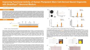 科学海报Improving Functional Activity of Human Pluripotent Stem Cell-Derived Neural Organoids with BrainPhys Neuronal Medium
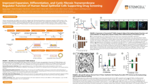 科学海报Improved Expansion, Differentiation, and Cystic Fibrosis Transmembrane Regulator Function of Human Nasal Epithelial Cells Supporting Drug Screening

 沪公网安备31010102008431号