Drowley L et al. (FEB 2016)
Stem cells translational medicine 5 2 164--74
Human Induced Pluripotent Stem Cell-Derived Cardiac Progenitor Cells in Phenotypic Screening: A Transforming Growth Factor-β Type 1 Receptor Kinase Inhibitor Induces Efficient Cardiac Differentiation.
Several progenitor cell populations have been reported to exist in hearts that play a role in cardiac turnover and/or repair. Despite the presence of cardiac stem and progenitor cells within the myocardium,functional repair of the heart after injury is inadequate. Identification of the signaling pathways involved in the expansion and differentiation of cardiac progenitor cells (CPCs) will broaden insight into the fundamental mechanisms playing a role in cardiac homeostasis and disease and might provide strategies for in vivo regenerative therapies. To understand and exploit cardiac ontogeny for drug discovery efforts,we developed an in vitro human induced pluripotent stem cell-derived CPC model system using a highly enriched population of KDR(pos)/CKIT(neg)/NKX2.5(pos) CPCs. Using this model system,these CPCs were capable of generating highly enriched cultures of cardiomyocytes under directed differentiation conditions. In order to facilitate the identification of pathways and targets involved in proliferation and differentiation of resident CPCs,we developed phenotypic screening assays. Screening paradigms for therapeutic applications require a robust,scalable,and consistent methodology. In the present study,we have demonstrated the suitability of these cells for medium to high-throughput screens to assess both proliferation and multilineage differentiation. Using this CPC model system and a small directed compound set,we identified activin-like kinase 5 (transforming growth factor-β type 1 receptor kinase) inhibitors as novel and potent inducers of human CPC differentiation to cardiomyocytes. Significance: Cardiac disease is a leading cause of morbidity and mortality,with no treatment available that can result in functional repair. This study demonstrates how differentiation of induced pluripotent stem cells can be used to identify and isolate cell populations of interest that can translate to the adult human heart. Two separate examples of phenotypic screens are discussed,demonstrating the value of this biologically relevant and reproducible technology. In addition,this assay system was able to identify novel and potent inducers of differentiation and proliferation of induced pluripotent stem cell-derived cardiac progenitor cells.
View Publication
产品类型:
产品号#:
70919
产品名:
Ma Z et al. (JUL 2015)
Nature communications 6 May 7413
Self-organizing human cardiac microchambers mediated by geometric confinement.
Tissue morphogenesis and organ formation are the consequences of biochemical and biophysical cues that lead to cellular spatial patterning in development. To model such events in vitro,we use PEG-patterned substrates to geometrically confine human pluripotent stem cell colonies and spatially present mechanical stress. Modulation of the WNT/β-catenin pathway promotes spatial patterning via geometric confinement of the cell condensation process during epithelial-mesenchymal transition,forcing cells at the perimeter to express an OCT4+ annulus,which is coincident with a region of higher cell density and E-cadherin expression. The biochemical and biophysical cues synergistically induce self-organizing lineage specification and creation of a beating human cardiac microchamber confined by the pattern geometry. These highly defined human cardiac microchambers can be used to study aspects of embryonic spatial patterning,early cardiac development and drug-induced developmental toxicity.
View Publication
Chang Q et al. (SEP 2002)
Infection and Immunity 70 9 4977--86
Structure-function relationships for human antibodies to pneumococcal capsular polysaccharide from transgenic mice with human immunoglobulin Loci.
To investigate the influence of antibody structure and specificity on antibody efficacy against Streptococcus pneumoniae,human monospecific antibodies (MAbs) to serotype 3 pneumococcal capsular polysaccharide (PPS-3) were generated from transgenic mice reconstituted with human immunoglobulin loci (XenoMouse mice) vaccinated with a PPS-3-tetanus toxoid conjugate and their molecular genetic structures,epitope specificities,and protective efficacies in normal and complement-deficient mice were determined. Nucleic acid sequence analysis of three MAbs (A7,1A2,and 7C5) revealed that they use two different V(H)3 genes (A7 and 1A2 both use V3-15) and three different V(kappa) gene segments. The MAbs were found to have similar affinities for PPS-3 but different epitope specificities and CDR3 regions. Both A7 and 7C5 had a lysine at the V(H)-D junction,whereas 1A2 had a threonine. Challenge experiments with serotype 3 S. pneumoniae in BALB/c mice revealed that both 10- and 1- micro g doses of A7 and 7C5 were protective,while only a 10- micro g dose of 1A2 was protective. Both A7 and 7C5 were also protective in mice lacking either an intact alternative (FB(-/-)) or classical (C4(-/-)) complement pathway,but 1A2 was not protective in either strain. Our data suggest that PPS-3 consists of epitopes that can elicit both highly protective and less protective antibodies and that the superior efficacies of certain antibodies may be a function of their structures and/or specificities. Further investigation of relationships between structure,specificity,and efficacy for defined MAbs to PPS may identify antibody features that might be useful surrogates for antibody (and vaccine) efficacy.
View Publication
产品类型:
产品号#:
03800
03801
03802
03803
03804
03805
03806
产品名:
ClonaCell™-HY杂交瘤试剂盒
ClonaCell™-HY培养基A
ClonaCell™-HY 培养基 B
ClonaCell™-HY 培养基 C
ClonaCell™-HY 培养基 D
ClonaCell™-HY 培养基 E
ClonaCell™-HY PEG
York D et al. (DEC 2016)
BMC Biotechnology 16 1 23
Generating aldehyde-tagged antibodies with high titers and high formylglycine yields by supplementing culture media with copper(II)
BACKGROUND The ability to site-specifically conjugate a protein to a payload of interest (e.g.,a fluorophore,small molecule pharmacophore,oligonucleotide,or other protein) has found widespread application in basic research and drug development. For example,antibody-drug conjugates represent a class of biotherapeutics that couple the targeting specificity of an antibody with the chemotherapeutic potency of a small molecule drug. While first generation antibody-drug conjugates (ADCs) used random conjugation approaches,next-generation ADCs are employing site-specific conjugation. A facile way to generate site-specific protein conjugates is via the aldehyde tag technology,where a five amino acid consensus sequence (CXPXR) is genetically encoded into the protein of interest at the desired location. During protein expression,the Cys residue within this consensus sequence can be recognized by ectopically-expressed formylglycine generating enzyme (FGE),which converts the Cys to a formylglycine (fGly) residue. The latter bears an aldehyde functional group that serves as a chemical handle for subsequent conjugation. RESULTS The yield of Cys conversion to fGly during protein production can be variable and is highly dependent on culture conditions. We set out to achieve consistently high yields by modulating culture conditions to maximize FGE activity within the cell. We recently showed that FGE is a copper-dependent oxidase that binds copper in a stoichiometric fashion and uses it to activate oxygen,driving enzymatic turnover. Building upon that work,here we show that by supplementing cell culture media with copper we can routinely reach high yields of highly converted protein. We demonstrate that cells incorporate copper from the media into FGE,which results in increased specific activity of the enzyme. The amount of copper required is compatible with large scale cell culture,as demonstrated in fed-batch cell cultures with antibody titers of 5 g textperiodcentered L(-1),specific cellular production rates of 75 pg textperiodcentered cell(-1) textperiodcentered d(-1),and fGly conversion yields of 95-98 %. CONCLUSIONS We describe a process with a high yield of site-specific formylglycine (fGly) generation during monoclonal antibody production in CHO cells. The conversion of Cys to fGly depends upon the activity of FGE,which can be ensured by supplementing the culture media with 50 uM copper(II) sulfate.
View Publication
产品类型:
产品号#:
03814
产品名:
ClonaCell™-TCS 培养基
Baatz JE et al. (JUL 2014)
In vivo (Athens,Greece) 28 4 411--423
Cryopreservation of viable human lung tissue for versatile post-thaw analyses and culture.
Clinical trials are currently used to test therapeutic efficacies for lung cancer,infections and diseases. Animal models are also used as surrogates for human disease. Both approaches are expensive and time-consuming. The utility of human biospecimens as models is limited by specialized tissue processing methods that preserve subclasses of analytes (e.g. RNA,protein,morphology) at the expense of others. We present a rapid and reproducible method for the cryopreservation of viable lung tissue from patients undergoing lobectomy or transplant. This method involves the pseudo-diaphragmatic expansion of pieces of fresh lung tissue with cryoprotectant formulation (pseudo-diaphragmatic expansion-cryoprotectant perfusion or PDX-CP) followed by controlled-rate freezing in cryovials. Expansion-perfusion rates,volumes and cryoprotectant formulation were optimized to maintain tissue architecture,decrease crystal formation and increase long-term cell viability. Rates of expansion of 4 cc/min or less and volumes ranging from 0.8-1.2 × tissue volume were well-tolerated by lung tissue obtained from patients with chronic obstructive pulmonary disease or idiopathic pulmonary fibrosis,showing minimal differences compared to standard histopathology. Morphology was greatly improved by the PDX-CP procedure compared to simple fixation. Fresh versus post-thawed lung tissue showed minimal differences in histology,RNA integrity numbers and post-translational modified protein integrity (2-dimensional differential gel electrophoresis). It was possible to derive numerous cell types,including alveolar epithelial cells,fibroblasts and stem cells,from the tissue for at least three months after cryopreservation. This new method should provide a uniform,cost-effective approach to the banking of biospecimens,with versatility to be amenable to any post-acquisition process applicable to fresh tissue samples.
View Publication
产品类型:
产品号#:
05850
05857
05870
05875
85850
85857
85870
85875
产品名:
mTeSR™1
mTeSR™1
Su YR et al. (AUG 2008)
Arteriosclerosis,thrombosis,and vascular biology 28 8 1439--46
Lentiviral transduction of apoAI into hematopoietic progenitor cells and macrophages: applications to cell therapy of atherosclerosis.
OBJECTIVE: We used genetically engineered mouse hematopoietic progenitor cells (HPCs) to investigate the therapeutic effects of human apoAI on atherosclerosis in apoE(-/-) mice. METHODS AND RESULTS: Lentiviral constructs expressing either human apoAI (LV-apoAI) or green fluorescent protein (LV-GFP) cDNA under a macrophage specific promoter (CD68) were generated and used for ex vivo transduction of mouse HPCs and macrophages. The transduction efficiency was textgreater25% for HPCs and textgreater70% for macrophages. ApoAI was found in the macrophage culture media,mostly associated with the HDL fraction. Interestingly,a significant increase in mRNA and protein levels for ATP binding cassette A1 (ABCA1) and ABCG1 were found in apoAI-expressing macrophages after acLDL loading. Expression of apoAI significantly increased cholesterol efflux in wild-type and apoE(-/-) macrophages. HPCs transduced with LV-apoAI ex vivo and then transplanted into apoE(-/-) mice caused a 50% reduction in atherosclerotic lesion area compared to GFP controls,without influencing plasma HDL-C levels. CONCLUSIONS: Lentiviral transduction of apoAI into HPCs reduces atherosclerosis in apoE(-/-) mice. Expression of apoAI in macrophages improves cholesterol trafficking in wild-type apoE-producing macrophages and causes upregulation of ABCA1 and ABCG1. These novel observations set the stage for a cell therapy approach to atherosclerosis regression,exploiting the cooperation between apoE and apoAI to maximize cholesterol exit from the plaque.
View Publication
Inagi R et al. (NOV 2007)
Nephrology,dialysis,transplantation : official publication of the European Dialysis and Transplant Association - European Renal Association 22 11 3311--7
Establishment of a sandwich ELISA for human megsin, a kidney-specific serine protease inhibitor.
BACKGROUND: We previously identified a novel serine protease inhibitor (serpin),megsin,which is predominantly expressed in the kidney. Megsin expression is up-regulated in human and experimental renal diseases associated with mesangial proliferation and expansion,suggesting that urinary megsin may be a novel diagnostic marker for some renal diseases. METHODS: We established a specific and sensitive sandwich enzyme-linked immunosorbent assay (ELISA) for megsin and measured urinary megsin of patients with various renal diseases. RESULTS: Megsin ELISA specifically detected megsin but not other serpins. The detection limit was 0.04 ng/ml,which allowed detection of urinary megsin in 3.6% of healthy individuals. The antigenic epitope in the urine detected by the ELISA was confirmed as megsin protein by time-of-flight mass spectrometry. Among patients with rapidly progressive glomerulonephritis (n = 18),55.6% were urinary megsin-positive,while 24.1% in IgA nephropathy (n = 112) and 15.1% in chronic non-IgA glomerulonephritis (n = 245) were urinary megsin-positive,respectively. Among patients with chronic renal failure due to unknown causes (n = 74),18.9% were positive for urinary megsin. In diabetic patients with or without nephropathy (n = 1073),12.3% were urinary megsin-positive,while positivity of urinary megsin in patients with non-renal diseases (n = 768) was equivalent (3.3%) to that of healthy individuals. Of note,when urinary megsin-positive patients with diabetic nephropathy (n = 71) were classified into four stages by their proteinuria and estimated glomerular filtration rate,urinary megsin excretion increased as the stage progressed up to stage 3A,suggesting correlation of that with mesangial expansion level. Urinary megsin decreased in the advanced stage,probably reflecting development of glomerulosclerosis. CONCLUSION: We established a high-sensitive megsin ELISA,which detects urinary megsin in some patients with renal diseases and in only a few healthy subjects. Megsin ELISA may be a novel diagnostic tool for renal diseases.
View Publication


 EasySep™小鼠TIL(CD45)正选试剂盒





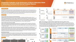 科学海报Comparative Evaluation of the Performance of Neuron Maturation Media in Generating Functional hPSC-Derived Forebrain Neurons
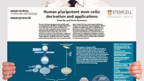 挂图Derivation and Applications of Human Pluripotent Stem Cells Overview of the derivation of human embryonic stem cells (hESCs) and induced pluripotent stem cells (iPSCs)发布日期: 11/26/2020

 沪公网安备31010102008431号