Conforti L et al. (JAN 2003)
Journal of immunology (Baltimore,Md. : 1950) 170 2 695--702
Hypoxia regulates expression and activity of Kv1.3 channels in T lymphocytes: a possible role in T cell proliferation.
T lymphocytes are exposed to hypoxia during their development and also when they migrate to hypoxic pathological sites such as tumors and wounds. Although hypoxia can affect T cell development and function,the mechanisms by which immune cells sense and respond to changes in O(2)-availability are poorly understood. K(+) channels encoded by the Kv1.3 subtype of the voltage-dependent Kv1 gene family are highly expressed in lymphocytes and are involved in the control of membrane potential and cell function. In this study,we investigate the sensitivity of Kv1.3 channels to hypoxia in freshly isolated human T lymphocytes and leukemic Jurkat T cells. Acute exposure to hypoxia (20 mmHg,2 min) inhibits Kv1.3 currents in both cell types by 20%. Prolonged exposure to hypoxia (1% O(2) for 24 h) selectively decreases Kv1.3 protein levels in Jurkat T cells by 47%,but not Kvbeta2 and SK2 Ca-activated K(+) channel subunit levels. The decrease in Kv1.3 protein levels occurs with no change in Kv1.3 mRNA expression and is associated with a significant decrease in K(+) current density. A decrease in Kv1.3 polypeptide levels similar to that obtained during hypoxia is produced by Kv1.3 channel blockage. Our results indicate that hypoxia produces acute and long-term inhibition of Kv1.3 channels in T lymphocytes. This effect could account for the inhibition of lymphocyte proliferation during hypoxia. Indeed,we herein present evidence showing that hypoxia selectively inhibits TCR-mediated proliferation and that this inhibition is associated with a decrease in Kv1.3 proteins.
View Publication
Frazer-Abel AA et al. (NOV 2004)
The Journal of pharmacology and experimental therapeutics 311 2 758--69
Nicotine activates nuclear factor of activated T cells c2 (NFATc2) and prevents cell cycle entry in T cells.
We used primary peripheral blood T cells,a population that exists in G(0) and can be stimulated to enter the cell cycle synchronously,to define more precisely the effects of nicotine on pathways that control cell cycle entry and progression. Our data show that nicotine decreased the ability of T cells to transit through the G(0)/G(1) boundary (acquire competence) and respond to progression signals. These effects were due to nuclear factor of activated T cells c2 (NFATc2)-dependent repression of cyclin-dependent kinase 4 (CDK4) expression. Growth arrest at the G(0)/G(1) boundary was further enforced by inhibition of cyclin D2 expression and by increased expression and stabilization of p27Kip1. Intriguingly,T cells from habitual users of tobacco products and from NFATc2-deficient mice constitutively expressed CDK4 and were resistant to the antiproliferative effects of nicotine. These results indicate that nicotine impairs T cell cycle entry through NFATc2-dependent mechanisms and suggest that,in the face of chronic nicotine exposure,selection may favor cells that can evade these effects. We postulate that cross talk between nicotinic acetylcholine receptors and growth factor receptor-activated pathways offers a novel mechanism by which nicotine may directly impinge on cell cycle progression. This offers insight into possible reasons that underlie the unique effects of nicotine on distinct cell types and identifies new targets that may be useful control tobacco-related diseases.
View Publication
产品类型:
产品号#:
19751
19751RF
产品名:
Wang X et al. (MAR 2009)
Journal of immunology (Baltimore,Md. : 1950) 182 6 3597--608
MEKK3 is essential for lymphopenia-induced T cell proliferation and survival.
T cell homeostasis is crucial for maintaining an efficient and balanced T cell immunity. The interaction between TCR and self peptide (sp) MHC ligands is known to be the key driving force in this process,and it is believed to be functionally and mechanistically different from that initiated by the antigenic TCR stimulation. Yet,very little is known about the downstream signaling events triggered by this TCR-spMHC interaction and how they differ from those triggered by antigenic TCR stimulation. In this study,we show that T cell conditional ablation of MEKK3,a Ser/Thr kinase in the MAPK cascade,causes a significant reduction in peripheral T cell numbers in the conditional knockout mice,but does not perturb thymic T cell development and maturation. Using an adoptive mixed transfer method,we show that MEKK3-deficient T cells are severely impaired in lymphopenia-induced cell proliferation and survival. Interestingly,the Ag-induced T cell proliferation proceeds normally in the absence of MEKK3. Finally,we found that the activity of ERK1/2,but not p38 MAPK,was attenuated during the lymphopenia-driven response in MEKK3-deficient T cells. Together,these data suggest that MEKK3 may play a crucial selective role for spMHC-mediated T cell homeostasis.
View Publication
产品类型:
产品号#:
18751
18751RF
产品名:
Tinoco R et al. (MAY 2016)
Immunity 44 5 1190--203
PSGL-1 Is an Immune Checkpoint Regulator that Promotes T Cell Exhaustion.
Chronic viruses and cancers thwart immune responses in humans by inducing T cell dysfunction. Using a murine chronic virus that models human infections,we investigated the function of the adhesion molecule,P-selectin glycoprotein ligand-1 (PSGL-1),that is upregulated on responding T cells. PSGL-1-deficient mice cleared the virus due to increased intrinsic survival of multifunctional effector T cells that had downregulated PD-1 as well as other inhibitory receptors. Notably,this response resulted in CD4(+)-T-cell-dependent immunopathology. Mechanistically,PSGL-1 ligation on exhausted CD8(+) T cells inhibited T cell receptor (TCR) and interleukin-2 (IL-2) signaling and upregulated PD-1,leading to diminished survival with TCR stimulation. In models of melanoma cancer in which T cell dysfunction occurs,PSGL-1 deficiency led to PD-1 downregulation,improved T cell responses,and tumor control. Thus,PSGL-1 plays a fundamental role in balancing viral control and immunopathology and also functions to regulate T cell responses in the tumor microenvironment.
View Publication
Goda C et al. (FEB 2006)
International immunology 18 2 233--40
Involvement of IL-32 in activation-induced cell death in T cells.
NK cell transcript 4 (NK4),now denoted as IL-32,was originally identified as a transcript whose expression was increased in activated NK cells. It has been very recently demonstrated that NK4 is secreted from several cells upon the stimulation of some inflammatory cytokines such as IL-18,IL-1beta,IFN-gamma and IL-12. Furthermore,NK4 induces production of tumor necrosis factor,macrophage inflammatory protein (MIP)-2 and IL-8 in monocytic cell lines,indicating that this factor would be involved in the inflammatory responses. Based on these findings,NK4 was renamed IL-32. However,the biological activities of IL-32 on other cell types remained undetermined. Furthermore,it was still argued whether IL-32 acts on cells from outside or inside the cells. In this article,we first report that expression of IL-32 was up-regulated in activated T cells and NK cells,and that IL-32beta was the predominantly expressed isoform in activated T cells. IL-32 was specifically expressed in T cells undergoing apoptosis and enforced expression of IL-32-induced apoptosis,whereas its down-regulation rescued the cells from apoptosis in HeLa cells. IL-32 existing in the supernatant would be derived from the cytoplasm of apoptotic cells. These results strongly indicated that IL-32 would be involved in activation-induced cell death in T cells,probably via its intracellular actions. Our present findings expand our understanding of the biological function of IL-32 and argue that IL-32 may act on cells,not only from the outside but also from the inside.
View Publication
产品类型:
产品号#:
15021
15061
15025
15065
产品名:
RosetteSep™人T细胞富集抗体混合物
RosetteSep™人T细胞富集抗体混合物
RosetteSep™人NK细胞富集抗体混合物
RosetteSep™人NK细胞富集抗体混合物
Makaroff LE et al. (MAR 2009)
Proceedings of the National Academy of Sciences of the United States of America 106 12 4799--804
Postthymic maturation influences the CD8 T cell response to antigen.
Complete T cell development requires postthymic maturation,and we investigated the influence of this ontological period on the CD8 T cell response to infection by comparing responses of mature CD8 T cells with those of recent thymic emigrants (RTEs). When activated with a noninflammatory stimulus or a bacterial or viral pathogen,CD8 RTEs generated a lower proportion of cytokine-producing effector cells and long-lived memory precursors compared with their mature counterparts. Although peripheral T cell maturation is complete within several weeks after thymic egress,RTE-derived memory cells continued to express inappropriate levels of memory cell markers and display an altered pattern of cytokine production,even 8 weeks after infection. When rechallenged,RTE-derived memory cells generated secondary effector cells that were phenotypically and functionally equivalent to those generated by their mature counterparts. The defects at the effector and memory stages were not associated with differences in the expression of T cell receptor-,costimulation-,or activation-associated cell surface markers yet were associated with lower Ly6C expression levels at the effector stage. This work demonstrates that the stage of postthymic maturation influences cell fate decisions and cytokine profiles of stimulated CD8 T cells,with repercussions that are apparent long after cells have progressed from the RTE compartment.
View Publication
产品类型:
产品号#:
19753
19753RF
产品名:
Lichterfeld M et al. (SEP 2004)
The Journal of experimental medicine 200 6 701--12
Loss of HIV-1-specific CD8+ T cell proliferation after acute HIV-1 infection and restoration by vaccine-induced HIV-1-specific CD4+ T cells.
Virus-specific CD8(+) T cells are associated with declining viremia in acute human immunodeficiency virus (HIV)1 infection,but do not correlate with control of viremia in chronic infection,suggesting a progressive functional defect not measured by interferon gamma assays presently used. Here,we demonstrate that HIV-1-specific CD8(+) T cells proliferate rapidly upon encounter with cognate antigen in acute infection,but lose this capacity with ongoing viral replication. This functional defect can be induced in vitro by depletion of CD4(+) T cells or addition of interleukin 2-neutralizing antibodies,and can be corrected in chronic infection in vitro by addition of autologous CD4(+) T cells isolated during acute infection and in vivo by vaccine-mediated induction of HIV-1-specific CD4(+) T helper cell responses. These data demonstrate a loss of HIV-1-specific CD8(+) T cell function that not only correlates with progressive infection,but also can be restored in chronic infection by augmentation of HIV-1-specific T helper cell function. This identification of a reversible defect in cell-mediated immunity in chronic HIV-1 infection has important implications for immunotherapeutic interventions.
View Publication


 EasySep™小鼠TIL(CD45)正选试剂盒





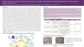 科学海报Workflow Solutions for Human T Cell Isolation and Expansion
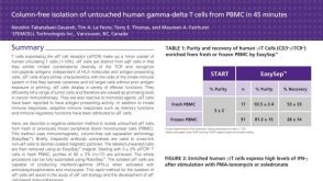 科学海报Immunomagnetic Cell Isolation of Human Gamma-Delta T Cells from PBMC

 沪公网安备31010102008431号