
 EasySep™小鼠TIL(CD45)正选试剂盒


搜索结果: 'easysep direct'
-
 技术公告Isolating Extracellular Vesicles from Urine with EasySep™ EV Human Positive Selection Kits
产品类型:
细胞类型:
其他细胞系
产品号#:
产品名:
发布日期: 01/01/2024 -
 科学海报Fast and Easy Isolation of CD27-Positive Human Memory B Cells Using EasySep™ Releasable RapidSpheres™
产品类型:
Conference:
AAI 2017
产品号#:
17864
产品名:
EasySep™人记忆B细胞分选试剂盒
-
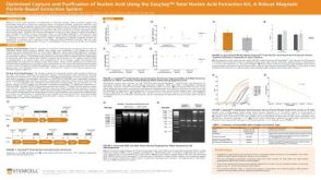
 技术公告Achieve Scalable, High-Quality Nucleic Acid Extractions with the EasySep™ Total Nucleic Acid Extraction Kit
产品类型:
细胞类型:
B细胞,NK细胞,T细胞,其他细胞系,单个核细胞,单核细胞,巨噬细胞,树突状细胞(DCs),淋巴细胞,癌细胞及细胞系,粒细胞及其亚群,肿瘤细胞
产品号#:
产品名:
发布日期: 09/11/2025 -
 技术公告Isolate Mouse CD45.1 or CD45.2 Positive Cells with EasySep™ Release Mouse Positive Selection Kits
产品类型:
细胞类型:
B细胞,NK细胞,T细胞,先天性淋巴细胞,单个核细胞,单核细胞,巨噬细胞,树突状细胞(DCs),浆细胞,淋巴细胞,白细胞,粒细胞及其亚群,调节性T细胞,造血干祖细胞,髓系细胞
产品号#:
产品名:
发布日期: 01/15/2025 -
 科学海报Rapid, one-step isolation of granulocytes directly from whole blood without RBC lysis, sedimentation or density spearation
产品类型:
Conference:
KEYSTONE 2016
产品号#:
19659
产品名:
EasySep™ Direct人Pan-粒细胞分选试剂盒
-

产品类型:
产品号#:
产品名:
-
 科学海报Immunomagnetic Cell Isolation of Untouched Lymphocyte Populations Directly from Whole Blood
产品类型:
Conference:
AAI 2014
产品号#:
19655
19661
19662
19663
19664
19674
18000
19659
19655RF
89671
89671RF
89684
89684RF
19671
19671RF
19684
19684RF
产品名:
EasySep™ Direct人总淋巴细胞分选试剂盒
EasySep™ Direct人T细胞分选试剂盒
EasySep™ Direct人CD4+ T细胞分选试剂盒
EasySep™ Direct人CD8+ T细胞分选试剂盒
EasySep™ Direct人B-CLL细胞分选试剂盒
EasySep™ Direct人B细胞分选试剂盒
EasySep™磁极
EasySep™ Direct人Pan-粒细胞分选试剂盒
RoboSep™ Direct人总淋巴细胞分选试剂盒
EasySep™ Direct HLA交叉配型T细胞分选试剂盒
RoboSep™ Direct HLA交叉配型T细胞分选试剂盒
EasySep™ Direct HLA交叉配型B细胞分选试剂盒
RoboSep™ Direct HLA交叉配型B细胞分选试剂盒
-
产品类型:
产品号#:
19854
19854RF
产品名:
EasySep™小鼠B细胞分选试剂盒
RoboSep™ 小鼠B细胞分选试剂盒
-
产品类型:
产品号#:
18387
18387RF
产品名:

 沪公网安备31010102008431号