M. Epeldegui et al. (jun 2019)
Scientific reports 9 1 9371
Elevated numbers of PD-L1 expressing B cells are associated with the development of AIDS-NHL.
The risk for non-Hodgkin lymphoma (NHL) is markedly increased in persons living with human immunodeficiency virus (HIV) infection,and remains elevated in those on anti-retroviral therapy (cART). Both the loss of immunoregulation of Epstein-Barr virus (EBV) infected cells,as well as chronic B-cell activation,are believed to contribute to the genesis of AIDS-related NHL (AIDS-NHL). However,the mechanisms that lead to AIDS-NHL have not been completely defined. A subset of B cells that is characterized by the secretion of IL10,as well as the expression of the programmed cell death ligand-1 (PD-L1/CD274),was recently described. These PD-L1+ B cells can exert regulatory function,including the dampening of T-cell activation,by interacting with the program cell death protein (PD1) on target cells. The role of PD-L1+ B cells in the development of AIDS-NHL has not been explored. We assessed B cell PD-L1 expression on B cells preceding AIDS-NHL diagnosis in a nested case-control study of HIV+ subjects who went on to develop AIDS-NHL,as well as HIV+ subjects who did not,using multi-color flow cytometry. Archival frozen viable PBMC were obtained from the UCLA Multicenter AIDS Cohort Study (MACS). It was seen that the number of CD19+CD24++CD38++and CD19+PD-L1+cells was significantly elevated in cases 1-4 years prior to AIDS-NHL diagnosis,compared to controls,raising the possibility that these cells may play a role in the etiology of AIDS-NHL. Interestingly,most PD-L1+ expression on CD19+ cells was seen on CD19+CD24++CD38++ cells. In addition,we showed that HIV can directly induce PD-L1 expression on B cells through interaction of virion-associated CD40L with CD40 on B cells.
View Publication
产品类型:
产品号#:
15024
15064
产品名:
RosetteSep™人B细胞富集抗体混合物
RosetteSep™人B细胞富集抗体混合物
R. J. Komban et al. ( 2019)
Nature communications 10 1 2423
Activated Peyer's patch B cells sample antigen directly from M cells in the subepithelial dome.
The germinal center (GC) reaction in Peyer's patches (PP) requires continuous access to antigens,but how this is achieved is not known. Here we show that activated antigen-specific CCR6+CCR1+GL7- B cells make close contact with M cells in the subepithelial dome (SED). Using in situ photoactivation analysis of antigen-specific SED B cells,we find migration of cells towards the GC. Following antigen injection into ligated intestinal loops containing PPs,40{\%} of antigen-specific SED B cells bind antigen within 2 h,whereas unspecifc cells do not,indicating B cell-receptor involvment. Antigen-loading is not observed in M cell-deficient mice,but is unperturbed in mice depleted of classical dendritic cells (DC). Thus,we report a M cell-B cell antigen-specific transporting pathway in PP that is independent of DC. We propose that this antigen transporting pathway has a critical role in gut IgA responses,and should be taken into account when developing mucosal vaccines.
View Publication
产品类型:
产品号#:
19854
19854RF
产品名:
EasySep™小鼠B细胞分选试剂盒
RoboSep™ 小鼠B细胞分选试剂盒
S. Omenetti et al. (jun 2019)
Immunity
The Intestine Harbors Functionally Distinct Homeostatic Tissue-Resident and Inflammatory Th17 Cells.
T helper 17 (Th17) cells are pathogenic in many inflammatory diseases,but also support the integrity of the intestinal barrier in a non-inflammatory manner. It is unclear what distinguishes inflammatory Th17 cells elicited by pathogens and tissue-resident homeostatic Th17 cells elicited by commensals. Here,we compared the characteristics of Th17 cells differentiating in response to commensal bacteria (SFB) to those differentiating in response to a pathogen (Citrobacter rodentium). Homeostatic Th17 cells exhibited little plasticity towards expression of inflammatory cytokines,were characterized by a metabolism typical of quiescent or memory T cells,and did not participate in inflammatory processes. In contrast,infection-induced Th17 cells showed extensive plasticity towards pro-inflammatory cytokines,disseminated widely into the periphery,and engaged aerobic glycolysis in addition to oxidative phosphorylation typical for inflammatory effector cells. These findings will help ensure that future therapies directed against inflammatory Th17 cells do not inadvertently damage the resident gut population.
View Publication
产品类型:
产品号#:
19752
19752RF
19052
19052RF
产品名:
EasySep™人CD4+ T细胞富集试剂盒
RoboSep™ 人CD4+ T细胞富集试剂盒含滤芯吸头
C. Onyilagha et al. (jun 2019)
Journal of immunology (Baltimore,Md. : 1950)
NK Cells Are Critical for Optimal Immunity to Experimental Trypanosoma congolense Infection.
NK cells are key innate immune cells that play critical roles in host defense. Although NK cells have been shown to regulate immunity to some infectious diseases,their role in immunity to Trypanosoma congolense has not been investigated. NK cells are vital sources of IFN-gamma and TNF-alpha; two key cytokines that are known to play important roles in resistance to African trypanosomes. In this article,we show that infection with T. congolense leads to increased levels of activated and functional NK cells in multiple tissue compartments. Systemic depletion of NK cells with anti-NK1.1 mAb led to increased parasitemia,which was accompanied by significant reduction in IFN-gamma production by immune cells in the spleens and liver of infected mice. Strikingly,infected NFIL3-/- mice (which genetically lack NK cell development and function) on the normally resistant background were highly susceptible to T. congolense infection. These mice developed fulminating and uncontrolled parasitemia and died significantly earlier (13 ± 1 d) than their wild-type control mice (106 ± 26 d). The enhanced susceptibility of NFIL3-/- mice to infection was accompanied by significantly impaired cytokine (IFN-gamma and TNF-alpha) response by CD3+ T cells in the spleens and liver. Adoptive transfer of NK cells into NFIL3-/- mice before infection rescued them from acute death in a perforin-dependent manner. Collectively,these studies show that NK cells are critical for optimal resistance to T. congolense,and its deficiency leads to enhanced susceptibility in infected mice.
View Publication
Houtenbos I et al. (JUL 2003)
Cancer immunology,immunotherapy : CII 52 7 455--62
Serum-free generation of antigen presenting cells from acute myeloid leukaemic blasts for active specific immunisation.
PURPOSE: Immunotherapy holds promise as a new strategy for the eradication of residual cells in acute myeloid leukaemia (AML). Leukaemic antigen presenting cells (APCs) combining optimal antigen presentation and tumour antigenicity could be used as potent T cell activators. For clinical purposes it is desirable to culture APCs under serum-free conditions. Therefore,we compared morphological,immunophenotypical and functional outcome of the serum-free culture of AML-APCs to their serum-enriched culture. METHODS: AML blasts (n=19) were cultured in the presence of either a cytokine mix or calcium ionophore (CI) for 14 and 2 days,respectively,in FCS-containing medium (FCS),StemSpan serum-free medium (SP) and CellGro serum-free medium (CG). After culture relative yields were calculated and immunophenotypic analysis of APC markers was performed. The mixed leukocyte reaction (MLR) was used to determine T cell stimulating capacity. RESULTS: Serum-free culture of AML-APCs resulted in comparable morphology,relative yields and immunophenotype to serum-enriched culture. By comparing both serum-free media we observed a trend towards a more mature phenotype of CI-cultured AML-APCs in SP. MLR showed that serum-free cultured cells have equal T cell stimulatory capacity in comparison with serum-enriched culture. CONCLUSION: These data show that the serum-free culture of AML-APCs is feasible and that these APCs are comparable to serum-enriched cultured AML-APCs with regard to morphological,immunophenotypical and functional characteristics. These AML-APCs are suitable for the development of active specific immunisation protocols which meet the criteria for good clinical practise (GCP).
View Publication
产品类型:
产品号#:
09600
09650
产品名:
StemSpan™ SFEM
StemSpan™ SFEM
Chen G-Q et al. (APR 2003)
Cancer research 63 8 1853--9
Methylated metabolites of arsenic trioxide are more potent than arsenic trioxide as apoptotic but not differentiation inducers in leukemia and lymphoma cells.
Treatment with arsenic trioxide (As(2)O(3)) by inducing apoptosis and partial differentiation of acute promyelocytic leukemia (APL) cells results in clinical remission in APL patients resistant to chemotherapy and all-trans-retinoic acid. As(2)O(3) (iAs(III)) is methylated in the liver to mono- and dimethylated metabolites,including methylarsonic acid,methylarsonous acid,dimethylarsinic acid,and dimethylarsinous acid. Methylated trivalent metabolites that are potent cytotoxins,genotoxins,and enzyme inhibitors may contribute to the in vivo therapeutic effect of iAs(III). Therefore,we compared the potency of iAs(III) and trivalent metabolites using chemical precursors of methylarsonous acid and dimethylarsinous acid to induce differentiation,growth inhibition,and apoptosis. Methylarsine oxide (MAs(III)O) and to a lesser extent iododimethylarsine were more potent growth inhibitors and apoptotic inducers than iAs(III) in NB4 cells,an APL cell line. This was also observed in K562 human leukemia,lymphoma cell lines,and in primary culture of chronic lymphocytic leukemia cells,but not human bone marrow progenitor cells. Apoptosis was associated with greater hydrogen peroxide accumulation and inhibition of glutathione peroxidase activity. MAs(III)O,in contrast to iAs(III),did not induce PML-retinoic acid receptor alpha degradation,or restore PML nuclear bodies or differentiation in NB4 cells. In a cocultivation experiment,hepatoma-derived HepG2 cells,but not NB4 cells,methylate radiolabeled iAs(III). Methylated metabolites released from HepG2 cells are preferentially accumulated by NB4 cells. This experimental model suggests that in vivo hepatic methylation of iAs(III) may contribute to As(2)O(3)-induced apoptosis but not differentiation of APL cells. MAs(III)O as an apoptotic inducer should be considered in the treatment of other hematologic malignancies like lymphoma.
View Publication
产品类型:
产品号#:
15024
15064
产品名:
RosetteSep™人B细胞富集抗体混合物
RosetteSep™人B细胞富集抗体混合物
Wognum AW et al. ( )
Archives of medical research 34 6 461--75
Identification and isolation of hematopoietic stem cells.
Hematopoietic stem cells (HSCs) are defined by their ability to repopulate all of the hematopoietic lineages in vivo and sustain the production of these cells for the life span of the individual. In the absence of reliable direct markers for HSCs,their identification and enumeration depends on functional long-term,multilineage,in vivo repopulation assays. The extremely low frequency of HSCs in any tissue and the absence of a specific HSC phenotype have made their purification and characterization a highly challenging goal. HSCs and primitive hematopoietic cells can be distinguished from mature blood cells by their lack of lineage-specific markers and presence of certain other cell-surface antigens,such as CD133 (for human cells) and c-kit and Sca-1 (for murine cells). Functional analyses of purified subpopulations of primitive hematopoietic cells have led to the development of several procedures for isolating cell populations that are highly enriched in cells with in vivo stem cell activity. Simplified methods for obtaining these cells at high yield have been important to the practical exploitation of such advances. This article reviews recent progress in identifying human and mouse HSCs and current techniques for their purification.
View Publication
产品类型:
产品号#:
18056
18056RF
产品名:
Baumann BC et al. (MAY 2004)
Journal of immunology (Baltimore,Md. : 1950) 172 10 6460--7
Lack of galactose-alpha-1,3-galactose expression on porcine endothelial cells prevents complement-induced lysis but not direct xenogeneic NK cytotoxicity.
The galactose-alpha-1,3-galactose (alphaGal) carbohydrate epitope is expressed on porcine,but not human cells,and therefore represents a major target for preformed human anti-pig natural Abs (NAb). Based on results from pig-to-primate animal models,NAb binding to porcine endothelial cells will likely induce complement activation,lysis,and hyperacute rejection in pig-to-human xenotransplantation. Human NK cells may also contribute to innate immune responses against xenografts,either by direct recognition of activating molecules on target cells or by FcgammaRIII-mediated xenogeneic Ab-dependent cellular cytotoxicity (ADCC). The present study addressed the question as to whether the lack of alphaGal protects porcine endothelial cells from NAb/complement-induced lysis,direct xenogeneic NK lysis,NAb-dependent ADCC,and adhesion of human NK cells under shear stress. Homologous recombination,panning,and limiting dilution cloning were used to generate an alphaGal-negative porcine endothelial cell line,PED2*3.51. NAb/complement-induced xenogeneic lysis of PED2*3.51 was reduced by an average of 86% compared with the alphaGal-positive phenotype. PED2*3.51 resisted NK cell-mediated ADCC with a reduction of lysis ranging from 30 to 70%. However,direct xenogeneic lysis of PED2*3.51,mediated either by freshly isolated or IL-2-activated human NK cells or the NK cell line NK92,was not reduced. Furthermore,adhesion of IL-2-activated human NK cells did not rely on alphaGal expression. In conclusion,removal of alphaGal leads to a clear reduction in complement-induced lysis and ADCC,but does not resolve adhesion of NK cells and direct anti-porcine NK cytotoxicity,indicating that alphaGal is not a dominant target for direct human NK cytotoxicity against porcine cells.
View Publication
产品类型:
产品号#:
05150
产品名:
MyeloCult™ H5100
Schlecht G et al. (SEP 2004)
Blood 104 6 1808--15
Murine plasmacytoid dendritic cells induce effector/memory CD8+ T-cell responses in vivo after viral stimulation.
Like their human counterparts,mouse plasmacytoid dendritic cells (pDCs) play a central role in innate immunity against viral infections,but their capacity to prime T cells in vivo remains unknown. We show here that virus-activated pDCs differentiate into antigen-presenting cells able to induce effector/memory CD8(+) T-cell responses in vivo against both epitopic peptides and endogenous antigen,whereas pDCs activated by synthetic oligodeoxynucleotides containing unmethylated cytosine-guanine motifs (CpG) acquire only the ability to recall antigen-experienced T-cell responses. We also show that immature pDCs are unable to induce effector or regulatory CD8(+) T-cell responses. Thus,murine pDCs take part in both innate and adaptive immune responses by directly priming naive CD8(+) T cells during viral infection.
View Publication
产品类型:
产品号#:
09600
09650
产品名:
StemSpan™ SFEM
StemSpan™ SFEM
Koka R et al. (SEP 2004)
Journal of immunology (Baltimore,Md. : 1950) 173 6 3594--8
Cutting edge: murine dendritic cells require IL-15R alpha to prime NK cells.
NK cells protect hosts against viral pathogens and transformed cells,and dendritic cells (DCs) play important roles in activating NK cells. We now find that murine IL-15Ralpha-deficient DCs fail to support NK cell cytolytic activity and elaboration of IFN-gamma,despite the fact that these DCs express normal levels of costimulatory molecules and IL-12. By contrast,IL-15Ralpha expression on NK cells is entirely dispensable for their activation by DCs. In addition,blockade with anti-IL-15Ralpha and anti-IL-2Rbeta but not anti-IL-2Ralpha-specific Abs prevents NK cell activation by wild-type DCs. Finally,presentation of IL-15 by purified IL-15Ralpha/Fc in trans synergizes with IL-12 to support NK cell priming. These findings suggest that murine DCs require IL-15Ralpha to present IL-15 in trans to NK cells during NK cell priming.
View Publication


 EasySep™小鼠TIL(CD45)正选试剂盒





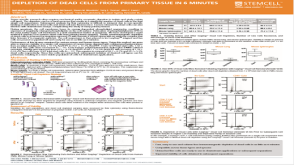 科学海报Depletion of Dead Cells from Primary Tissue in 6 Minutes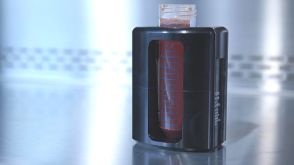

 沪公网安备31010102008431号