Inoue S et al. (AUG 2006)
Cancer research 66 15 7741--7
Inhibitory effects of B cells on antitumor immunity.
B-cell functions in antitumor immunity are not well understood. In this study,we evaluated the role of B cells in the development of antitumor immunity using Friend murine leukemia virus gag-expressing mouse EL-4 (EL-4 gag),D5 mouse melanoma,or MCA304 mouse sarcoma cells. To screen tumors for susceptibility to B-cell-deficient immune environments,spleen cells from naive C57BL/6 [wild-type (WT)] and B-cell knockout (BKO) mice were cultured with irradiated tumor cells in vitro. When cells were stimulated with EL-4 gag or D5 (but not MCA304 tumors),IFN-gamma production from CD8 T cells and natural killer cells was markedly decreased in WT compared with BKO cultures. IFN-gamma production was correlated with CD40 ligand expression on the tumor and inversely with interleukin-10 (IL-10) production by B cells. Sorted WT B cells produced more IL-10 than CD40 knockout (CD40KO) B cells when cocultured with EL-4 gag or D5 (but not MCA304). IFN-gamma production by BKO cells was reduced by the addition of sorted naive WT B cells (partially by CD40KO B cells) or recombinant mouse IL-10. In vivo tumor progression mirrored in vitro studies in that WT mice were unable to control tumor growth whereas EL-4 gag and D5 tumors (but not MCA304) were eliminated in BKO mice. Robust in vivo antitumor CTLs developed only in BKO tumor-challenged mice. Our studies provide the first mechanistic basis for the concept that B-cell depletion could therapeutically enhance antitumor immune responses to certain tumors by decreasing IL-10 production from B cells.
View Publication
产品类型:
产品号#:
18754
18754RF
产品名:
Suto A et al. (JUN 2008)
The Journal of experimental medicine 205 6 1369--79
Development and characterization of IL-21-producing CD4+ T cells.
It has recently been shown that interleukin (IL)-21 is produced by Th17 cells,functions as an autocrine growth factor for Th17 cells,and plays critical roles in autoimmune diseases. In this study,we investigated the differentiation and characteristics of IL-21-producing CD4(+) T cells by intracellular staining. Unexpectedly,we found that under Th17-polarizing conditions,the majority of IL-21-producing CD4(+) T cells did not produce IL-17A and -17F. We also found that IL-6 and -21 potently induced the development of IL-21-producing CD4(+) T cells without the induction of IL-4,IFN-gamma,IL-17A,or IL-17F production. On the other hand,TGF-beta inhibited IL-6- and IL-21-induced development of IL-21-producing CD4(+) T cells. IL-2 enhanced the development of IL-21-producing CD4(+) T cells under Th17-polarizing conditions. Finally,IL-21-producing CD4(+) T cells exhibited a stable phenotype of IL-21 production in the presence of IL-6,but retained the potential to produce IL-4 under Th2-polarizing conditions and IL-17A under Th17-polarizing conditions. These results suggest that IL-21-producing CD4(+) T cells exhibit distinct characteristics from Th17 cells and develop preferentially in an IL-6-rich environment devoid of TGF-beta,and that IL-21 functions as an autocrine growth factor for IL-21-producing CD4(+) T cells.
View Publication
产品类型:
产品号#:
21000
20119
20155
19752
19752RF
产品名:
RoboSep™- S
RoboSep™ 吸头组件抛光剂
RoboSep™分选管套装(9个塑料管)
Ellestad KK et al. (JUL 2009)
Journal of immunology (Baltimore,Md. : 1950) 183 1 298--309
Early life exposure to lipopolysaccharide suppresses experimental autoimmune encephalomyelitis by promoting tolerogenic dendritic cells and regulatory T cells.
The rising incidence of autoimmune diseases such as multiple sclerosis (MS) in developed countries might be due to a more hygienic environment,particularly during early life. To investigate this concept,we developed a model of neonatal exposure to a common pathogen-associated molecular pattern,LPS,and determined its impact on experimental autoimmune encephalomyelitis (EAE). Mice exposed to LPS at 2 wk of age showed a delayed onset and diminished severity of myelin oligodendrocyte glycoprotein (MOG)-induced EAE,induced at 12 wk,compared with vehicle-exposed animals. Spinal cord transcript levels of CD3epsilon and F4/80 were lower in LPS- compared with PBS-exposed EAE animals with increased IL-10 levels in the LPS-exposed group. Splenic CD11c(+) cells from LPS-exposed animals exhibited reduced MHC class II and CD83 expression but increased levels of CD80 and CD86 both before and during EAE. MOG-treated APC from LPS-exposed animals stimulated less T lymphocyte proliferation but increased expansion of CD4(+)FoxP3(+) T cells compared with APC from PBS-exposed animals. Neuropathological studies disclosed reduced myelin and axonal loss in spinal cords from LPS-exposed compared with PBS-exposed animals with EAE,and this neuroprotective effect was associated with an increased number of CD3(+)FoxP3(+) immunoreactive cells. Analyses of human brain tissue revealed that FoxP3 expression was detected in lymphocytes,albeit reduced in MS compared with non-MS patients' brains. These findings support the concept of early-life microbial exposure influencing the generation of neuroprotective regulatory T cells and may provide insights into new immunotherapeutic strategies for MS.
View Publication
产品类型:
产品号#:
18758
18758RF
18768
18768RF
产品名:
H.-W. Wu et al. (may 2019)
Clinical cancer research : an official journal of the American Association for Cancer Research
Anti-CD105 Antibody Eliminates Tumor Microenvironment Cells and Enhances Anti-GD2 Antibody Immunotherapy of Neuroblastoma with Activated Natural Killer Cells.
Purpose: We determined whether elimination of CD105+ cells in the tumor microenvironment (TME) with anti-CD105 antibodies enhanced anti-disialoganglioside (GD2) antibody dinutuximab therapy of neuroblastoma when combined with activated natural killer (aNK) cells.Experimental Design: The effect of MSCs and monocytes on antibody-dependent cellular cytotoxicity (ADCC) mediated by dinutuximab with aNK cells against neuroblastoma cells was determined in vitro. ADCC with anti-CD105 mAb TRC105 and aNK cells against MSCs,monocytes,and endothelial cells,which express CD105,was evaluated. Anti-neuroblastoma activity in immunodeficient NSG mice of dinutuximab with aNK cells without or with anti-CD105 mAbs was determined using neuroblastoma cell lines and a patient-derived xenograft.Results: ADCC mediated by dinutuximab with aNK cells against neuroblastoma cells in vitro was suppressed by addition of MSCs and monocytes,and dinutuximab with aNK cells was less effective against neuroblastomas formed with coinjected MSCs and monocytes in NSG mice than against those formed by tumor cells alone. Anti-CD105 antibody TRC105 with aNK cells mediated ADCC against MSCs,monocytes,and endothelial cells. Neuroblastomas formed in NSG mice by two neuroblastoma cell lines or a patient-derived xenograft coinjected with MSCs and monocytes were most effectively treated with dinutuximab and aNK cells when anti-human (TRC105) and anti-mouse (M1043) CD105 antibodies were added,which depleted human MSCs and murine endothelial cells and macrophages from the TME.Conclusions: Immunotherapy of neuroblastoma with anti-GD2 antibody dinutuximab and aNK cells is suppressed by CD105+ cells in the TME,but suppression is overcome by adding anti-CD105 antibodies to eliminate CD105+ cells.
View Publication
产品类型:
产品号#:
19359
19359RF
18000
100-0697
产品名:
EasySep™人单核细胞分选试剂盒
RoboSep™ 人单核细胞分选试剂盒
EasySep™磁极
EasySep™人单核细胞分选试剂盒
Guilliams M et al. (MAR 2010)
Blood 115 10 1958--68
Skin-draining lymph nodes contain dermis-derived CD103(-) dendritic cells that constitutively produce retinoic acid and induce Foxp3(+) regulatory T cells.
Small intestinal CD103(+) dendritic cells (DCs) have the selective ability to promote de novo generation of regulatory T cells via the production of retinoic acid (RA). Considering that aldehyde dehydrogenase (ALDH) activity controls the production of RA,we used a flow cytometry-based assay to measure ALDH activity at the single-cell level and to perform a comprehensive analysis of the RA-producing DC populations present in lymphoid and nonlymphoid mouse tissues. RA-producing DCs were primarily of the tissue-derived,migratory DC subtype and can be readily found in the skin and in the lungs as well as in their corresponding draining lymph nodes. The RA-producing skin-derived DCs were capable of triggering the generation of regulatory T cells,a finding demonstrating that the presence of RA-producing,tolerogenic DCs is not restricted to the intestinal tract as previously thought. Unexpectedly,the production of RA by skin DCs was restricted to CD103(-) DCs,indicating that CD103 expression does not constitute a universal" marker for RA-producing mouse DCs. Finally�
View Publication
产品类型:
产品号#:
01700
01705
01702
产品名:
ALDEFLUOR™ 试剂盒
ALDEFLUOR™ DEAB试剂, 1.5 mM, 1 mL
ALDEFLUOR™检测缓冲液
Glinka Y et al. (JUL 2008)
Journal of leukocyte biology 84 1 302--10
Neuropilin-1 is a receptor for transforming growth factor beta-1, activates its latent form, and promotes regulatory T cell activity.
Neuropilin-1 (Nrp1) is a multifunctional protein,identified principally as a receptor for the class 3 semaphorins and members of the vascular endothelial growth factor (VEGF) family,but it is capable of other interactions. It is a marker of regulatory T cells (Tr),which often carry Nrp1 and latency-associated peptide (LAP)-TGF-beta1 (the latent form). The signaling TGF-beta1 receptors bind only active TGF-beta1,and we hypothesized that Nrp1 binds the latent form. Indeed,we found that Nrp1 is a high-affinity receptor for latent and active TGF-beta1. Free LAP,LAP-TGF-beta1,and active TGF-beta1 all competed with VEGF165 for binding to Nrp1. LAP has a basic,arginine-rich C-terminal motif similar to VEGF and peptides that bind to the b1 domain of Nrp1. A C-terminal LAP peptide (QSSRHRR) bound to Nrp1 and inhibited the binding of VEGF and LAP-TGF-beta1. We also analyzed the effects of Nrp1/LAP-TGF-beta1 coexpression on T cell function. Compared with Nrp1(-) cells,sorted Nrp1+ T cells had a much greater capacity to capture LAP-TGF-beta1. Sorted Nrp1(-) T cells captured soluble Nrp1-Fc,and this increased their ability to capture LAP-TGF-beta1. Conventional CD4+CD25(-)Nrp1(-) T cells coated with Nrp1-Fc/LAP-TGF-beta1 acquired strong Tr activity. Moreover,LAP-TGF-beta was activated by Nrp1-Fc and also by a peptide of the b2 domain of Nrp1 (RKFK; similar to a thrombospondin-1 peptide). Breast cancer cells,which express Nrp1,also captured and activated LAP-TGF-beta1 in a Nrp1-dependent manner. Thus,Nrp1 is a receptor for TGF-beta1,activates its latent form,and is relevant to Tr activity and tumor biology.
View Publication
产品类型:
产品号#:
19752
19752RF
产品名:
Collins SM et al. (DEC 2013)
Cancer immunology,immunotherapy : CII 62 12 1841--9
Elotuzumab directly enhances NK cell cytotoxicity against myeloma via CS1 ligation: evidence for augmented NK cell function complementing ADCC.
Elotuzumab is a monoclonal antibody in development for multiple myeloma (MM) that targets CS1,a cell surface glycoprotein expressed on MM cells. In preclinical models,elotuzumab exerts anti-MM efficacy via natural killer (NK)-cell-mediated antibody-dependent cellular cytotoxicity (ADCC). CS1 is also expressed at lower levels on NK cells where it acts as an activating receptor. We hypothesized that elotuzumab may have additional mechanisms of action via ligation of CS1 on NK cells that complement ADCC activity. Herein,we show that elotuzumab appears to induce activation of NK cells by binding to NK cell CS1 which promotes cytotoxicity against CS1(+) MM cells but not against autologous CS1(+) NK cells. Elotuzumab may also promote CS1-CS1 interactions between NK cells and CS1(+) target cells to enhance cytotoxicity in a manner independent of ADCC. NK cell activation appears dependent on differential expression of the signaling intermediary EAT-2 which is present in NK cells but absent in primary,human MM cells. Taken together,these data suggest elotuzumab may enhance NK cell function directly and confer anti-MM efficacy by means beyond ADCC alone.
View Publication
产品类型:
产品号#:
18387
18387RF
产品名:
Mathers AR et al. (JAN 2009)
Journal of immunology (Baltimore,Md. : 1950) 182 2 921--33
Differential capability of human cutaneous dendritic cell subsets to initiate Th17 responses.
Human skin-migratory dendritic cells (DCs) have the ability to prime and bias Th1 and Th2 CD4+ T lymphocytes. However,whether human cutaneous DCs are capable of initiating proinflammatory Th17 responses remains undetermined. We report that skin-migratory DCs stimulate allogeneic naive CD4+ T cells that differentiate simultaneously into two distinct effector Th17 and Th1 populations capable of homing to the skin,where they induce severe cutaneous damage. Skin-migratory Langerhans cells (smiLCs) were the main cutaneous DC subset capable of inducing Th17 responses dependent on the combined effects of IL-15 and stabilized IL-6,which resulted in IL-6 trans-signaling of naive CD4+ T cells. Different from smiLCs,purified skin-migratory dermal DCs did not synthesize IL-15 and were unable to bias Th17 responses. Nevertheless,these dermal DCs were capable of differentiating Th17 cells in mixed leukocyte cultures supplemented with IL-15 and stabilized IL-6. Overall,our data demonstrate that human epidermal smiLCs induce Th17 responses by mechanisms different from those previously described and highlight the need to target clinical treatments based on these variations.
View Publication

 EasySep™小鼠TIL(CD45)正选试剂盒


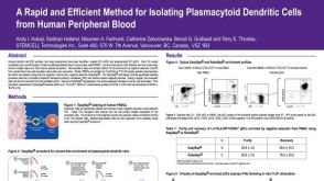 科学海报Isolation of Plasmacytoid Dendritic Cells from Peripheral Blood
 技术公告Isolating Extracellular Vesicles from Urine with EasySep™ EV Human Positive Selection Kits
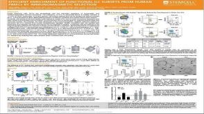 科学海报Efficient Enrichment of Functional ILC Subsets from Human PBMCs by Immunomagnetic Selection

 沪公网安备31010102008431号