
 EasySep™小鼠TIL(CD45)正选试剂盒


搜索结果: 'EasySep'
-
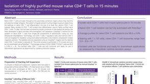 科学海报Immunomagnetic Cell Isolation of Highly Purified Mouse Naïve CD4+ T Cells in 15 Minutes
产品类型:
Conference:
CSI 2013
产品号#:
19765
19765RF
18000
产品名:
EasySep™小鼠Naïve CD4+ T细胞分选试剂盒
RoboSep™ 小鼠Naïve CD4+ T细胞分选试剂盒
EasySep™磁极
-
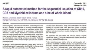 科学海报Isolation of CD19, CD3 and Myeloid Cells from one Tube of Whole Blood
产品类型:
Conference:
AAI 2007
产品号#:
21000
20155
20119
18081
18081RF
18084
18084RF
18683
18683RF
产品名:
RoboSep™- S
RoboSep™分选管套装(9个塑料管)
RoboSep™ 吸头组件抛光剂
-
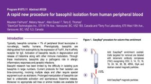 科学海报Basophil Isolation from Human Peripheral Blood
产品类型:
Conference:
AAI 2008
产品号#:
19069
19069RF
产品名:
-
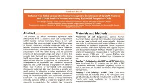 科学海报Column-Free Enrichment of EPCAM Positive and CD49F Positive Human Mammary Epithelial Progenitor Cells
产品类型:
Conference:
AACR 2003
产品号#:
18356
18356RF
18359
产品名:
-
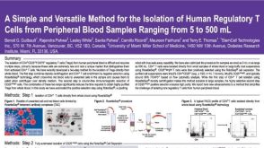 科学海报Isolation of Human Regulatory T Cells from Peripheral Blood Samples
产品类型:
Conference:
KEYSTONE 2007
产品号#:
21000
20155
20119
15862
15862RF
产品名:
RoboSep™- S
RoboSep™分选管套装(9个塑料管)
RoboSep™ 吸头组件抛光剂
-
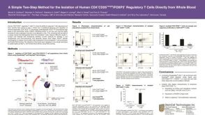 科学海报Isolation of Human CD4+CD25+Bright/Foxp3+ Regulatory T Cells Directly from Whole Blood
产品类型:
Conference:
ASH 2005,BSI 2005
产品号#:
15862
15862RF
产品名:
-
-
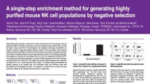 科学海报Cell Enrichment to Obtain Mouse NK Cell Populations By Negative Selection
产品类型:
Conference:
AAI 2008,CSI 2008
产品号#:
产品名:


 沪公网安备31010102008431号