M. Epeldegui et al. (jun 2019)
Scientific reports 9 1 9371
Elevated numbers of PD-L1 expressing B cells are associated with the development of AIDS-NHL.
The risk for non-Hodgkin lymphoma (NHL) is markedly increased in persons living with human immunodeficiency virus (HIV) infection,and remains elevated in those on anti-retroviral therapy (cART). Both the loss of immunoregulation of Epstein-Barr virus (EBV) infected cells,as well as chronic B-cell activation,are believed to contribute to the genesis of AIDS-related NHL (AIDS-NHL). However,the mechanisms that lead to AIDS-NHL have not been completely defined. A subset of B cells that is characterized by the secretion of IL10,as well as the expression of the programmed cell death ligand-1 (PD-L1/CD274),was recently described. These PD-L1+ B cells can exert regulatory function,including the dampening of T-cell activation,by interacting with the program cell death protein (PD1) on target cells. The role of PD-L1+ B cells in the development of AIDS-NHL has not been explored. We assessed B cell PD-L1 expression on B cells preceding AIDS-NHL diagnosis in a nested case-control study of HIV+ subjects who went on to develop AIDS-NHL,as well as HIV+ subjects who did not,using multi-color flow cytometry. Archival frozen viable PBMC were obtained from the UCLA Multicenter AIDS Cohort Study (MACS). It was seen that the number of CD19+CD24++CD38++and CD19+PD-L1+cells was significantly elevated in cases 1-4 years prior to AIDS-NHL diagnosis,compared to controls,raising the possibility that these cells may play a role in the etiology of AIDS-NHL. Interestingly,most PD-L1+ expression on CD19+ cells was seen on CD19+CD24++CD38++ cells. In addition,we showed that HIV can directly induce PD-L1 expression on B cells through interaction of virion-associated CD40L with CD40 on B cells.
View Publication
产品类型:
产品号#:
15024
15064
产品名:
RosetteSep™人B细胞富集抗体混合物
RosetteSep™人B细胞富集抗体混合物
Schlecht G et al. (SEP 2004)
Blood 104 6 1808--15
Murine plasmacytoid dendritic cells induce effector/memory CD8+ T-cell responses in vivo after viral stimulation.
Like their human counterparts,mouse plasmacytoid dendritic cells (pDCs) play a central role in innate immunity against viral infections,but their capacity to prime T cells in vivo remains unknown. We show here that virus-activated pDCs differentiate into antigen-presenting cells able to induce effector/memory CD8(+) T-cell responses in vivo against both epitopic peptides and endogenous antigen,whereas pDCs activated by synthetic oligodeoxynucleotides containing unmethylated cytosine-guanine motifs (CpG) acquire only the ability to recall antigen-experienced T-cell responses. We also show that immature pDCs are unable to induce effector or regulatory CD8(+) T-cell responses. Thus,murine pDCs take part in both innate and adaptive immune responses by directly priming naive CD8(+) T cells during viral infection.
View Publication
产品类型:
产品号#:
09600
09650
产品名:
StemSpan™ SFEM
StemSpan™ SFEM
Blanco J et al. (DEC 2004)
The Journal of biological chemistry 279 49 51305--14
High level of coreceptor-independent HIV transfer induced by contacts between primary CD4 T cells.
Cell-to-cell virus transmission is one of the most efficient mechanisms of human immunodeficiency virus (HIV) spread,requires CD4 and coreceptor expression in target cells,and may also lead to syncytium formation and cell death. Here,we show that in addition to this classical coreceptor-mediated transmission,the contact between HIV-producing cells and primary CD4 T cells lacking the appropriate coreceptor induced the uptake of HIV particles by target cells in the absence of membrane fusion or productive HIV replication. HIV uptake by CD4 T cells required cellular contacts mediated by the binding of gp120 to CD4 and intact actin cytoskeleton. HIV antigens taken up by CD4 T cells were rapidly endocytosed to trypsin-resistant compartments inducing a partial disappearance of CD4 molecules from the cell surface. Once the cellular contact was stopped,captured HIV were released as infectious particles. Electron microscopy revealed that HIV particles attached to the surface of target cells and accumulated in large (0.5-1.0 microm) intracellular vesicles containing 1-14 virions,without any evidence for massive clathrin-mediated HIV endocytosis. The capture of HIV particles into trypsin-resistant compartments required the availability of the gp120 binding site of CD4 but was independent of the intracytoplasmic tail of CD4. In conclusion,we describe a novel mechanism of HIV transmission,activated by the contact of infected and uninfected primary CD4 T cells,by which HIV could exploit CD4 T cells lacking the appropriate coreceptor as an itinerant virus reservoir.
View Publication
产品类型:
产品号#:
15022
15062
产品名:
RosetteSep™人CD4+ T细胞富集抗体混合物
RosetteSep™人CD4+ T细胞富集抗体混合物
Franç et al. (SEP 2009)
Blood 114 13 2632--8
Mesenchymal stromal cells cross-present soluble exogenous antigens as part of their antigen-presenting cell properties.
Recent studies involving bone marrow mesenchymal stromal cells (MSCs) demonstrated that interferon (IFN)-gamma stimulation induces major histocompatibility complex (MHC) class II-mediated antigen presentation in MSCs both in vitro and in vivo. Concordantly,we investigated the ability of MSCs to present extracellular antigen through their MHC class I molecules,a process known as cross-presentation. Using an in vitro antigen presentation assay,we demonstrated that murine MSCs can cross-present soluble ovalbumin (OVA) to naive CD8(+) T cells from OT-I mice. Cross-presentation by MSC was proteasome dependent and partly dependent on transporter associated with antigen-processing molecules. Pretreatment of MSC with IFN-gamma increased cross-presentation by up-regulating antigen processing and presentation. However,although the transcription of the transporter associated with antigen processing-1 molecules and the immunoproteasome subunit LMP2 induced by IFN-gamma was inhibited by transforming growth factor-beta,the overall cross-presentation capacity of MSCs remained unchanged after transforming growth factor-beta treatment. These observations were validated in vivo by performing an immune reconstitution assay in beta(2)-microglobulin(-/-) mice and show that OVA cross-presentation by MSCs induces the proliferation of naive OVA-specific CD8(+) T cells. In conclusion,we demonstrate that MSCs can cross-present exogenous antigen and induce an effective CD8(+) T-cell immune response,a property that could be exploited as a therapeutic cell-based immune biopharmaceutic for the treatment of cancer or infectious diseases.
View Publication
产品类型:
产品号#:
19753
19753RF
产品名:
Hou TZ et al. ( 2015)
The Journal of Immunology 194 5 2148--2159
A Transendocytosis Model of CTLA-4 Function Predicts Its Suppressive Behavior on Regulatory T Cells
Manipulation of the CD28/CTLA-4 pathway is at the heart of a number of immunomodulatory approaches used in both autoimmunity and cancer. Although it is clear that CTLA-4 is a critical regulator of T cell responses,the immunological contexts in which CTLA-4 controls immune responses are not well defined. In this study,we show that whereas CD80/CD86-dependent activation of resting human T cells caused extensive T cell proliferation and robust CTLA-4 expression,in this context CTLA-4 blocking Abs had no impact on the response. In contrast,in settings where CTLA-4(+) cells were present as regulators�
View Publication
Ammirati E et al. (DEC 2008)
Arteriosclerosis,thrombosis,and vascular biology 28 12 2305--11
Expansion of T-cell receptor zeta dim effector T cells in acute coronary syndromes.
OBJECTIVE: The T-cell receptor zeta (TCR zeta)-chain is a master sensor and regulator of lymphocyte responses. Loss of TCR zeta-chain expression has been documented during infectious and inflammatory diseases and defines a population of effector T cells (TCR zeta(dim) T cells) that migrate to inflamed tissues. We assessed the expression and functional correlates of circulating TCR zeta(dim) T cells in coronary artery disease. METHODS AND RESULTS: We examined the expression of TCR zeta-chain by flow cytometry in 140 subjects. Increased peripheral blood CD4(+) TCR zeta(dim) T cells were found in patients with acute coronary syndromes (ACS,n=66; median 5.3%,interquartile 2.6 to 9.1% of total CD4(+) T cells; Ptextless0.0001) compared to chronic stable angina (CSA,n=32; 1.6%; 1.0 to 4.1%) and controls (n=42; 1.5%; 0.5 to 2.9%). Such increase was significantly greater in ACS patients with elevated levels of C-reactive protein,and it persisted after the acute event. Moreover,TCR zeta(dim) cells were also more represented within CD8(+) T cell,NK,and CD4(+)CD28(null) T cell subsets in ACS compared to CSA and controls. Finally,CD4(+) and CD8(+) TCR zeta(dim) T cells isolated from ACS displayed an enhanced transendothelial migratory capacity. CONCLUSIONS: TCR zeta(dim) T cells,an effector T-cell subset with transendothelial migratory ability,are increased in ACS,and may be implicated in coronary instability.
View Publication
产品类型:
产品号#:
19051
19051RF
产品名:
EasySep™人T细胞富集试剂盒
RoboSep™ 人T细胞富集试剂盒含滤芯吸头
Hagness M et al. ( 2012)
The Journal of Immunology 188 11 5459--66
Kinetics and activation requirements of contact-dependent immune suppression by human regulatory T cells
Naturally occurring regulatory T cells (Tregs) maintain self tolerance by dominant suppression of potentially self-reactive T cells in peripheral tissues. However,the activation requirements,the temporal aspects of the suppressive activity,and mode of action of human Tregs are subjects of controversy. In this study,we show that Tregs display significant variability in the suppressive activity ex vivo as 54% of healthy blood donors examined had fully suppressive Tregs spontaneously,whereas in the remaining donors,anti-CD3/CD2/CD28 stimulation was required for Treg suppressive activity. Furthermore,anti-CD3/CD2/CD28 stimulation for 6 h and subsequent fixation in paraformaldehyde rendered the Tregs fully suppressive in all donors. The fixation-resistant suppressive activity of Tregs operated in a contact-dependent manner that was not dependent on APCs,but could be fully obliterated by trypsin treatment,indicating that a cell surface protein is directly involved. By add-back of active,fixed Tregs at different time points after activation of responding T cells,the responder cells were susceptible to Treg-mediated immune suppression up to 24 h after stimulation. This defines a time window in which effector T cells are susceptible to Treg-mediated immune suppression. Lastly,we examined the effect of a set of signaling inhibitors that perturb effector T cell activation and found that none of the examined inhibitors affected Treg activation,indicating pathway redundancy or that Treg activation proceeds by signaling mechanisms distinct from those of effector T cells.
View Publication


 EasySep™小鼠TIL(CD45)正选试剂盒




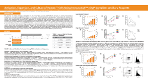 科学海报Activation, Expansion, and Culture of Human T Cells Using ImmunoCult™ cGMP-Compliant Ancillary Reagents
 实验方案Gene Editing Human Peripheral Blood Natural Killer Cells Using the CellPore™ Transfection System
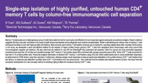 科学海报Cell Isolation of Human CD4+ Memory T Cells By Column-Free Immunomagnetic Cell Separation

 沪公网安备31010102008431号