Diversification of human plasmacytoid predendritic cells in response to a single stimulus.
Innate immune cells adjust to microbial and inflammatory stimuli through a process termed environmental plasticity,which links a given individual stimulus to a unique activated state. Here,we report that activation of human plasmacytoid predendritic cells (pDCs) with a single microbial or cytokine stimulus triggers cell diversification into three stable subpopulations (P1-P3). P1-pDCs (PD-L1+CD80-) displayed a plasmacytoid morphology and specialization for type I interferon production. P3-pDCs (PD-L1-CD80+) adopted a dendritic morphology and adaptive immune functions. P2-pDCs (PD-L1+CD80+) displayed both innate and adaptive functions. Each subpopulation expressed a specific coding- and long-noncoding-RNA signature and was stable after secondary stimulation. P1-pDCs were detected in samples from patients with lupus or psoriasis. pDC diversification was independent of cell divisions or preexisting heterogeneity within steady-state pDCs but was controlled by a TNF autocrine and/or paracrine communication loop. Our findings reveal a novel mechanism for diversity and division of labor in innate immune cells.
View Publication
NAP-2 Secreted by Human NK Cells Can Stimulate Mesenchymal Stem/Stromal Cell Recruitment.
Strategies for improved homing of mesenchymal stem cells (MSCs) to a place of injury are being sought and it has been shown that natural killer (NK) cells can stimulate MSC recruitment. Here,we studied the chemokines behind this recruitment. Assays were performed with bone marrow human MSCs and NK cells freshly isolated from healthy donor buffy coats. Supernatants from MSC-NK cell co-cultures can induce MSC recruitment but not to the same extent as when NK cells are present. Antibody arrays and ELISA assays confirmed that NK cells secrete RANTES (CCL5) and revealed that human NK cells secrete NAP-2 (CXCL7),a chemokine that can induce MSC migration. Inhibition with specific antagonists of CXCR2,a receptor that recognizes NAP-2,abolished NK cell-mediated MSC recruitment. This capacity of NK cells to produce chemokines that stimulate MSC recruitment points toward a role for this immune cell population in regulating tissue repair/regeneration.
View Publication
产品类型:
产品号#:
19055
19055RF
产品名:
EasySep™人NK细胞富集试剂盒
RoboSep™ 人NK细胞富集试剂盒含滤芯吸头
Mian MF et al. (JUL 2010)
Molecular therapy : the journal of the American Society of Gene Therapy 18 7 1379--88
FimH can directly activate human and murine natural killer cells via TLR4.
Although the importance of natural killer (NK) cells in innate immune responses against tumors or viral infections are well documented,their ability to directly recognize pathogens is less well defined. We have recently reported FimH,a bacterial fimbrial protein,as a novel Toll-like receptor (TLR)4 ligand that potently induces antiviral responses. Here,we investigated whether FimH either directly or indirectly can activate human and murine NK cells. We demonstrate that FimH potently activates both human and murine NK cells in vitro to induce cytokines [interferon (IFN)-gamma and tumor necrosis factor (TNF)-alpha] and cytotoxicity. Importantly,NK cells directly recognize FimH-expressing pathogens as FimH(+),but not FimH(-),bacteria were able to activate human NK cells. FimH activation of NK cells required TLR4 and MyD88 signaling,as NK cells from both TLR4(-/-) and MyD88(-/-) mice as well as human NK-92 cells,which lack TLR4,were all unresponsive to FimH. In addition,TLR4 neutralization significantly abrogated the response of human NK cells to FimH. Activation of purified NK cells by FimH was independent of lipopolysaccharide (LPS) or other bacterial contaminations. These data demonstrate for the first time that highly purified NK cells directly recognize and respond to FimH via TLR4-MyD88 pathways to aid innate protection against cancer or microbial infections.
View Publication
产品类型:
产品号#:
19055
19055RF
产品名:
EasySep™人NK细胞富集试剂盒
RoboSep™ 人NK细胞富集试剂盒含滤芯吸头
Brusko TM et al. (JAN 2010)
PloS one 5 7 e11726
Human antigen-specific regulatory T cells generated by T cell receptor gene transfer.
BACKGROUND: Therapies directed at augmenting regulatory T cell (Treg) activities in vivo as a systemic treatment for autoimmune disorders and transplantation may be associated with significant off-target effects,including a generalized immunosuppression that may compromise beneficial immune responses to infections and cancer cells. Adoptive cellular therapies using purified expanded Tregs represents an attractive alternative to systemic treatments,with results from animal studies noting increased therapeutic potency of antigen-specific Tregs over polyclonal populations. However,current methodologies are limited in terms of the capacity to isolate and expand a sufficient quantity of endogenous antigen-specific Tregs for therapeutic intervention. Moreover,FOXP3+ Tregs fall largely within the CD4+ T cell subset and are thus routinely MHC class II-specific,whereas class I-specific Tregs may function optimally in vivo by facilitating direct tissue recognition. METHODOLOGY/PRINCIPAL FINDINGS: To overcome these limitations,we have developed a novel means for generating large numbers of antigen-specific Tregs involving lentiviral T cell receptor (TCR) gene transfer into in vitro expanded polyclonal natural Treg populations. Tregs redirected with a high-avidity class I-specific TCR were capable of recognizing the melanoma antigen tyrosinase in the context of HLA-A*0201 and could be further enriched during the expansion process by antigen-specific reactivation with peptide loaded artificial antigen presenting cells. These in vitro expanded Tregs continued to express FOXP3 and functional TCRs,and maintained the capacity to suppress conventional T cell responses directed against tyrosinase,as well as bystander T cell responses. Using this methodology in a model tumor system,murine Tregs designed to express the tyrosinase TCR effectively blocked antigen-specific effector T cell (Teff) activity as determined by tumor cell growth and luciferase reporter-based imaging. CONCLUSIONS/SIGNIFICANCE: These results support the feasibility of class I-restricted TCR transfer as a promising strategy to redirect the functional properties of Tregs and provide for a more efficacious adoptive cell therapy.
View Publication
产品类型:
产品号#:
15022
15062
15621
15661
产品名:
RosetteSep™人CD4+ T细胞富集抗体混合物
RosetteSep™人CD4+ T细胞富集抗体混合物
RosetteSep™人CD3去除抗体混合物
RosetteSep™人CD3去除抗体混合物
Tsang JY-S et al. (JUL 2006)
Journal of leukocyte biology 80 1 145--51
Altered proximal T cell receptor (TCR) signaling in human CD4+CD25+ regulatory T cells.
CD4+CD25+ regulatory T cells play an important role in peripheral tolerance. Upon T cell receptor (TCR)-mediated activation,the cells fail to proliferate but are induced to have a suppressor function. The intracellular signaling events that lead to their responses have not been elucidated. In this study,we have examined the proximal TCR signaling events in freshly isolated human CD4+CD25+ regulatory T cells after TCR ligation. In contrast to CD4+CD25- T cells,TCR ligation of CD4+CD25+ regulatory T cells by anti-CD3 cross-linking resulted in a lower calcium influx and extracellular signal-regulated kinase 1/2 phosphorylation. Examination of the CD3zeta chain phosphorylation status indicated that CD4+CD25+ regulatory T cells have poor phosphorylation of the protein and consequently,reduced recruitment of zeta-associated protein-70 to the TCR immunoreceptor tyrosine motif. The adaptor protein,Src homology 2 domain-containing leukocyte phosphoprotein of 76 kDa,which relays signals to downstream signaling components,also showed reduced phosphorylation,which correlated with reduced VAV guanine nucleotide exchange factors association. Consistent with other findings,the defect is accompanied with impaired actin cap formation,implicating a failure of actin remodeling of the cells. Together,our results demonstrate that CD4+CD25+ regulatory T cells have altered TCR proximal signaling pathways,which could be critical for inducing the distinct behavior of these cells.
View Publication
产品类型:
产品号#:
15022
15062
产品名:
RosetteSep™人CD4+ T细胞富集抗体混合物
RosetteSep™人CD4+ T细胞富集抗体混合物
Darce JR et al. (MAY 2007)
Journal of immunology (Baltimore,Md. : 1950) 178 9 5612--22
Divergent effects of BAFF on human memory B cell differentiation into Ig-secreting cells.
B cell-activating factor belonging to the TNF family (BAFF) plays a critical role in B cell maturation,yet its precise role in B cell differentiation into Ig-secreting cells (ISCs) remains unclear. In this study,we find that upon isolation human naive and memory B (MB) cells have prebound BAFF on their surface,whereas germinal center (GC) B cells lack detectable levels of prebound BAFF. We attribute their lack of prebound BAFF to cell activation,because we demonstrate that stimulation of naive and MB cells results in the loss of prebound BAFF. Furthermore,the absence of prebound BAFF on GC B cells is not related to a lack of BAFF-binding receptors or an inability to bind exogenous BAFF. Instead,our data suggest that accessibility to soluble BAFF is limited within GCs,perhaps to prevent skewing of the conventional B cell differentiation program. In support of this concept,whereas BAFF significantly enhances ISC differentiation in response to T cell-dependent activation,we report for the first time the ability of BAFF to considerably attenuate ISC differentiation of MB cells in response to CpG stimulation,a form of T cell-independent activation. Our data suggest that BAFF may be providing regulatory signals during specific T cell-independent events,which protect the balance between MB cells and ISCs outside GCs. Taken together,these data define a complex role for BAFF in humoral immune responses and show for the first time that BAFF can also play an inhibitory role in B cell differentiation.
View Publication
产品类型:
产品号#:
21000
20119
20155
19054
19054RF
产品名:
RoboSep™- S
RoboSep™ 吸头组件抛光剂
RoboSep™分选管套装(9个塑料管)
EasySep™人B细胞富集试剂盒
RoboSep™ 人B细胞富集试剂盒含滤芯吸头
Abdelwahab SF et al. (DEC 2003)
Proceedings of the National Academy of Sciences of the United States of America 100 25 15006--10
HIV-1-suppressive factors are secreted by CD4+ T cells during primary immune responses.
CD4+ T cells are required for immunity against many viral infections,including HIV-1 where a positive correlation has been observed between strong recall responses and low HIV-1 viral loads. Some HIV-1-specific CD4+ T cells are preferentially infected with HIV-1,whereas others escape infection by unknown mechanisms. One possibility is that some CD4+ T cells are protected from infection by the secretion of soluble HIV-suppressive factors,although it is not known whether these factors are produced during primary antigen-specific responses. Here,we show that soluble suppressive factors are produced against CXCR4 and CCR5 isolates of HIV-1 during the primary immune response of human CD4+ T cells. This activity requires antigenic stimulation of naïve CD4+ T cells. One anti-CXCR4 factor is macrophage-derived chemokine (chemokine ligand 22,CCL22),and anti-CCR5 factors include macrophage inflammatory protein-1 alpha (CCL3),macrophage inflammatory protein-1 beta (CCL4),and RANTES (regulated upon activation of normal T cells expressed and secreted) (CCL5). Intracellular staining confirms that CD3+CD4+ T cells are the source of the prototype HIV-1-inhibiting chemokines CCL22 and CCL4. These results show that CD4+ T cells secrete an evolving HIV-1-suppressive activity during the primary immune response and that this activity is comprised primarily of CC chemokines. The data also suggest that production of such factors should be considered in the design of vaccines against HIV-1 and as a mechanism whereby the host can control infections with this virus.
View Publication
产品类型:
产品号#:
09500
09600
09650
19155
19155RF
产品名:
BIT 9500血清替代物
StemSpan™ SFEM
StemSpan™ SFEM
Fang H et al. (APR 2005)
Journal of immunology (Baltimore,Md. : 1950) 174 8 4966--71
Anthrax lethal toxin blocks MAPK kinase-dependent IL-2 production in CD4+ T cells.
Anthrax lethal toxin (LT) is a critical virulence factor that cleaves and inactivates MAPK kinases (MAPKKs) in host cells and has been proposed as a therapeutic target in the treatment of human anthrax infections. Despite the potential use of anti-toxin agents in humans,the standard activity assays for anthrax LT are currently based on cytotoxic actions of anthrax LT that are cell-,strain-,and species-specific,which have not been demonstrated to occur in human cells. We now report that T cell proliferation and IL-2 production inversely correlate with anthrax LT levels in human cell assays. The model CD4+ T cell tumor line,Jurkat,is a susceptible target for the specific protease action of anthrax LT. Anthrax LT cleaves and inactivates MAPKKs in Jurkat cells,whereas not affecting proximal or parallel TCR signal transduction pathways. Moreover,anthrax LT specifically inhibits PMA/ionomycin- and anti-CD3-induced IL-2 production in Jurkat cells. An inhibitor of the protease activity of anthrax LT completely restores IL-2 production by anthrax LT-treated Jurkat cells. Anthrax LT acts on primary CD4+ T cells as well,cleaving MAPKKs and leading to a 95% reduction in anti-CD3-induced proliferation and IL-2 production. These findings not only will be useful in the development of new human cell-based bioassays for the activity of anthrax LT,but they also suggest new mechanisms that facilitate immune evasion by Bacillus anthracis. Specifically,anthrax LT inhibits IL-2 production and proliferative responses in CD4+ T cells,thereby blocking functions that are pivotal in the regulation of immune responses.
View Publication
产品类型:
产品号#:
15022
15062
产品名:
RosetteSep™人CD4+ T细胞富集抗体混合物
RosetteSep™人CD4+ T细胞富集抗体混合物
Lu Q et al. (DEC 2014)
PLoS ONE 9 12 e114949
Negligible immunogenicity of induced pluripotent stem cells derived from human skin fibroblasts
Human induced pluripotent stem cells (hiPSCs) have potential applications in cell replacement therapy and regenerative medicine. However,limited information is available regarding the immunologic features of iPSCs. In this study,expression of MHC and T cell co-stimulatory molecules in hiPSCs,and the effects on activation,proliferation and cytokine production in allogeneic human peripheral blood mononuclear cells were examined. We found that no-integrate hiPSCs had no MHC-II and T cell co-stimulatory molecules expressions but had moderate level of MHC-I and HLA-G expressions. In contrast to human skin fibroblasts (HSFs) which significantly induced allogeneic T cell activation and proliferation,hiPSCs failed to induce allogeneic CD45+ lymphocyte and CD8+ T cell activation and proliferation but could induce a low level of allogeneic CD4+ T cell proliferation. Unlike HSFs which induced allogeneic lymphocytes to produce high levels of IFN-γ,TNF-α and IL-17,hiPSCs only induced allogeneic lymphocytes to produce IL-2 and IL-10,and promote IL-10-secreting regulatory T cell (Treg) generation. Our study suggests that the integration-free hiPSCs had low or negligible immunogenicity,which may result from their induction of IL-10-secreting Treg.
View Publication
产品类型:
产品号#:
05850
05857
05870
05875
85850
85857
85870
85875
产品名:
mTeSR™1
mTeSR™1
E. Drent et al. (jul 2019)
Clinical cancer research : an official journal of the American Association for Cancer Research 25 13 4014--4025
Combined CD28 and 4-1BB Costimulation Potentiates Affinity-tuned Chimeric Antigen Receptor-engineered T Cells.
PURPOSE Targeting nonspecific,tumor-associated antigens (TAA) with chimeric antigen receptors (CAR) requires specific attention to restrict possible detrimental on-target/off-tumor effects. A reduced affinity may direct CAR-engineered T (CAR-T) cells to tumor cells expressing high TAA levels while sparing low expressing normal tissues. However,decreasing the affinity of the CAR-target binding may compromise the overall antitumor effects. Here,we demonstrate the prime importance of the type of intracellular signaling on the function of low-affinity CAR-T cells. EXPERIMENTAL DESIGN We used a series of single-chain variable fragments (scFv) with five different affinities targeting the same epitope of the multiple myeloma-associated CD38 antigen. The scFvs were incorporated in three different CAR costimulation designs and we evaluated the antitumor functionality and off-tumor toxicity of the generated CAR-T cells in vitro and in vivo. RESULTS We show that the inferior cytotoxicity and cytokine secretion mediated by CD38 CARs of very low-affinity (Kd {\textless} 1.9 × 10⁻⁶ mol/L) bearing a 4-1BB intracellular domain can be significantly improved when a CD28 costimulatory domain is used. Additional 4-1BB signaling mediated by the coexpression of 4-1BBL provided the CD28-based CD38 CAR-T cells with superior proliferative capacity,preservation of a central memory phenotype,and significantly improved in vivo antitumor function,while preserving their ability to discriminate target antigen density. CONCLUSIONS A combinatorial costimulatory design allows the use of very low-affinity binding domains (Kd {\textless} 1 mumol/L) for the construction of safe but also optimally effective CAR-T cells. Thus,very-low-affinity scFvs empowered by selected costimulatory elements can enhance the clinical potential of TAA-targeting CARs.
View Publication

 EasySep™小鼠TIL(CD45)正选试剂盒


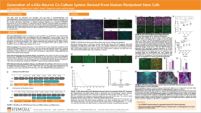 科学海报Generation of a Glia-Neuron Co-Culture System Derived From Human Pluripotent Stem Cells
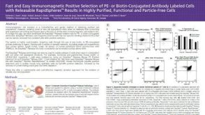 科学海报Positive Selection of PE- or Biotin-Conjugated Antibody Labeled Cells with Releasable Rapidspheres™
 沪公网安备31010102008431号