Tomihara K et al. (JUN 2010)
Journal of immunology (Baltimore,Md. : 1950) 184 11 6151--60
Antigen-specific immunity and cross-priming by epithelial ovarian carcinoma-induced CD11b(+)Gr-1(+) cells.
Both innate and adaptive immune systems are considered important for cancer prevention,immunosurveillance,and control of cancer progression. It is known that,although both systems initially eliminate emerging tumor cells efficiently,tumors eventually escape immune attack by a variety of mechanisms,including differentiation and recruitment of immunosuppressive CD11b(+)Gr-1(+) myeloid suppressor cells into the tumor microenvironment. However,we show that CD11b(+)Gr-1(+) cells found in ascites of epithelial ovarian cancer-bearing mice at advanced stages of disease are immunostimulatory rather than being immunosuppressive. These cells consist of a homogenous population of cells that morphologically resemble neutrophils. Moreover,like dendritic cells,immunostimulatory CD11b(+)Gr-1(+) cells can strongly cross-prime,augmenting the proliferation of functional CTLs via signaling through the expression of costimulatory molecule CD80. Adoptive transfer of these immunostimulatory CD11b(+)Gr-1(+) cells from ascites of ovarian cancer-bearing mice results in the significant regression of s.c. tumors even without being pulsed with exogenous tumor Ag prior to adoptive transfer. We now show for the first time that adaptive immune responses against cancer can be augmented by these cancer-induced granulocyte-like immunostimulatory myeloid (CD11b(+)Gr-1(+)) cells,thereby mediating highly effective antitumor immunity in an adoptive transfer model of immunity.
View Publication
The Intestine Harbors Functionally Distinct Homeostatic Tissue-Resident and Inflammatory Th17 Cells.
T helper 17 (Th17) cells are pathogenic in many inflammatory diseases,but also support the integrity of the intestinal barrier in a non-inflammatory manner. It is unclear what distinguishes inflammatory Th17 cells elicited by pathogens and tissue-resident homeostatic Th17 cells elicited by commensals. Here,we compared the characteristics of Th17 cells differentiating in response to commensal bacteria (SFB) to those differentiating in response to a pathogen (Citrobacter rodentium). Homeostatic Th17 cells exhibited little plasticity towards expression of inflammatory cytokines,were characterized by a metabolism typical of quiescent or memory T cells,and did not participate in inflammatory processes. In contrast,infection-induced Th17 cells showed extensive plasticity towards pro-inflammatory cytokines,disseminated widely into the periphery,and engaged aerobic glycolysis in addition to oxidative phosphorylation typical for inflammatory effector cells. These findings will help ensure that future therapies directed against inflammatory Th17 cells do not inadvertently damage the resident gut population.
View Publication
Goda C et al. (FEB 2006)
International immunology 18 2 233--40
Involvement of IL-32 in activation-induced cell death in T cells.
NK cell transcript 4 (NK4),now denoted as IL-32,was originally identified as a transcript whose expression was increased in activated NK cells. It has been very recently demonstrated that NK4 is secreted from several cells upon the stimulation of some inflammatory cytokines such as IL-18,IL-1beta,IFN-gamma and IL-12. Furthermore,NK4 induces production of tumor necrosis factor,macrophage inflammatory protein (MIP)-2 and IL-8 in monocytic cell lines,indicating that this factor would be involved in the inflammatory responses. Based on these findings,NK4 was renamed IL-32. However,the biological activities of IL-32 on other cell types remained undetermined. Furthermore,it was still argued whether IL-32 acts on cells from outside or inside the cells. In this article,we first report that expression of IL-32 was up-regulated in activated T cells and NK cells,and that IL-32beta was the predominantly expressed isoform in activated T cells. IL-32 was specifically expressed in T cells undergoing apoptosis and enforced expression of IL-32-induced apoptosis,whereas its down-regulation rescued the cells from apoptosis in HeLa cells. IL-32 existing in the supernatant would be derived from the cytoplasm of apoptotic cells. These results strongly indicated that IL-32 would be involved in activation-induced cell death in T cells,probably via its intracellular actions. Our present findings expand our understanding of the biological function of IL-32 and argue that IL-32 may act on cells,not only from the outside but also from the inside.
View Publication
产品类型:
产品号#:
15021
15061
15025
15065
产品名:
RosetteSep™人T细胞富集抗体混合物
RosetteSep™人T细胞富集抗体混合物
RosetteSep™人NK细胞富集抗体混合物
RosetteSep™人NK细胞富集抗体混合物
Schumann K et al. (AUG 2015)
Proceedings of the National Academy of Sciences of the United States of America 112 33 10437--42
Generation of knock-in primary human T cells using Cas9 ribonucleoproteins.
T-cell genome engineering holds great promise for cell-based therapies for cancer,HIV,primary immune deficiencies,and autoimmune diseases,but genetic manipulation of human T cells has been challenging. Improved tools are needed to efficiently knock out" genes and "knock in" targeted genome modifications to modulate T-cell function and correct disease-associated mutations. CRISPR/Cas9 technology is facilitating genome engineering in many cell types�
View Publication
产品类型:
产品号#:
17952
17952RF
100-0696
产品名:
EasySep™人CD4+ T细胞分选试剂盒
RoboSep™ 人CD4+ T细胞分选试剂盒
EasySep™人CD4+ T细胞分离试剂盒
Roybal KT et al. (FEB 2016)
Cell 164 4 770--9
Precision Tumor Recognition by T Cells With Combinatorial Antigen-Sensing Circuits.
T cells can be re-directed to kill cancer cells using chimeric antigen receptors (CARs) or T cell receptors (TCRs). This approach,however,is constrained by the rarity of tumor-specific single antigens. Targeting antigens also found on bystander tissues can cause life-threatening adverse effects. A powerful way to enhance ON-target activity of therapeutic T cells is to engineer them to require combinatorial antigens. Here,we engineer a combinatorially activated T cell circuit in which a synthetic Notch receptor for one antigen induces the expression of a CAR for a second antigen. These dual-receptor AND-gate T cells are only armed and activated in the presence of dual antigen tumor cells. These T cells show precise therapeutic discrimination in vivo-sparing single antigen bystander" tumors while efficiently clearing combinatorial antigen "disease" tumors. This type of precision dual-receptor circuit opens the door to immune recognition of a wider range of tumors. VIDEO ABSTRACT."
View Publication
产品类型:
产品号#:
15022
15062
15023
15063
产品名:
RosetteSep™人CD4+ T细胞富集抗体混合物
RosetteSep™人CD4+ T细胞富集抗体混合物
RosetteSep™人CD8+ T细胞富集抗体混合物
RosetteSep™人CD8+ T细胞富集抗体混合物
Watkins NA et al. (MAY 2009)
Blood 113 19 e1--9
A HaemAtlas: characterizing gene expression in differentiated human blood cells.
Hematopoiesis is a carefully controlled process that is regulated by complex networks of transcription factors that are,in part,controlled by signals resulting from ligand binding to cell-surface receptors. To further understand hematopoiesis,we have compared gene expression profiles of human erythroblasts,megakaryocytes,B cells,cytotoxic and helper T cells,natural killer cells,granulocytes,and monocytes using whole genome microarrays. A bioinformatics analysis of these data was performed focusing on transcription factors,immunoglobulin superfamily members,and lineage-specific transcripts. We observed that the numbers of lineage-specific genes varies by 2 orders of magnitude,ranging from 5 for cytotoxic T cells to 878 for granulocytes. In addition,we have identified novel coexpression patterns for key transcription factors involved in hematopoiesis (eg,GATA3-GFI1 and GATA2-KLF1). This study represents the most comprehensive analysis of gene expression in hematopoietic cells to date and has identified genes that play key roles in lineage commitment and cell function. The data,which are freely accessible,will be invaluable for future studies on hematopoiesis and the role of specific genes and will also aid the understanding of the recent genome-wide association studies.
View Publication
Carrera Silva EA et al. ( 2017)
Blood 130 17 1898--1902
CD207+CD1a+ cells circulate in pediatric patients with active Langerhans cell histiocytosis.
Langerhans cell histiocytosis (LCH) is a rare disease with an unknown etiology characterized by heterogeneous lesions containing CD207+CD1a+ cells that can arise in almost any tissue and cause significant morbidity and mortality. Precursors of pathological Langerhans cells have yet to be defined. Our aim was to identify circulating CD207+CD1a+ cells and their inducers in LCH. Expression of CD207 and CD1a in the blood myeloid compartment as well as thymic stromal lymphopoietin (TSLP) and transforming growth factor β (TGF-β) plasma levels were measured in 22 pediatric patients with active disease (AD) or nonactive disease (NAD). In patients with AD vs those with NAD,the myeloid compartment showed an increased CD11b (CD11bhigh plus CD11b+) fraction (39.7 ± 3.6 vs 18.6 ± 1.9),a higher percentage of circulating CD11bhighCD11c+CD207+ cells (44.5 ± 11.3 vs 3.2 ± 0.5),and the presence of CD11chighCD207+CD1a+ cells (25.0 ± 9.1 vs 2.3 ± 0.5). Blood CD207+CD1a+ cells were not observed in adult controls or umbilical cord. Increased TSLP and TGF-β levels were detected in patients with AD. Interestingly,plasma from patients with AD induces CD207 expression on CD14+ monocytes. We conclude that CD207+CD1a+ cells are circulating in patients with active LCH,and TSLP and TGF-β are potential drivers of Langerhans-like cells in vivo.
View Publication


 EasySep™小鼠TIL(CD45)正选试剂盒





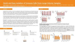 科学海报Quick and Easy Isolation of Immune Cells From Large-Volume Samples
 技术公告Co-Culturing Colorectal Organoids and T Cells using IntestiCult™ and ImmunoCult™
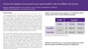 科学海报Immunomagnetic Cell Isolation of Human Gamma-Delta T Cells from PBMC
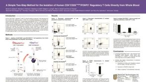 科学海报Isolation of Human CD4+CD25+Bright/Foxp3+ Regulatory T Cells Directly from Whole Blood
 沪公网安备31010102008431号